

fig6
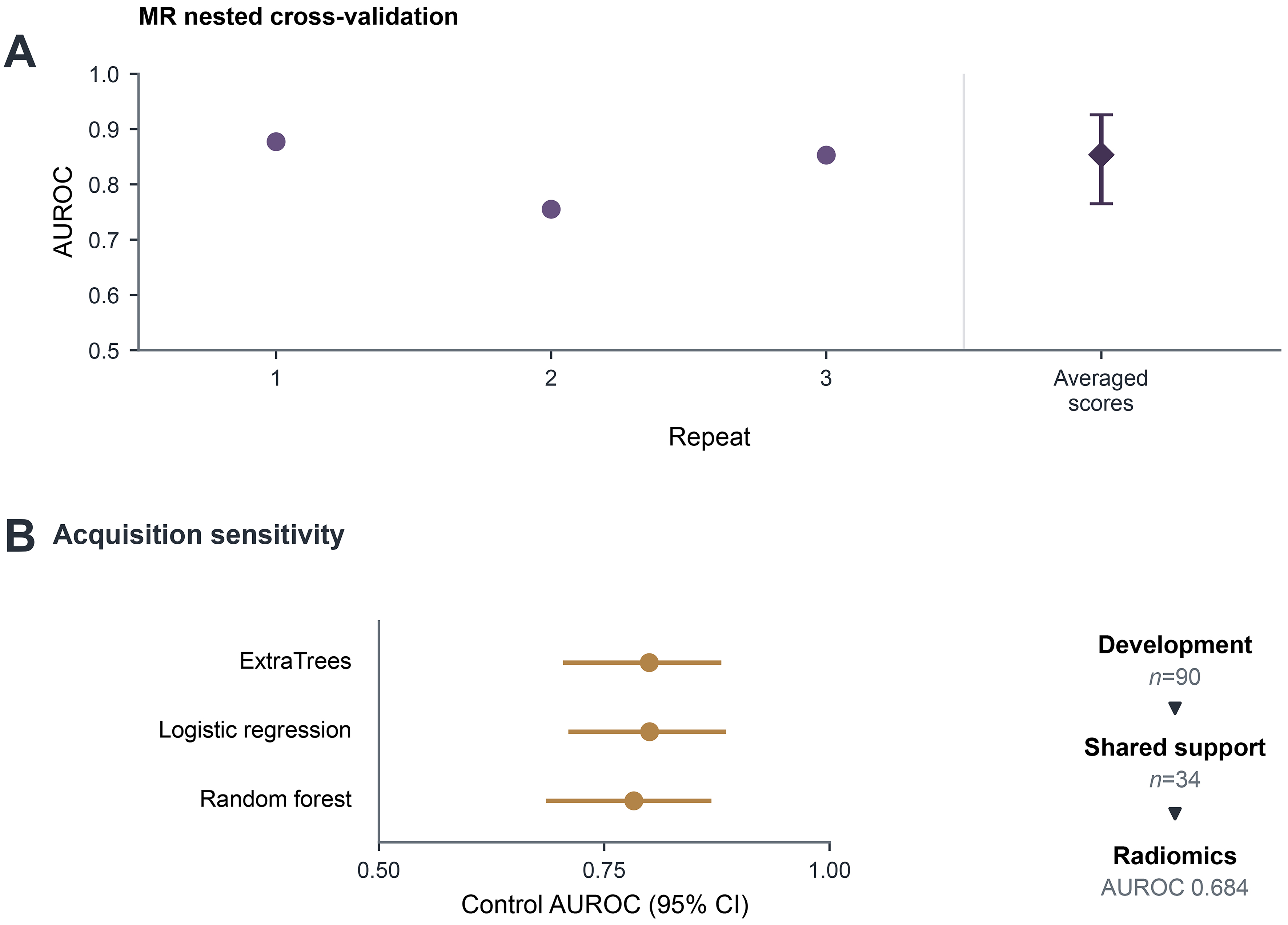
Figure 6. Sensitivity and robustness analyses. (A) Nested CV using 107 original radiomic features from 90 MR development patients, with three repeats of five outer folds. Circles show repeat-specific AUROCs. The diamond and interval show AUROC from each patient’s mean out-of-fold probability across repeats (0.854; bootstrap 95%CI: 0.765-0.926); (B) acquisition-only control AUROCs with 95%CIs, alongside the reduction to 34 patients whose acquisition signatures contain both study labels; nested radiomics AUROC in this subset was 0.684. The separate 16-patient MR holdout was excluded, but 14 development patients overlapped the original 33-patient holdout. These MR analyses use a separate feature representation and do not independently validate the original whole-tumor-plus-habitat model. Contour sensitivity, feature-selection stability, learning curves, and signature-level support are shown in Supplementary Figures 5-7. The study labels do not establish clinically verified infection status. AUROC: Area under the receiver operating characteristic curve; CI: confidence interval; CV: cross-validation; MR: magnetic resonance.