

fig1
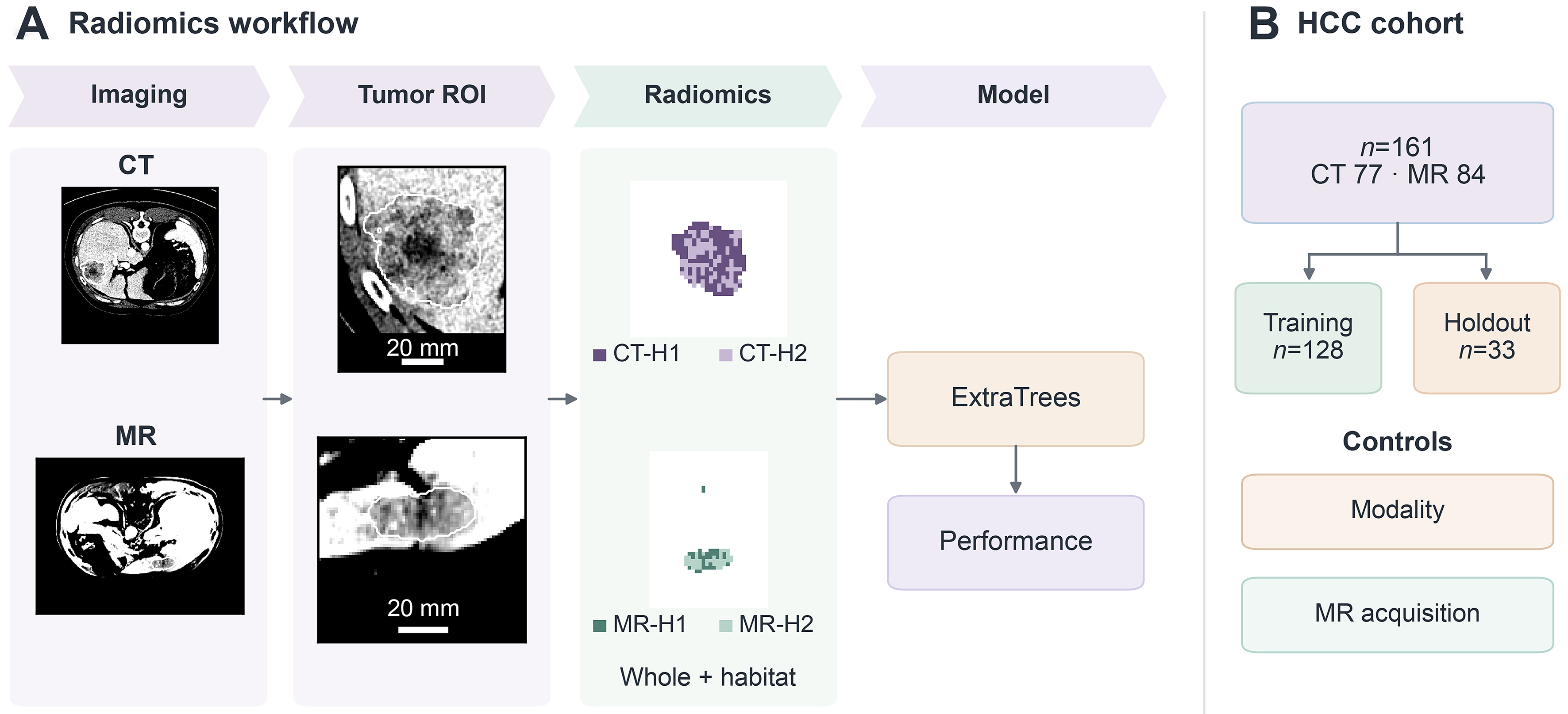
Figure 1. Study design. (A) CT and MR slices, tumor ROIs and two-cluster (K = 2) habitat maps illustrate whole-tumor and habitat feature extraction. Each patient contributes their available modality. Habitats were fitted separately by modality using patient-balanced training voxels; label-invariant summaries were combined with whole-tumor features for an ExtraTrees classifier. CT-H1/CT-H2 and MR-H1/MR-H2 are image-defined labels with no assumed cross-modality or histologic equivalence. ROI crops carry 20-mm bars derived from source spacing; maps retain their own processing grids without an assumed raw-to-map registration or scale bar. Display-case selection, intensity rescaling, and map cropping are described in Supplementary Methods; (B) the cohort comprised 161 patients with HCC, including 77 CT and 84 MR patients, split into training (n = 128) and internal holdout (n = 33) sets without shared patients. Modality-only and MR acquisition-only controls were compared with the image model on the same holdout. The endpoint is study-documented binary Cs status: labels 0 and 1 do not establish clinically verified absence or presence of active infection. The holdout was reused in exploratory analyses. CT: Computed tomography; Cs: Clonorchis sinensis; HCC: hepatocellular carcinoma; K: number of clusters; MR: magnetic resonance; ROI: region of interest; H1/H2: habitat 1/habitat 2.