



Pseudocirrhosis: pathophysiology, clinical mimics, and its role in hepatocellular carcinoma diagnosis and management
 0
0 
Abstract
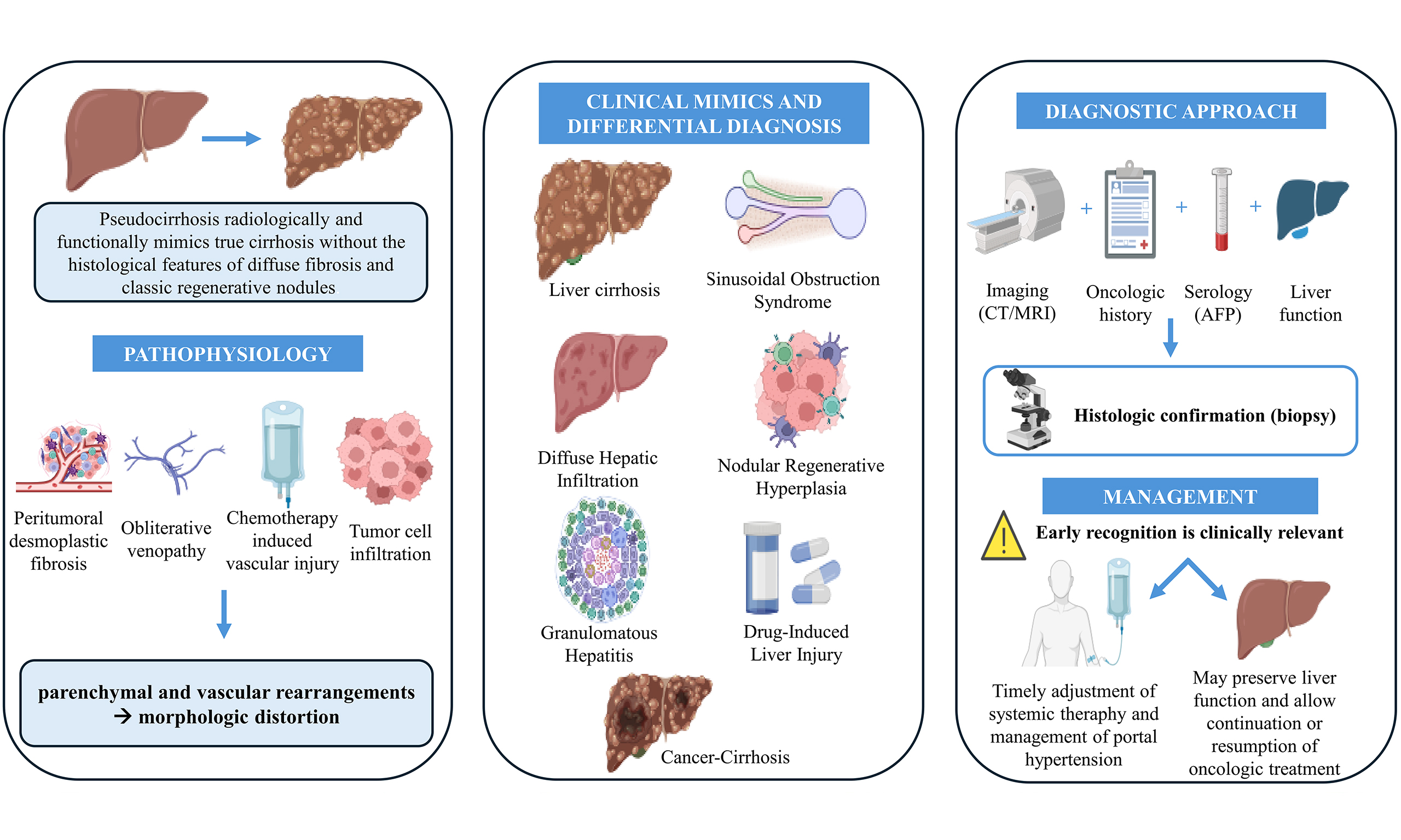
Pseudocirrhosis is an uncommon but clinically relevant condition that radiologically and functionally mimics true liver cirrhosis, despite the absence of the typical histological features of diffuse fibrosis and classic regenerative nodules. It most frequently occurs in patients with metastatic malignancies, particularly breast cancer, usually in the absence of known pre-existing chronic liver disease, and may progress rapidly, leading to morphologic liver distortion and clinically significant portal hypertension. Its pathophysiology is multifactorial, involving peritumoral desmoplastic fibrosis, chemotherapy-induced vascular injury with nodular regenerative hyperplasia and sinusoidal obstruction, as well as intra-sinusoidal or diffuse tumour cell infiltration with obliterative venopathy and endothelial injury. These mechanisms result in parenchymal and vascular rearrangements, often associated with portal hypertension, resembling those observed in chronic liver disease. Distinguishing pseudocirrhosis from similar conditions is critical to avoid misdiagnosis and inappropriate management. Several hepatic disorders share overlapping clinical and imaging findings, including vascular and granulomatous liver diseases, chronic drug-induced hepatotoxicity, and infiltrative malignancies. Accurate interpretation requires careful integration of oncologic history, treatment exposure, and specific imaging patterns, since standardized diagnostic criteria are lacking. One of the most insidious differential diagnoses is infiltrative hepatocellular carcinoma, which may exhibit diffuse parenchymal involvement, portal hypertension, and surface nodularity like pseudocirrhosis. When imaging is inconclusive and the diagnosis would influence clinical strategy, histologic confirmation is warranted, particularly when curative-intent or liver-directed therapies are being contemplated.
Keywords
INTRODUCTION
Pseudocirrhosis is an uncommon but clinically relevant entity that radiologically and clinically mimics liver cirrhosis in the absence of diffuse fibrosis and classic regenerative nodules[1]. It is most frequently observed in patients with metastatic malignancies, particularly breast carcinoma, usually without known pre-existing chronic liver disease, and may evolve rapidly, leading to portal hypertension and liver failure. Historically, the term hepar lobatum carcinomatosum was introduced in 1924 to describe lobulated hepatic contours at autopsy in a woman with breast cancer[2]. Decades later, in 1994, Young et al. proposed the term pseudocirrhosis based on clinical and radiologic features that reproduce the cirrhosis-like liver appearance in oncologic contexts[3]. Although the exact mechanisms are not fully defined, several processes likely contribute, including sinusoidal or intra-sinusoidal neoplastic infiltration, portal venous thrombosis, perisinusoidal fibrosis, alterations of intrahepatic lymphatics, and nodular regeneration[4]. These changes can end in clinically significant portal hypertension with ascites, varices, and splenomegaly[5].
Pseudocirrhosis has been described primarily in metastatic breast cancer, with additional reports in pancreatic, thyroid, oesophageal, and gastroesophageal neoplasia[6-8]. Standardized diagnostic criteria are lacking, and diagnosis is predominantly imaging-based. Early recognition and close monitoring are essential to anticipate complications of portal hypertension, including variceal bleeding and hepatic encephalopathy, which are associated with poor clinical outcomes and reduced survival[9,10].
Unlike cirrhosis, pseudocirrhosis can progress over a short time window (approximately 1-3 months). Typical imaging findings include surface nodularity, capsular retraction, segmental volume loss, and caudate lobe hypertrophy[11-14]. The differential diagnosis is broad and includes, among others, vascular liver disease, granulomatous liver disease and chronic drug-induced hepatotoxicity[15].
Therefore, this narrative review aims not only to summarize the pathophysiology and imaging features of pseudocirrhosis, but also to propose a practical diagnostic framework for clinicians facing a cirrhosis-like liver morphology in oncologic patients, with particular attention to the differential diagnosis with true cirrhosis and infiltrative hepatocellular carcinoma (HCC).
PATHOPHYSIOLOGY
The pathophysiology of pseudocirrhosis is multifactorial and not yet fully elucidated. Neoplastic cells may infiltrate the liver via arterial or portal routes, forming capillary-like networks or directly obstructing hepatic sinusoids[16,17]. Although classically recognized in hematologic malignancies, diffuse intra-sinusoidal spread can also occur in solid tumours, particularly in poorly differentiated breast cancer[18].
Three non-mutually exclusive mechanisms are commonly proposed: desmoplastic retraction, fibrotic scarring, and capsular retraction occurred in response to neoplastic infiltration or as a consequence of effective cytotoxic therapy around regressed metastases; nodular regenerative hyperplasia (NRH) secondary to chemotherapy-related ischemia and sinusoidal injury; sinusoidal or venular obstruction due to microscopic neoplastic infiltration or chemotherapy-related vascular injury[14,19-22] [Figure 1].
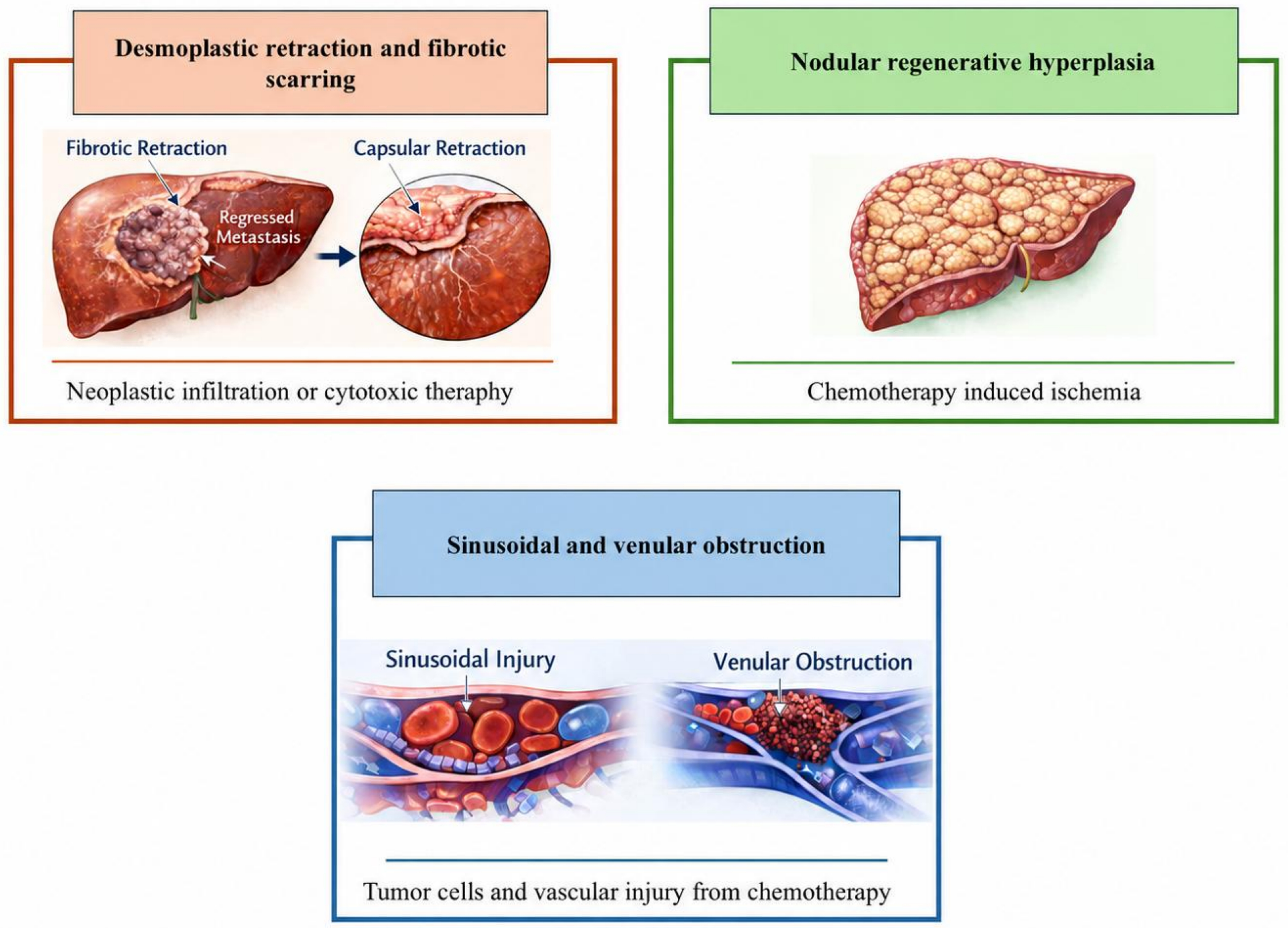
Figure 1. Schematic representation of the main mechanisms involved in pseudocirrhosis. Desmoplastic retraction and fibrotic scarring may occur after neoplastic infiltration or cytotoxic therapy, leading to capsular retraction and distortion of liver morphology. Chemotherapy-induced ischemic injury may promote nodular regenerative hyperplasia, while sinusoidal injury, venular obstruction, tumour cell infiltration, and chemotherapy-related vascular damage may contribute to intrahepatic vascular remodeling and portal hypertension. The figure was created in BioRender.com.
Peritumoral desmoplastic fibrosis is considered a major driver, representing a fibrotic stromal response to tumour infiltration that leads to capsular retraction, marked lobulation, and lobar atrophy, often predominant in the right lobe[11,23]. Hypoxia, tissue injury, and fibroblast activation, mediated by cytokines and growth factors from tumour cells or injured hepatocytes, promote collagen deposition in peritumoral regions[24,25].
NRH represents a compensatory response to subacute or chronic vascular injury and has been associated with systemic chemotherapy, chronic immunosuppressants (including thiopurines), or antiretroviral agents[26]. It is characterized histologically by diffuse small regenerative nodules in the absence of bridging fibrosis and can cause clinically relevant non-cirrhotic portal hypertension[5,15,20,27]. Finally, intrahepatic venous outflow impairment may arise from external compression or intra sinusoidal obstruction by metastases, mimicking aspects of Budd-Chiari pathophysiology[28]. For instance, in metastatic breast carcinoma, several reports have documented extensive sinusoidal dissemination of tumour cells with associated stromal desmoplasia[29], a pattern that is typically associated with rapid clinical deterioration and poor prognosis [Table 1]. Lastly, certain chemotherapeutic agents (e.g. oxaliplatin, taxanes) directly injure sinusoidal endothelium and contribute to the morphologic and functional alterations observed in pseudocirrhosis[19,40].
Literature review of pseudocirrhosis cases in metastatic breast carcinoma with sinusoidal tumour cell infiltration and stromal desmoplasia: diagnostic approach and prognosis
| First author | Diagnosis | Outcome from the admission |
| Borja et al. (1975)[30] | Histology: autopsy | Death in 10 days |
| Nascimento et al. (2001) - case 1[29] | Histology: transjugular liver biopsy | Death in 28 days |
| Nascimento et al. (2001) - case 2[29] | Histology: percutaneous liver biopsy | Death in 28 days |
| Mitchell et al. (2001)[31] | Histology: transjugular liver biopsy | Death in 21 days |
| Nakajima et al. (2005)[32] | Histology: laparoscopic liver biopsy | Death in few days |
| Fournier et al. (2010)[33] | Histology: transjugular liver biopsy | Unknown |
| Graber et al. (2010)[34] | Histology: transjugular liver biopsy | Unknown |
| Jüngst et al. (2013)[35] | Histology: transjugular liver biopsy | Death in 30 days |
| Hidalgo-Blanco et al. (2018)[36] | Histology: percutaneous liver biopsy | Death in 30 days |
| Knouse et al. (2019)[37] | Histology: transjugular liver biopsy | Death in a few days |
| Millard et al. (2019)[38] | Histology: percutaneous liver biopsy | Unknown |
| Hoshina et al. (2021)[39] | Histology: laparoscopic liver biopsy | Death in 30 days |
| Takata et al. (2022)[24] | Histology: percutaneous liver biopsy | Death in a few days |
CLINICAL MIMICS OF PSEUDOCIRRHOSIS
From a clinical standpoint, pseudocirrhosis should be approached as part of a stepwise differential diagnosis rather than as an isolated radiologic label. The first diagnostic question is whether the cirrhosis-like morphology reflects pre-existing or newly recognized chronic liver disease. In patients with established risk factors for HCC, infiltrative tumour must be actively excluded, particularly when alpha-fetoprotein (AFP) is markedly elevated. Conversely, pseudocirrhosis becomes more likely when a rapidly evolving nodular liver contour, capsular retraction, segmental volume loss, and portal hypertension develop in a patient with metastatic disease, especially after systemic therapy, in the absence of prior chronic liver disease and without typical HCC imaging features.
Several other clinical conditions may mimic pseudocirrhosis, both radiologically and functionally, despite having distinct aetiologies and pathophysiologic mechanisms. A proper differential diagnosis is essential to avoid misclassification and inappropriate management, particularly in oncologic patients [Figure 2 and Table 2].
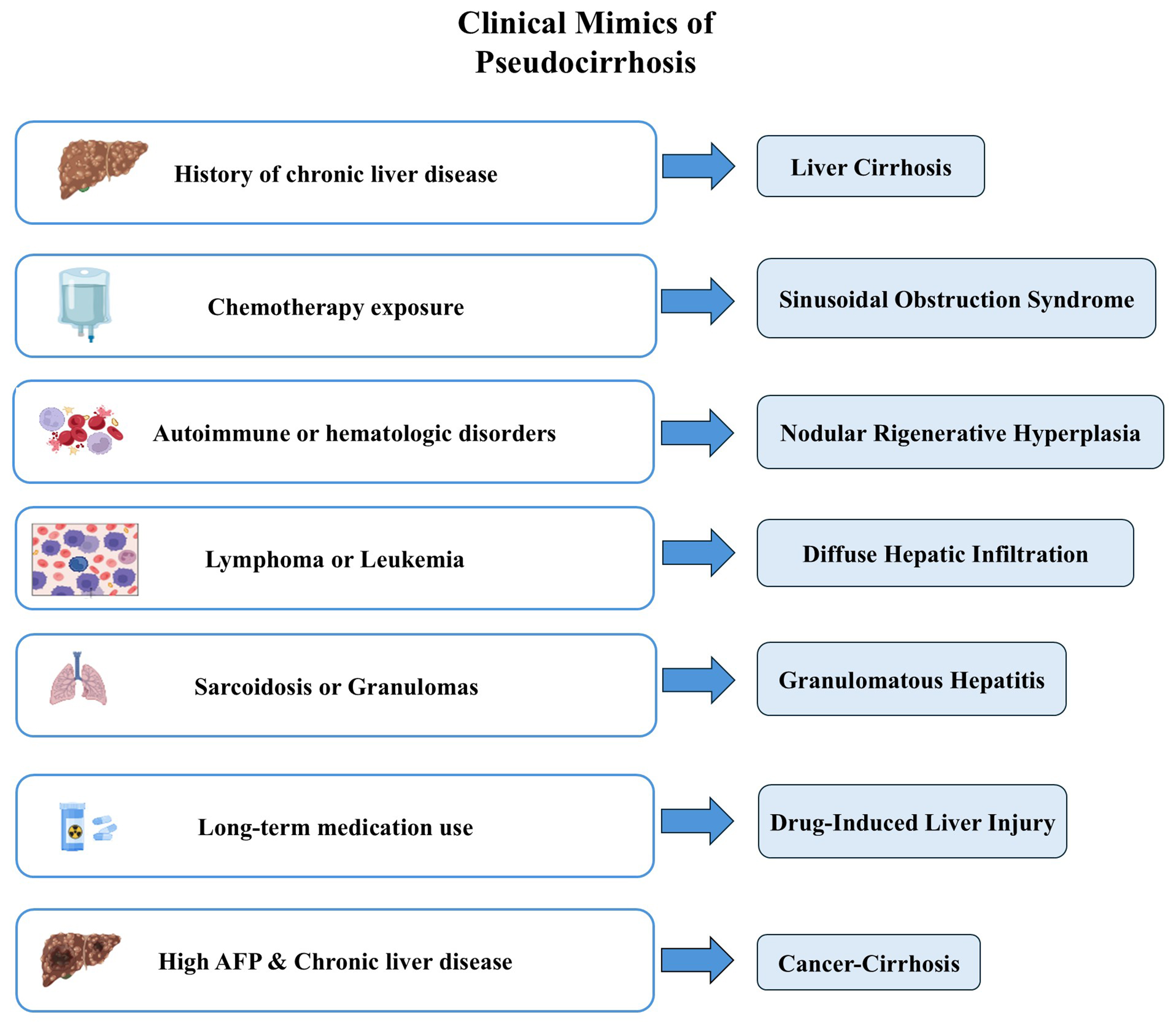
Figure 2. Clinical conditions that may mimic pseudocirrhosis according to the main clinical context or predisposing factor. Chronic liver disease may indicate true liver cirrhosis, chemotherapy exposure may suggest sinusoidal obstruction syndrome, autoimmune or hematologic disorders may be associated with nodular regenerative hyperplasia, and lymphoma or leukaemia may cause diffuse hepatic infiltration. Granulomatous disease, chronic drug exposure, and elevated AFP in patients with chronic liver disease should prompt consideration of granulomatous hepatitis, drug-induced liver injury, and Cancer-Cirrhosis, respectively. The figure was created in BioRender.com. AFP: Alpha-fetoprotein.
Differential diagnosis between pseudocirrhosis and its major clinical and radiologic mimics
| Condition | Typical clinical setting | Key imaging findings | Portal hypertension | Laboratory clues | Distinguishing features |
| Pseudocirrhosis[1,11-14,19-22,41] | Metastatic malignancy with liver involvement, with or without systemic treatment | Capsular retraction, lobulated contour, segmental volume loss, caudate hypertrophy, rapid morphologic evolution | Frequent and often rapidly progressive | Variable liver dysfunction, thrombocytopenia, and tumour-specific biomarker abnormalities | Cirrhosis-like morphology in the absence of diffuse bridging fibrosis and classic regenerative nodules, possible association with desmoplastic fibrosis, sinusoidal tumour infiltration, and chemotherapy-related vascular injury |
| Liver cirrhosis[4,11,13] | Known chronic liver disease, recognized liver disease risk factors, or newly diagnosed chronic liver disease | Diffuse nodular contour, regenerative nodules, volume redistribution with caudate hypertrophy, slowly progressive morphologic remodeling | Typical and slowly progressive | Variable liver dysfunction and thrombocytopenia | Established cirrhotic architecture with bridging fibrosis and regenerative nodules |
| Sinusoidal obstruction syndrome[18,28,42-45] | Oxaliplatin-based chemotherapy, hematopoietic stem cell transplantation, or hematologic malignancies | Hepatomegaly, heterogeneous enhancement, periportal oedema, usually without capsular retraction or segmental volume loss | Frequent and potentially acute | Cholestatic liver test abnormalities, thrombocytopenia, and variable liver dysfunction | Sinusoidal endothelial injury with congestion and venular obliteration in the absence of metastatic infiltration |
| Nodular regenerative hyperplasia[26,46-51] | Autoimmune disease, hematologic disorders, immunosuppressive or cytotoxic therapy, and chemotherapy exposure | Mild surface nodularity, usually without marked lobar distortion or capsular retraction | Possible and usually non-cirrhotic | Usually preserved liver synthetic function with possible thrombocytopenia | Diffuse micronodular regenerative transformation in the absence of bridging fibrosis or fibrous septa |
| Infiltrative haematological malignancies[32,34,38,52] | Lymphoma, leukaemia, myeloproliferative disorders | Diffuse hepatic infiltration, hepatomegaly, miliary nodules, usually without segmental atrophy or capsular retraction | Possible | Elevated LDH, cytopenias, systemic inflammatory markers | Diffuse sinusoidal or portal infiltration by malignant haematologic cells |
| Granulomatous liver disease[53-56] | Sarcoidosis and other granulomatous disorders | Coarse nodular hepatic echotexture, parenchymal heterogeneity, without marked retractive changes, lobar collapse, or segmental volume loss | Possible in advanced disease | Elevated ACE, cholestatic liver test abnormalities | Non-caseating granulomas or granulomatous inflammation |
| Drug-induced liver injury[11,21,41,57] | Long-term exposure to methotrexate, tamoxifen, antiretroviral agents, or other hepatotoxic drugs | Diffuse steatosis, periportal changes, or chronic fibrotic remodeling, usually without segmental remodeling, capsular retraction, or lobar volume loss | Possible in advanced disease | Variable liver dysfunction according to injury pattern | Steatosis, steatohepatitis, pericellular fibrosis, vascular injury, depending on the causative agent |
| Infiltrative hepatocellular carcinoma[58-65] | Usually advanced chronic liver disease or cirrhosis | Diffuse or permeative infiltration, subtle or patchy arterial enhancement, heterogeneous washout, hepatobiliary phase hypointensity, and portal vein tumour thrombosis | Frequent and often progressive | AFP often markedly elevated with variable liver dysfunction | Diffuse malignant hepatocellular infiltration with vascular invasion |
Liver cirrhosis
Among all mimics, cirrhosis remains the most challenging to distinguish, as it most closely resembles pseudocirrhosis on imaging, showing lobulated contours, retractive changes, and signs of portal hypertension[4,11,13]. Moreover, liver dysfunction and clinically significant portal hypertension may occur in both entities, but in pseudocirrhosis they are often linked to tumour burden or therapeutic toxicity rather than to progressive hepatic architectural collapse[41]. Crucially, although cirrhosis can occasionally be undiagnosed, it is typically a known pre-existing condition, diagnosed based on clinical history and previous investigations, whereas pseudocirrhosis is usually identified at the time of diagnosis, often as a new and unexpected finding during oncologic follow-up.
Sinusoidal obstruction syndrome
Another important condition in the differential diagnosis of pseudocirrhosis is sinusoidal obstruction syndrome (SOS), also known as veno-occlusive disease. This entity most commonly arises because of chemotherapy-induced endothelial injury, particularly following oxaliplatin-based regimens in colorectal cancer or conditioning regimens in hematologic malignancies such as leukaemia and after hematopoietic stem cell transplantation[35,42-44]. Clinically, SOS may manifest with ascites, splenomegaly, and portal hypertension, features also frequently observed in pseudocirrhosis. However, SOS is not caused by metastatic infiltration, and it typically lacks imaging findings such as retractive changes or segmental volume loss, which are more characteristic of pseudocirrhosis. On imaging, the liver may appear enlarged, with heterogeneous enhancement and periportal oedema, rather than lobulated or atrophic[18]. Contrast-enhanced ultrasound (CEUS) has been explored in the evaluation of SOS. Some studies have reported that CEUS can detect diffuse or geographic enhancement patterns in the hepatic parenchyma, with scattered hypoechoic areas[45]. However, these findings are not specific, and CEUS remains of limited diagnostic value in differentiating SOS from pseudocirrhosis, as both conditions may lack distinctive enhancement patterns or washout[45].
Nodular regenerative hyperplasia
Although NRH may also be encountered in the setting of pseudocirrhosis, it is a distinct clinicopathologic entity that can arise across a broad spectrum of other systemic disorders and itself mimic pseudocirrhosis, thereby entering the differential diagnosis.
NRH has been associated with a range of systemic conditions, including hematologic disorders[46], autoimmune diseases[47], long-term exposure to immunosuppressive and cytotoxic agents (such as azathioprine or 6-thioguanine)[41] and to chemotherapy for solid tumours and hematologic malignancies[41,48]. Histologically, NRH is defined by diffuse transformation into small regenerative nodules without fibrous septa, clearly distinguishing it from both cirrhosis and pseudocirrhosis[49]. Clinically, it may remain silent or manifest with non-cirrhotic portal hypertension, including splenomegaly or variceal bleeding[49]. When present, imaging abnormalities are typically mild and lack distinguishing features (such as marked surface irregularity or lobar volume loss), thus making NRH difficult to identify radiologically without clinical suspicion[50]. Surface nodularity and signs of portal hypertension may be observed, but there is no segmental volume loss, caudate hypertrophy, or lobular contour retraction, which are more typical of pseudocirrhosis. On contrast-enhanced studies, NRH usually demonstrates homogeneous or mildly heterogeneous parenchymal enhancement, without nodular collapse or retractive changes[49,50]. CEUS has been reported to show non-specific findings in NRH, and nodules are often not visualized or not differentiable from background liver tissue[51]. To date, no characteristic enhancement pattern has been validated, and CEUS remains of limited utility in diagnosing NRH[51].
Infiltrative haematological malignancies
Infiltrative hematologic malignancies, such as lymphoma and leukaemia, can diffusely involve the liver parenchyma without forming discrete masses, leading to hepatomegaly, irregular hepatic contours, and portal hypertension[38]. These may present radiologically as diffuse hepatic infiltration, homogeneous enlargement, or a miliary pattern of multiple small nodules, particularly in lymphoma[38]. However, these infiltrative patterns generally lack the lobar volume loss, contour retraction, or post-treatment fibrosis that characterize pseudocirrhosis[52]. Hepatic morphology is often preserved in early stages, and diagnosis relies on clinical and laboratory findings and evidence of underlying hematologic disease[32,34].
Granulomatous disease
Hepatic granulomatous diseases, including sarcoidosis, may also mimic pseudocirrhosis through chronic lobular inflammation, periportal granuloma formation, and secondary fibrosis[53,54]. These conditions can present with portal hypertension, capsular surface irregularities, and distortion of liver morphology[53,55]. However, they typically do not exhibit segmental volume loss, caudate lobe hypertrophy, or parenchymal collapse, which are more typical of pseudocirrhosis secondary to metastatic disease or chemotherapy[3,11,12]. Imaging may show coarse nodular hepatic echotexture and signs of portal hypertension but lacks the retractive features or treated-lesion sequelae of pseudocirrhosis[11,13]. Final diagnosis often requires correlation with clinical features, laboratory markers and, in selected cases, histological confirmation through liver biopsy[36,56].
Drug-induced liver injury
In this context, drug-induced liver injury (DILI) should enter the differential diagnosis of pseudocirrhosis only when it results in insidious, chronic architectural damage, effectively representing an evolving cirrhotic process that may be clinically unexpected. This scenario deeply differs from the predominantly vascular, reactive mechanism of pseudocirrhosis, in which chemotherapy-related sinusoidal injury and NRH underlie the cirrhosis-like hepatic remodelling. DILI may occur in patients exposed to long-term treatment. Agents most frequently implicated include methotrexate, tamoxifen, and certain antiretrovirals, which can induce diffuse architectural remodelling or periportal fibrosis, simulating some features of pseudocirrhosis[41]. Methotrexate, widely used in autoimmune diseases and malignancies, has been associated with hepatic steatosis, fibrosis, and, in some cases, cirrhosis[57]. Histologically, it may cause macrovesicular steatosis, hepatocyte ballooning, and pericellular fibrosis[11,57]. Tamoxifen, a selective estrogen receptor modulator used in breast cancer, has been associated with the development of metabolic-associated steatotic liver disease (MASLD) and, in some cases, with progression to steatohepatitis and fibrosis. Imaging typically shows diffuse hepatic steatosis without lobular distortion, surface nodularity, or lobar atrophy, and therefore lacks the morphologic features that would raise suspicion for pseudocirrhosis; however, if drug-induced liver disease silently progresses to steatohepatitis and advanced fibrosis, it may eventually enter the differential diagnosis as an insidious, evolving cirrhotic process[21]. Antiretroviral agents, particularly didanosine and stavudine, have been linked to mitochondrial toxicity, hepatic steatosis, and rare cases of fibrosis[49]. Imaging findings may include diffuse steatosis or periportal changes, but without the hallmark features of pseudocirrhosis such as retractive changes, surface irregularity, or segmental volume loss[11].
In all these scenarios of DILI, the parenchymal changes tend to be diffuse and evenly distributed, without the segmental remodelling and volume distortion typical of pseudocirrhosis. These features, together with a suggestive pharmacologic history, aid in distinguishing drug-induced hepatotoxicity from pseudocirrhosis[11].
PSEUDOCIRRHOSIS AND HCC
HCC, the most common primary malignancy of the liver, typically arises in the context of chronic liver disease and cirrhosis. The nodular and massive growth patterns of HCC are usually radiologically remarkable and often fulfill classic imaging criteria on dynamic contrast-enhanced computed tomography or magnetic resonance imaging (MRI). Conversely, the infiltrative subtype of HCC, accounting for 7%-20% of all cases, is notoriously difficult to detect and may present as a diffuse alteration of the hepatic parenchyma without a dominant lesion[58]. This growth pattern can involve entire lobes or even the whole liver and is often accompanied by portal vein tumour thrombosis and aggressive biological behaviour[15,59,60].
Thus, the presence of HCC risk factors changes the diagnostic hierarchy. In a patient with chronic liver disease or cirrhosis, a new diffuse hepatic distortion should not be attributed to pseudocirrhosis until infiltrative HCC has been reasonably excluded. The diagnostic intersection between these two entities represents a critical clinical scenario where imaging features, oncologic history, and liver function all converge to guide decision-making. From a radiologic point of view, infiltrative HCC tends to show subtle or patchy arterial enhancement, often with heterogeneous washout and hypo-intensity in the hepatobiliary phase, particularly when using hepatobiliary contrast agents[61,62]. Diffusion-weighted MRI may increase lesion detectability in these cases[63]. In particular, hepatobiliary contrast-enhanced MRI may improve lesion conspicuity in infiltrative HCC, especially in patients with diffuse or heterogeneous parenchymal distortion. These features, however, are not pathognomonic, and similar findings can be seen in cases of pseudocirrhosis with underlying diffuse metastatic disease. Importantly, while infiltrative HCC and pseudocirrhosis may both present with nodular liver contour, capsular retraction, segmental atrophy, and signs of portal hypertension, they arise from deeply different processes[3,14,35].
Serologic markers, especially AFP, have limited diagnostic accuracy and are no longer recommended as stand-alone tests for the diagnosis of HCC in major international guidelines. When AFP is used as a diagnostic marker, low cut-offs (e.g. 20 ng/mL) provide only moderate sensitivity but poor specificity, whereas higher thresholds of 200-400 ng/mL markedly increase specificity at the expense of very low sensitivity. In most series, AFP values ≥ 200-400 ng/mL have shown specificities close to or above 95%-99% for HCC in patients with chronic liver disease[59,64,65]. Despite these limitations, very high AFP concentrations often carry important diagnostic information. Markedly elevated AFP values, in the hundreds or thousands of ng/mL, are strongly associated with advanced, biologically aggressive HCC and with features such as large tumour burden and portal vein invasion[66,67]. In particular, infiltrative HCC frequently presents with elevated AFP levels, and published series report median values in the several-hundred ng/mL range, with a substantial proportion of patients exceeding conventional diagnostic thresholds[59]. By contrast, in pseudocirrhosis related to metastatic breast cancer or other non-AFP-producing primaries, AFP levels are typically normal or only mildly elevated. Thus, while normal AFP does not exclude infiltrative HCC, a strikingly elevated AFP level in a cirrhosis-like liver should strongly favour the diagnosis of HCC over pseudocirrhosis in the appropriate clinical context[58].
PROPOSED DIAGNOSTIC APPROACH TO PSEUDOCIRRHOSIS
Clinicians should suspect pseudocirrhosis when a patient with metastatic cancer, particularly with liver involvement, develops a rapid change in hepatic morphology characterized by surface nodularity, capsular retraction, segmental volume loss, caudate lobe hypertrophy, and signs of portal hypertension. The suspicion is strengthened by the absence of previous chronic liver disease, a close temporal relationship with systemic therapy, and discordance between improving or stable tumour burden and worsening portal hypertensive features. Serial comparison with prior imaging is essential, because rapid evolution over weeks to a few months is one of the most useful clues distinguishing pseudocirrhosis from conventional cirrhosis.
Distinguishing between infiltrative HCC and pseudocirrhosis can be challenging; a structured diagnostic algorithm can help guide the stepwise evaluation and multidisciplinary decision-making process in this context [Figure 3].
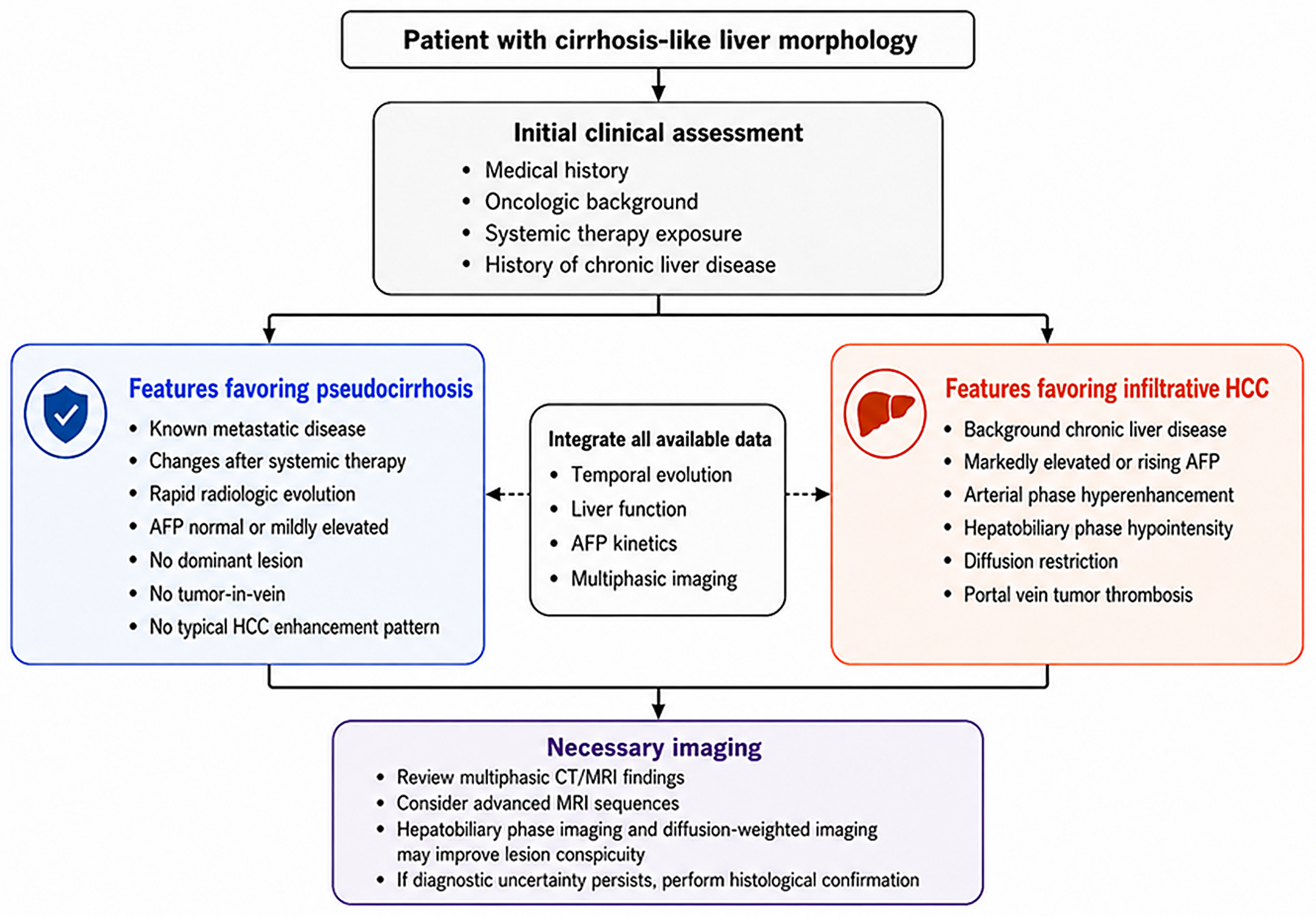
Figure 3. Diagnostic approach to pseudocirrhosis vs. infiltrative HCC. The figure was created in BioRender.com. AFP: Alpha-fetoprotein; CT: computed tomography; HCC: hepatocellular carcinoma; MRI: magnetic resonance imaging.
Obviously, the key starting point is the patient’s medical history. If a patient with known metastatic disease develops a liver morphology resembling cirrhosis, especially after systemic therapy, the diagnosis of pseudocirrhosis should be favored if the radiological changes are rapid, AFP is normal or only slightly elevated, and no dominant lesions, tumor-in-vein, or typical enhancement patterns are identified. Conversely, infiltrative HCC should be strongly considered when parenchymal distortion is associated with markedly elevated or progressively rising AFP levels, hyperenhancement in the arterial phase, hypointensity in the hepatobiliary phase, diffusion restriction, tumor thrombosis of the portal vein, but especially a history of chronic liver disease.
In this context, a multimodal approach is essential. Histologic confirmation via biopsy should be considered when imaging is inconclusive, when tumour markers and imaging are discordant, when occult sinusoidal metastasis is suspected despite apparently negative imaging, or when the result would change oncologic management. Infiltrative HCC, due to its aggressive nature and poor prognosis, in addition to the frequently observed associated chronic liver disease, is typically not amenable to curative treatment. Pseudocirrhosis, although potentially associated with signs of portal hypertension and hepatic failure, does not always carry the same oncologic implications[3,68]. In patients with ascites, portal hypertension, coagulopathy, or thrombocytopenia, the transjugular route should generally be preferred. Thus, multidisciplinary discussion involving hepatologists, oncologists, radiologists, and pathologists is recommended before discontinuing potentially effective antitumour therapy or before assigning the patient to best supportive care.
MANAGEMENT AND TREATMENT OF PSEUDOCIRRHOSIS
There is no disease-specific treatment for pseudocirrhosis, and management should be individualized according to the dominant mechanism, severity of portal hypertension, liver function, and oncologic status. When chemotherapy-related sinusoidal injury, NRH, or treatment-induced desmoplastic retraction is suspected, temporary interruption, dose reduction, or modification of the implicated systemic therapy should be discussed. Supportive treatment should follow the principles used for portal hypertension and be individualized according to liver function, tumour burden, and life expectancy.
In highly selected patients with symptomatic malignant pseudocirrhosis and refractory portal hypertension, transjugular intrahepatic portosystemic shunt (TIPS) has been reported as a feasible palliative option, although evidence remains limited and patient selection should be multidisciplinary[69].
The prognosis becomes particularly poor when pseudocirrhosis is complicated by clinically significant portal hypertension. In a systematic review and meta-analysis including 389 patients[70], portal hypertension was observed in most patients reported in case reports and case series, with ascites and oesophageal varices being the most frequent manifestations; the median time from pseudocirrhosis diagnosis to death was only 2 months in case reports and case series, whereas observational studies reported median times to death ranging from 3.6 to 8.5 months. Contemporary breast cancer cohorts confirm this adverse prognosis, with median overall survival after pseudocirrhosis diagnosis of approximately 7.6-7.9 months[19]. The presence of clinically significant portal hypertension may further identify a subgroup with particularly poor outcomes: in one cohort, patients with endoscopically evident oesophageal or gastric varices had a median overall survival of 5 months, compared with 13 months in those without varices, and median survival after documentation of varices was only 2 months[71]. However, these estimates should be interpreted cautiously because survival is strongly influenced by tumour subtype, tumour burden, liver function, availability of further oncologic therapy, and heterogeneous definitions of portal hypertension across studies.
Although pseudocirrhosis is often associated with poor prognosis, partial or complete regression has been reported in selected cases, especially when the process is driven by treatment-related injury or scarring around regressed metastases rather than uncontrolled diffuse tumour infiltration[6]. Therefore, early recognition is clinically relevant because timely adjustment of systemic therapy and management of portal hypertension may preserve liver function and allow continuation or resumption of oncologic treatment.
CONCLUSIONS
Pseudocirrhosis is often a rapidly progressive condition with a poor prognosis, reflecting both the underlying malignancy and treatment-related liver injury. Early recognition allows for closer monitoring of hepatic function, timely adjustment or withdrawal of potentially hepatotoxic systemic therapies, and earlier institution of supportive measures before overt hepatic failure precludes further oncologic options. Supportive care includes management of ascites, endoscopic treatment of varices, and management of hepatic encephalopathy, while in severe cases advanced interventions such as TIPS may be considered for refractory portal hypertension. In selected patients, partial regression of the pseudo-cirrhotic morphology after modification or discontinuation of systemic therapy has been reported, but this appears to be the exception rather than the rule.
At the same time, in patients with a cirrhosis-like liver and equivocal imaging findings, early histologic confirmation plays a pivotal role. Indeed, only a definite diagnosis reached by liver histology can safely justify active tumour-directed treatment, including rescue chemotherapy regimens, before progressive liver failure irreversibly limits these options.
Finally, current evidence on pseudocirrhosis remains weak and largely based on case reports, small retrospective cohorts, and heterogeneous systematic reviews. Diagnostic definitions vary across studies, histologic confirmation is available only in a minority of cases, and the relative contribution of different pathophysiological mechanisms is often difficult to identify. These limitations currently prevent the development of validated diagnostic criteria or evidence-based management algorithms. Future studies should aim to establish prospective multicentre registries with standardized imaging criteria, systematic comparison with previous imaging, predefined definitions of portal hypertension, integrated radiology-pathology correlation whenever biopsy is feasible and particular attention to modern systemic therapies.
DECLARATIONS
Acknowledgments
The graphical abstract was created in BioRender.com.
Authors’ contributions
Conceptualized the study and drafted the manuscript: Gallo P, Flagiello V, Di Pasquale G, Falcomatà A
Contributed to drafting the manuscript: Terracciani F, Mingiacchi A, Lerose F, Mastrostefano B, dell’Unto C, Galati G
Supervised the study and critically revised the manuscript for important intellectual content: Picardi A, Vespasiani-Gentilucci U
All authors read and approved the final manuscript.
Availability of data and materials
Not applicable.
AI and AI-assisted tools statement
During the preparation of this manuscript, the AI tool ChatGPT (version GPT-5.5 Thinking, released 2026-04-23) was used solely for figure editing. The tool did not influence the study design, data collection, analysis, interpretation, or the scientific content of the work. All authors take full responsibility for the accuracy, integrity, and final content of the manuscript.
Financial support and sponsorship
None.
Conflicts of interest
All authors declared that there are no conflicts of interest.
Ethical approval and consent to participate
Not applicable.
Consent for publication
Not applicable.
Copyright
© The Author(s) 2026.
REFERENCES
1. Oliai C, Douek ML, Rhoane C, et al. Clinical features of pseudocirrhosis in metastatic breast cancer. Breast Cancer Res Treat. 2019;177:409-17.
3. Young ST, Paulson EK, Washington K, Gulliver DJ, Vredenburgh JJ, Baker ME. CT of the liver in patients with metastatic breast carcinoma treated by chemotherapy: findings simulating cirrhosis. AJR Am J Roentgenol. 1994;163:1385-8.
4. Engelman D, Moreau M, Lepida A, Zaouak Y, Paesmans M, Awada A. Metastatic breast cancer and pseudocirrhosis: an unknown clinical entity. ESMO Open. 2020;5:e000695.
5. Manenti A, Caramaschi S, Manco G, Bonetti LR. Neoplastic pseudocirrhosis pathophysiological deepening. Dig Dis Sci. 2024;69:4493-4.
6. Kang SP, Taddei T, McLennan B, Lacy J. Pseudocirrhosis in a pancreatic cancer patient with liver metastases: a case report of complete resolution of pseudocirrhosis with an early recognition and management. World J Gastroenterol. 2008;14:1622-4.
7. Harry BL, Smith ML, Burton JR Jr, Dasari A, Eckhardt SG, Diamond JR. Medullary thyroid cancer and pseudocirrhosis: case report and literature review. Curr Oncol. 2012;19:e36-41.
8. Gounder J, Frankel A, Ngai S. Isolated serosal liver infiltration mimicking cirrhosis in gastroesophageal junction adenocarcinoma: a report of 2 cases. Radiol Case Rep. 2026;21:436-9.
9. Lee SL, Chang ED, Na SJ, et al. Pseudocirrhosis of breast cancer metastases to the liver treated by chemotherapy. Cancer Res Treat. 2014;46:98-103.
10. Phillips E, Sethi M, Vasanthakumar S, et al. The clinical features and outcomes of pseudocirrhosis in breast cancer. Cancers. 2024;16:2822.
11. Adike A, Karlin N, Menias C, Carey EJ. Pseudocirrhosis: a case series and literature review. Case Rep Gastroenterol. 2016;10:381-91.
12. Jeong WK, Choi SY, Kim J. Pseudocirrhosis as a complication after chemotherapy for hepatic metastasis from breast cancer. Clin Mol Hepatol. 2013;19:190-4.
14. Qayyum A, Lee GK, Yeh BM, Allen JN, Venook AP, Coakley FV. Frequency of hepatic contour abnormalities and signs of portal hypertension at CT in patients receiving chemotherapy for breast cancer metastatic to the liver. Clin Imaging. 2007;31:6-10.
15. Jha P, Poder L, Wang ZJ, Westphalen AC, Yeh BM, Coakley FV. Radiologic mimics of cirrhosis. AJR Am J Roentgenol. 2010;194:993-9.
16. Gibert-Ramos A, Sanfeliu-Redondo D, Aristu-Zabalza P, et al. The hepatic sinusoid in chronic liver disease: the optimal milieu for cancer. Cancers. 2021;13:5719.
17. Yang M, Zhang C. The role of liver sinusoidal endothelial cells in cancer liver metastasis. Am J Cancer Res. 2021;11:1845-60.
18. Parikh P, Rose T, Jeong D, et al. Diffuse intrasinusoidal hepatic metastasis from breast cancer: Multimodality imaging with pathology correlation. Radiol Case Rep. 2021;16:1005-9.
19. Huppert LA, Walker Z, Li M, et al. Clinical characteristics and outcomes in patients with metastatic breast cancer and pseudocirrhosis: a single center retrospective cohort study. Breast Cancer Res Treat. 2023;197:137-48.
20. Sadlik G, Anderson RC, Lei X, Cen SY, Duddalwar VA, Fong TL. Pseudocirrhosis: a case series with clinical and radiographic correlation and review of the literature. Dig Dis Sci. 2024;69:1004-14.
21. Sass DA, Clark K, Grzybicki D, Rabinovitz M, Shaw-Stiffel TA. Diffuse desmoplastic metastatic breast cancer simulating cirrhosis with severe portal hypertension: a case of “pseudocirrhosis”. Dig Dis Sci. 2007;52:749-52.
22. Vuppalanchi R, Saxena R, Storniolo AMV, Chalasani N. Pseudocirrhosis and liver failure in patients with metastatic breast cancer after treatment with palbociclib. Hepatology. 2017;65:1762-4.
23. Zeina AR, Abu-Mouch S, Mari A. Pseudocirrhosis in metastatic breast cancer. Isr Med Assoc J. 2017;19:328.
24. Takata K, Mogi A, Yamauchi R, Shakado S, Hirai F. Pseudocirrhosis due to desmoplastic response to chemotherapy in breast cancer liver metastases. Cureus. 2022;14:e25321.
25. Allison KH, Fligner CL, Parks WT. Radiographically occult, diffuse intrasinusoidal hepatic metastases from primary breast carcinomas: a clinicopathologic study of 3 autopsy cases. Arch Pathol Lab Med. 2004;128:1418-23.
26. Reshamwala PA, Kleiner DE, Heller T. Nodular regenerative hyperplasia: not all nodules are created equal. Hepatology. 2006;44:7-14.
27. Gentilucci UV, Gallo P, Perrone G, et al. Non-cirrhotic portal hypertension with large regenerative nodules: a diagnostic challenge. World J Gastroenterol. 2011;17:2580-4.
28. Valla DC, Cazals-Hatem D. Sinusoidal obstruction syndrome. Clin Res Hepatol Gastroenterol. 2016;40:378-85.
29. Nascimento AB, Mitchell DG, Rubin R, Weaver E. Diffuse desmoplastic breast carcinoma metastases to the liver simulating cirrhosis at MR imaging: report of two cases. Radiology. 2001;221:117-21.
30. Borja ER, Hori JM, Pugh RP. Metastatic carcinomatosis of the liver mimicking cirrhosis: case report and review of the literature. Cancer. 1975;35:445-9.
31. Mitchell ML, Filippone MD, Wozniak TF. Metastatic carcinomatous cirrhosis and hepatic hemosiderosis in a patient heterozygous for the H63D genotype. Arch Pathol Lab Med. 2001;125:1084-7.
32. Nakajima T, Sekoguchi S, Nishikawa T, et al. Multifocal intraportal invasion of breast carcinoma diagnosed by laparoscopy-assisted liver biopsy. World J Gastroenterol. 2005;11:2360-3.
33. Fournier C, Tisman G, Kleinman R, Park Y, Macdonald WD. Clinical evidence for overcoming capecitabine resistance in a woman with breast cancer terminating in radiologically occult micronodular pseudo-cirrhosis with portal hypertension: a case report. J Med Case Rep. 2010;4:112.
34. Graber I, Dumortier J, Poncet G, Queneau PE, Mathevet P, Scoazec JY. Hepar lobatum carcinomatosum revealing an occult metastatic lobular carcinoma of the breast. Ann Diagn Pathol. 2010;14:438-42.
35. Jüngst C, Krämer J, Schneider G, Lammert F, Zimmer V. Subacute liver failure by pseudocirrhotic metastatic breast cancer infiltration. Ann Hepatol. 2013;12:834-6.
36. Hidalgo-Blanco A, Aguirresarobe-Gil de San Vicente M, Aresti S, de Miguel E, Cabriada-Nuno JL. Pseudocirrhosis in metastatic breast cancer. Gastroenterol Hepatol. 2018;41:111-3.
37. Knouse P, Hancock C, Iwaz S, Kaiser P. Metastatic carcinomatosis cirrhosis: a rare pattern of metastasis. Cureus. 2019;11:e3876.
38. Millard T, Gupta A, Brenin C, Marshall P, Dillon P. Radiographically occult carcinomatous spread of breast cancer to the liver: a challenging case. Case Rep Oncol Med. 2019;2019:4935615.
39. Hoshina H, Takei H, Nakamura M, Nishimoto F, Hanamura S. Carcinomatous cirrhosis as radiographically occult liver metastases of breast cancer: a systematic literature review. Cancer Treat Res Commun. 2021;28:100388.
40. Aloysius MM, Zaitoun AM, Beckingham IJ, et al. The pathological response to neoadjuvant chemotherapy with FOLFOX-4 for colorectal liver metastases: a comparative study. Virchows Arch. 2007;451:943-8.
41. Aoyagi T, Takabe K, Tamanuki T, Matsubara H, Matsuzaki H. Pseudocirrhosis after chemotherapy in breast cancer, case reports. Breast Cancer. 2018;25:614-8.
42. Rubbia-Brandt L, Audard V, Sartoretti P, et al. Severe hepatic sinusoidal obstruction associated with oxaliplatin-based chemotherapy in patients with metastatic colorectal cancer. Ann Oncol. 2004;15:460-6.
43. Carreras E. Veno-occlusive disease of the liver after hemopoietic cell transplantation. Eur J Haematol. 2000;64:281-91.
44. European Association for the Study of the Liver. Electronic address: [email protected], European Association for the Study of the Liver. EASL Clinical Practice Guidelines on vascular diseases of the liver. J Hepatol. 2026;84:399-456.
45. Zhang Y, Yan Y, Song B. Noninvasive imaging diagnosis of sinusoidal obstruction syndrome: a pictorial review. Insights Imaging. 2019;10:110.
47. Morris JM, Oien KA, McMahon M, et al. Nodular regenerative hyperplasia of the liver: survival and associated features in a UK case series. Eur J Gastroenterol Hepatol. 2010;22:1001-5.
48. Wanless IR. Micronodular transformation (nodular regenerative hyperplasia) of the liver: a report of 64 cases among 2,500 autopsies and a new classification of benign hepatocellular nodules. Hepatology. 1990;11:787-97.
49. Hartleb M, Gutkowski K, Milkiewicz P. Nodular regenerative hyperplasia: evolving concepts on underdiagnosed cause of portal hypertension. World J Gastroenterol. 2011;17:1400-9.
50. Ames JT, Federle MP, Chopra K. Distinguishing clinical and imaging features of nodular regenerative hyperplasia and large regenerative nodules of the liver. Clin Radiol. 2009;64:1190-5.
51. Höpfner M, Scheel AH, Braun M, Rüschoff J, Löser C. Unusual nodular regenerative hyperplasia mimicking liver metastases in CEUS. Ultraschall Med. 2014;35:199-202.
52. Cervoni JP, Dobrin A, Sailley N, et al. [Hepar lobatum carcinomatusum: a rare cause of portal hypertension complicating hepatic metastases in breast cancer]. Gastroenterol Clin Biol. 2008;32:740-4.
53. Ibrahim AM, Bhandari B, Soriano PK, et al. Hepatic involvement in systemic sarcoidosis. Am J Case Rep. 2018;19:1212-5.
54. Ebert EC, Kierson M, Hagspiel KD. Gastrointestinal and hepatic manifestations of sarcoidosis. Am J Gastroenterol. 2008;103:3184-92.
55. Malhotra A, Naniwadekar A, Sood G. Hepatobiliary and pancreatic: cirrhosis secondary to hepatic sarcoidosis. J Gastroenterol Hepatol. 2008;23:1942.
56. Ungprasert P, Carmona EM, Crowson CS, Matteson EL. Diagnostic utility of angiotensin-converting enzyme in sarcoidosis: a population-based study. Lung. 2016;194:91-5.
57. Marsh RL, Kelly S, Mumtaz K, Kaffenberger J. Utility and limitations of transient elastography to monitor hepatic steatosis, hepatic fibrosis, and methotrexate-associated hepatic disease in psoriasis: a systematic review. J Clin Aesthet Dermatol. 2021;14:24-8.
58. Reynolds AR, Furlan A, Fetzer DT, et al. Infiltrative hepatocellular carcinoma: what radiologists need to know. Radiographics. 2015;35:371-86.
59. Kneuertz PJ, Demirjian A, Firoozmand A, et al. Diffuse infiltrative hepatocellular carcinoma: assessment of presentation, treatment, and outcomes. Ann Surg Oncol. 2012;19:2897-907.
60. Jang ES, Yoon JH, Chung JW, et al. Survival of infiltrative hepatocellular carcinoma patients with preserved hepatic function after treatment with transarterial chemoembolization. J Cancer Res Clin Oncol. 2013;139:635-43.
61. Park YS, Lee CH, Kim BH, et al. Using Gd-EOB-DTPA-enhanced 3-T MRI for the differentiation of infiltrative hepatocellular carcinoma and focal confluent fibrosis in liver cirrhosis. Magn Reson Imaging. 2013;31:1137-42.
62. Lim S, Kim YK, Park HJ, Lee WJ, Choi D, Park MJ. Infiltrative hepatocellular carcinoma on gadoxetic acid-enhanced and diffusion-weighted MRI at 3.0T. J Magn Reson Imaging. 2014;39:1238-45.
63. Rosenkrantz AB, Lee L, Matza BW, Kim S. Infiltrative hepatocellular carcinoma: comparison of MRI sequences for lesion conspicuity. Clin Radiol. 2012;67:e105-11.
64. Farinati F, Marino D, De Giorgio M, et al. Diagnostic and prognostic role of alpha-fetoprotein in hepatocellular carcinoma: both or neither? Am J Gastroenterol. 2006;101:524-32.
65. Zhang J, Chen G, Zhang P, et al. The threshold of alpha-fetoprotein (AFP) for the diagnosis of hepatocellular carcinoma: a systematic review and meta-analysis. PLoS One. 2020;15:e0228857.
66. Peng SY, Chen WJ, Lai PL, Jeng YM, Sheu JC, Hsu HC. High alpha-fetoprotein level correlates with high stage, early recurrence and poor prognosis of hepatocellular carcinoma: significance of hepatitis virus infection, age, p53 and beta-catenin mutations. Int J Cancer. 2004;112:44-50.
67. Mehta N, Fidelman N, Sarkar M, Yao FY. Factors associated with outcomes and response to therapy in patients with infiltrative hepatocellular carcinoma. Clin Gastroenterol Hepatol. 2013;11:572-8.
68. Demirjian A, Peng P, Geschwind JF, et al. Infiltrating hepatocellular carcinoma: seeing the tree through the forest. J Gastrointest Surg. 2011;15:2089-97.
69. Shreve LA, O'Leary C, Clark TWI, Stavropoulos SW, Soulen MC. Transjugular intrahepatic portosystemic shunt for the management of symptomatic malignant pseudocirrhosis. J Gastrointest Oncol. 2022;13:279-87.
70. Villani R, Di Cosimo F, Sangineto M, Romano AD, Serviddio G. Pseudocirrhosis and portal hypertension in patients with metastatic cancers: a systematic review and meta-analysis. Sci Rep. 2022;12:19865.
Cite This Article
 Open Access
How to Cite
Download Citation
Export Citation File:
Type of Import
Tips on Downloading Citation
Citation Manager File Format
Type of Import
Direct Import: When the Direct Import option is selected (the default state), a dialogue box will give you the option to Save or Open the downloaded citation data. Choosing Open will either launch your citation manager or give you a choice of applications with which to use the metadata. The Save option saves the file locally for later use.
Indirect Import: When the Indirect Import option is selected, the metadata is displayed and may be copied and pasted as needed.
About This Article
Copyright

Data & Comments
Data
0










Comments
Comments must be written in English. Spam, offensive content, impersonation, and private information will not be permitted. If any comment is reported and identified as inappropriate content by OAE staff, the comment will be removed without notice. If you have any queries or need any help, please contact us at [email protected].