

Staged anophthalmic socket reconstruction with an expanded forehead island flap pedicled on the superficial temporal artery: a case report
 0
Abstract
Reconstruction of a severely contracted anophthalmic socket is difficult when both soft-tissue volume and prosthesis retention are inadequate. We report a 26-year-old man with persistent right orbital depression and conjunctival sac stenosis after enucleation for retinoblastoma and subsequent autologous rib orbital plasty. Reconstruction was performed in two stages: a 100 mL forehead tissue expander was expanded to 245 mL over 3.5 months, followed by transfer of a 4 cm × 4 cm expanded forehead island flap based on the superficial temporal artery (STA) and accompanying vein. The flap was passed through a subcutaneous tunnel after scar release. Perfusion was satisfactory, and socket contour and prosthesis retention improved by 1 month. Computed tomography (CT) volumetry at 10 months showed an increase in right orbital volume from 26.99 to 32.33 cm3 (5.34 cm3). Autologous fat grafting to the upper eyelid was performed 1 year after flap transfer to refine the periorbital contour. At 2 years, the patient maintained a favorable clinical appearance and satisfactory prosthetic eye fitting; this later appearance reflects the combined procedures and does not establish long-term volumetric stability of the flap alone. This case demonstrates the technical feasibility of an expanded STA-pedicled forehead flap in a selected patient, while additional cases and longer objective follow-up are required.
Keywords


 Download PDF
INTRODUCTION
Anophthalmic socket reconstruction remains a formidable challenge in ophthalmic plastic and reconstructive surgery, holding great significance for patients with socket contracture deformities caused by trauma, tumor excision, or congenital anophthalmia[1-4]. Restoring the anatomical structure and functional integrity of the socket is crucial for both the aesthetic appearance and psychosocial well-being of the patient.
Currently, various surgical strategies have been developed to restore soft tissue volume in the socket, which can be broadly categorized into pedicled flaps and free tissue transfers[5,6]. Among these, the superficial temporal artery (STA) island flap is widely used for repairing various facial[7], eyelid, and periorbital tissue defects[8,9] due to its reliable blood supply and adequate vascular pedicle length. However, as reported in previous socket reconstructions[10], conventional unexpanded STA island flaps often face the problem of insufficient tissue volume when addressing large defects. To acquire adequate tissue, surgeons may be forced to extend the flap into the hair-bearing scalp, which inevitably introduces hair into the reconstructed cavity, compromising both functional and aesthetic outcomes[11,12]. To avoid transferring hair-bearing tissue into the socket, previous studies utilized expanded forehead flaps for socket reconstruction, successfully generating ample, thin, and hairless tissue[13]. However, traditional expanded forehead flaps are typically pedicled on the supratrochlear or supraorbital arteries. While demonstrating excellent color and texture matching, they inevitably leave highly visible scars at the anterior forehead donor site postoperatively, constituting a significant aesthetic defect in the highly demanding periorbital region[11,14]. Free forearm flaps can provide thin and pliable tissue suitable for socket lining reconstruction, but they require microvascular anastomosis, sacrifice major forearm vessels, and demand high surgical expertise, which restricts their application in routine clinical practice[15,16].
Prepared in accordance with the Case Report Guidelines (CARE)[17], this case report describes a two-stage reconstructive strategy combining forehead tissue expansion with an STA-pedicled island flap to address these limitations. The intended advantages were to increase available flap area and pliability, improve contour matching, avoid microvascular anastomosis, and place the donor-site incision within the temporal hairline. Because STA-based island flaps have previously been reported for eyelid, facial, and selected socket reconstruction, the main contribution of this case is best framed as a combined technical application rather than as a wholly new vascular concept.
CASE REPORT
Preoperative assessment
A 26-year-old male presented with a pronounced right anophthalmic socket depression. His medical history was significant for right eye enucleation at 10 years of age for retinoblastoma. One year before the present reconstruction, autologous rib orbital plasty had been performed at our institution. Examination before that procedure documented right periorbital and temporozygomatic depression, marked facial asymmetry, and a narrow, shallow conjunctival sac. The operative record documented harvest of approximately 8 cm of the right seventh rib, which was divided into three pieces and placed along the inferior orbital rim and zygomatic region, the lateral orbital rim and temporal region, and the superior orbital rim and brow. Soft-tissue depression and conjunctival sac stenosis persisted after this skeletal augmentation. No additional bony orbital procedure, soft-tissue flap transfer, or soft-tissue filling procedure had been performed before the present staged reconstruction. The patient reported difficulty retaining the ocular prosthesis, with inferior tilting or spontaneous dislodgement occurring more than twice daily.
Preoperative examination before the present staged reconstruction demonstrated marked collapse of the right periorbital contour [Figure 1]. Residual conjunctival tissue was present, but the conjunctival sac remained narrow and shallow, with shortened fornices that did not support a standard prosthetic shell. The prior rib plasty had augmented the orbital rims and adjacent facial contour; the remaining reconstructive requirements were judged to be predominantly soft-tissue volume replacement and release of conjunctival sac contracture. Preoperative computed tomography (CT) volumetry calculated the right orbital volume to be 26.99 cm3. Based on the estimated defect dimensions, residual socket depth, and the amount of soft-tissue augmentation required, a 4 cm × 4 cm flap was planned. Systemic evaluation was unremarkable. Bilateral STA pulsations were palpable, and no comorbidity was identified that would be expected to compromise flap viability.
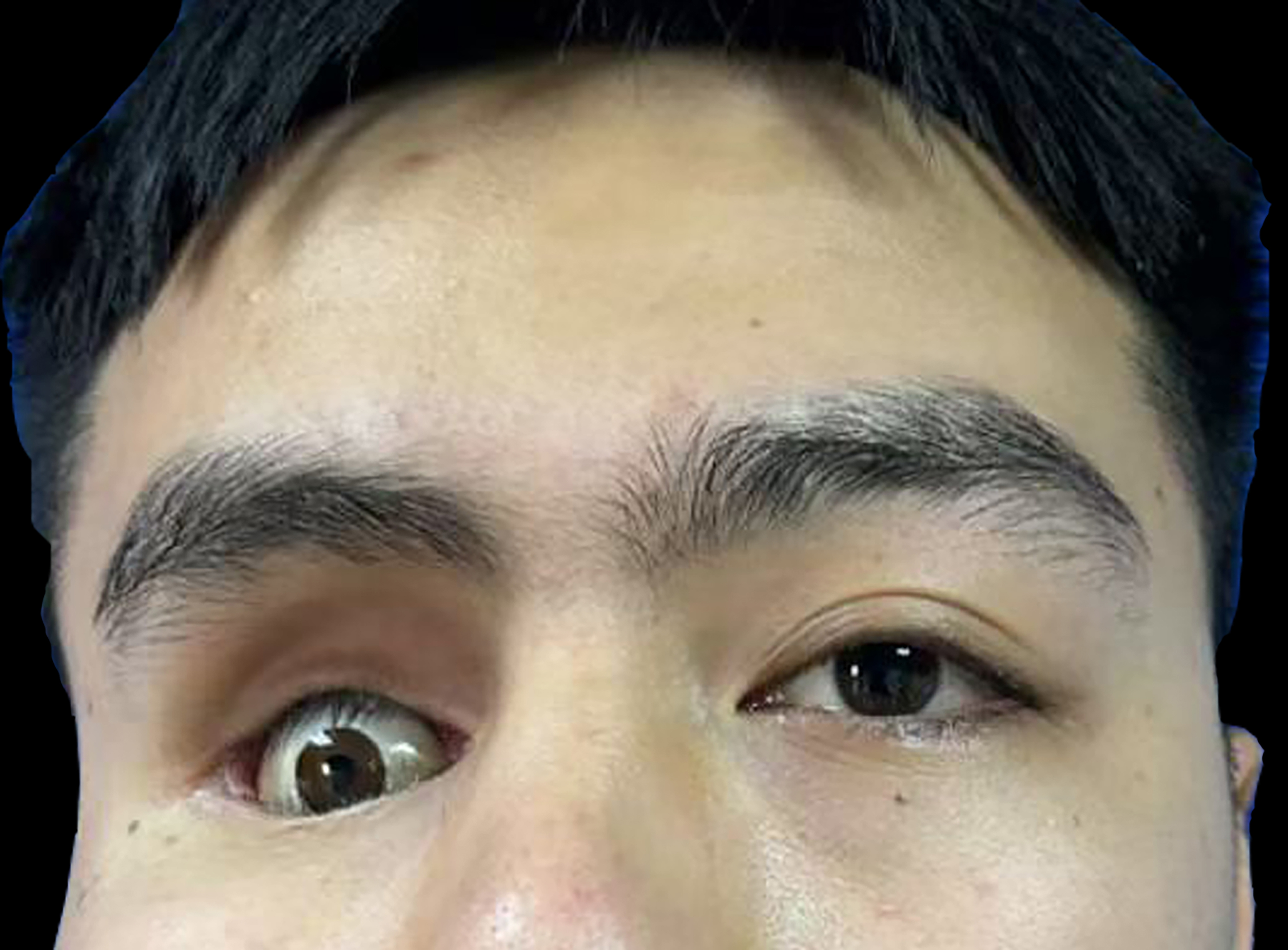
Figure 1. Preoperative appearance showing severe depression of the right anophthalmic socket and periorbital contour.
CT volumetric assessment
Preoperative and 10-month postoperative CT datasets were exported in Digital Imaging and Communications in Medicine (DICOM) format and analyzed with Mimics software (Materialise, Leuven, Belgium). The right orbital region was segmented on consecutive CT slices, and the contours were reviewed and corrected in axial, coronal, and sagittal views. The orbital rim defined the anterior limit, while the bony orbital walls and apex defined the remaining boundaries. Mimics generated a three-dimensional (3D) reconstruction from the segmented region and calculated the enclosed volume. The same anatomical boundaries and segmentation workflow were applied to both examinations.
Surgical technique
Stage I Surgery - Tissue Expander Implantation. Under general anesthesia, routine disinfection and draping were performed. A 100 mL spherical tissue expander was selected after integrity testing. The pocket was designed in the ipsilateral forehead near the hairline. A curved hairline incision was made, and dissection was carried deep to the galea aponeurotica and frontalis muscle to create the pocket. The expander was implanted, a closed suction drain was placed, and the incision was closed in layers with a pressure dressing. Expansion was performed gradually to a final volume of 245 mL over 3.5 months [Figure 2].
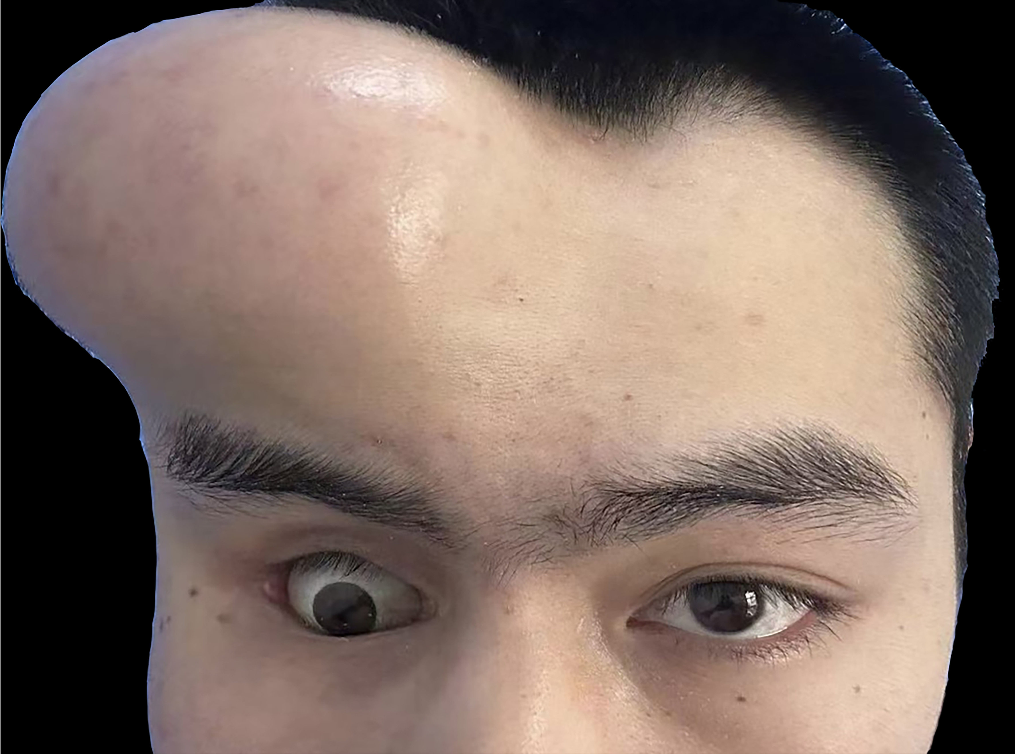
Figure 2. Stage I surgery: The tissue expander (implanted in the ipsilateral forehead) at the end of the 3.5-month expansion period.
Stage II Surgery - Flap Harvest and Transfer. Preoperative Doppler ultrasonography was used to map the STA course and its relationship to the planned flap. Under general anesthesia, the original incision was reopened, the expander was removed, and the capsule was dissected with meticulous hemostasis. A transverse incision was made in the contracted conjunctival sac to release scar tissue, and the socket dimensions were reassessed. STA pulsation was confirmed preauricularly and correlated with the Doppler markings.
A skin incision was made within the temporal hairline to carefully expose the STA and its accompanying superficial temporal vein (STV). A 4 cm × 4 cm island flap was designed within the expanded forehead tissue. A subcutaneous tunnel was created via blunt dissection from the preauricular region to the lateral canthus. The island flap was transferred into the socket through the tunnel, ensuring the vascular pedicle was neither kinked nor twisted. Flap perfusion was excellent; after confirming a normal blood supply, the flap was sutured in place and dressed, carefully avoiding any pressure on the pedicle [Figure 3].
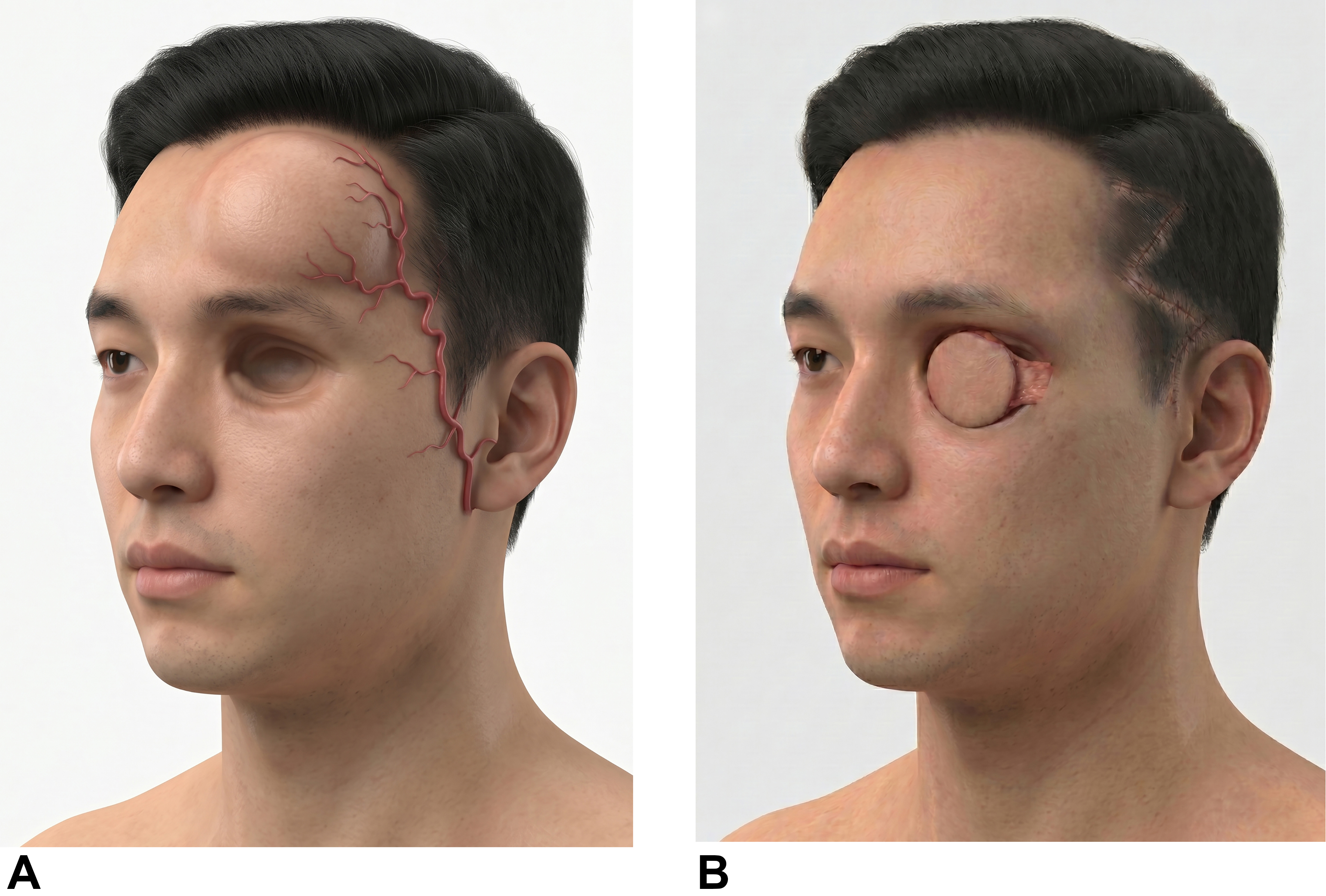
Figure 3. Schematic illustration of the two-stage reconstructive strategy. (A) Following the first stage of tissue expansion, the forehead skin is fully expanded, and the anatomical course of the STA is meticulously mapped; (B) During the second stage, the expanded STA-pedicled island flap is transposed into the anophthalmic socket via a subcutaneous tunnel to restore soft-tissue volume. The donor site is closed directly, leaving a concealed zigzag scar within the temporal hairline. STA: Superficial temporal artery.
Postoperative management
Broad-spectrum antibiotics were routinely administered for 2 days postoperatively. The flap color, capillary refill, and blood perfusion were closely monitored. The socket was kept clean, and pressure on the flap was avoided. Dressings were changed regularly based on the healing process, and sutures were removed 1 week postoperatively.
Outcomes
At the 1-month postoperative follow-up, the socket contour showed early improvement [Figure 4]. Prosthesis retention improved, and the patient no longer reported spontaneous dislodgement during routine activities. At 10 months, before any fat grafting, CT volumetry showed an increase in right orbital volume from 26.99 to 32.33 cm3 [Figure 5], a net increase of 5.34 cm3. Autologous fat grafting to the upper eyelid was then performed 1 year after flap transfer to refine the residual periorbital contour. At 2 years, the patient maintained a favorable clinical appearance and satisfactory prosthetic eye fitting without recurrent spontaneous dislodgement [Figure 6]. The 2-year appearance therefore reflects the combined effects of the flap and subsequent fat grafting. No CT volumetric assessment was performed at 2 years. The forehead donor site healed without a conspicuous anterior forehead scar because the incision was concealed within the hairline, and no visible eyebrow distortion was observed.
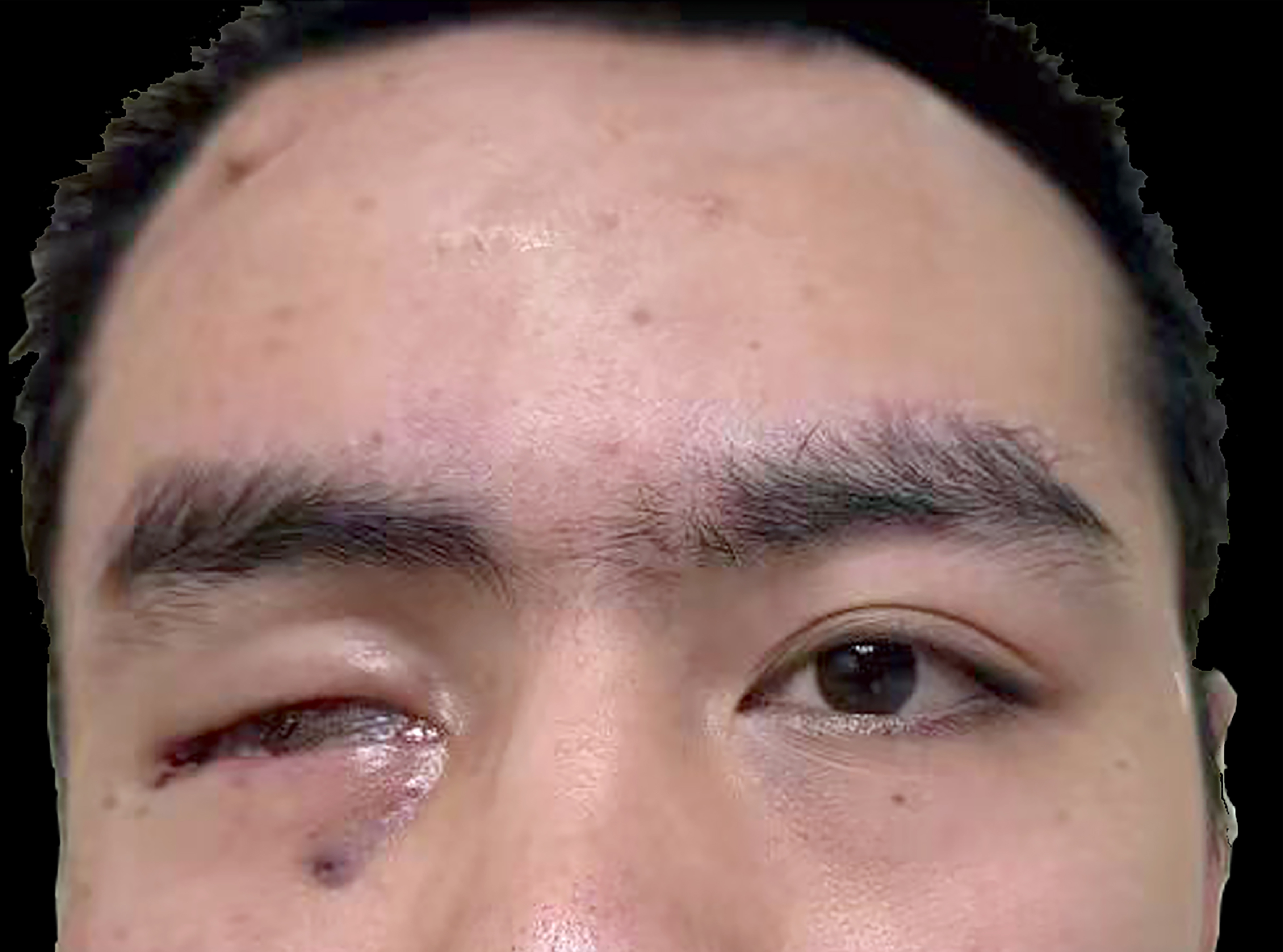
Figure 4. Early postoperative appearance at the 1-month follow-up, demonstrating preliminary improvement in the right periorbital contour and prosthesis retention.
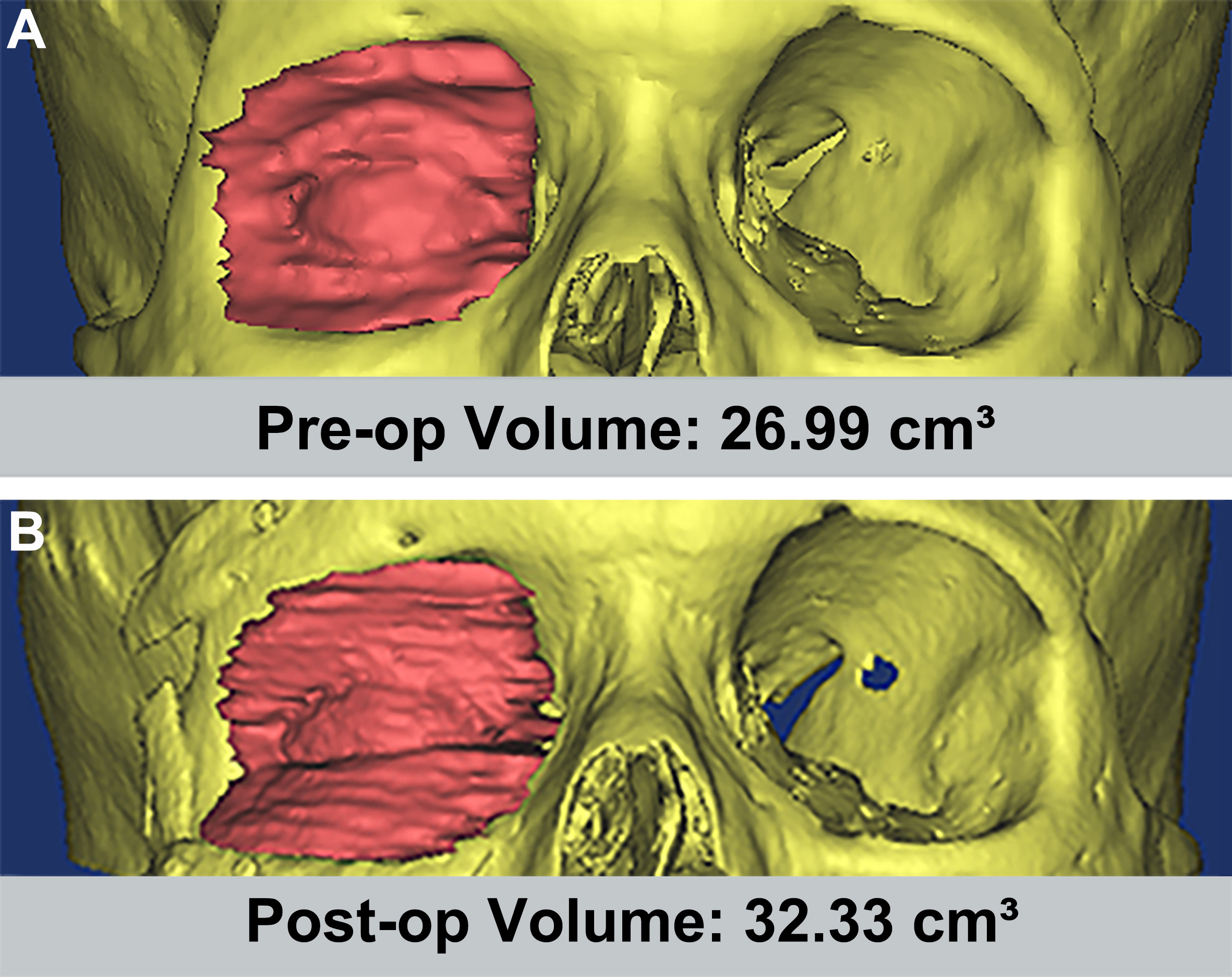
Figure 5. CT volumetry of the right orbit. (A) Preoperative 3D reconstruction showing a volume of 26.99 cm3; (B) 3D reconstruction at 10 months after flap transfer, before autologous fat grafting, showing a volume of 32.33 cm3 and a net increase of 5.34 cm3. CT: Computed tomography; 3D: three-dimensional; Pre-op: preoperative; Post-op: postoperative.

Figure 6. Facial appearance at 2 years after flap transfer. Autologous fat grafting to the upper eyelid had been performed 1 year after flap transfer to refine the periorbital contour; therefore, the appearance shown reflects both procedures.
Patient perspective
The patient reported that improved prosthesis stability reduced daily inconvenience and social embarrassment. This subjective improvement has been consistently reported by the patient throughout the 2-year follow-up period.
DISCUSSION
Anophthalmic socket reconstruction holds significant clinical importance for patients with anophthalmic socket syndrome, traumatic orbital deformities, and congenital anophthalmia. Beyond the aesthetic restoration of the periorbital contour, reconstructive surgery provides a stable anatomical foundation for wearing an ocular prosthesis, which is closely related to the patient’s psychological rehabilitation and quality of life[18-20]. Furthermore, for patients with concurrent eyelid malposition or laxity, adjunctive eyelid procedures such as lateral canthal suspension may be required[21]. Despite continuous advancements in surgical techniques, achieving optimal outcomes in socket reconstruction remains challenging due to the complexity of the orbital 3D structure, the highly demanding periorbital aesthetics, and the dual requirements for both cosmetic appearance and function.
The main technical value of this approach is the incorporation of preoperative forehead tissue expansion into an STA-pedicled regional flap strategy. Free flaps such as radial forearm or dorsalis pedis flaps can provide reliable lining and volume in complex orbital reconstruction, but they require microvascular anastomosis, may increase operative burden, and can create donor-site morbidity. In contrast, staged expansion permits recruitment of adjacent forehead tissue before flap transfer and may improve the available flap area and pliability for socket contouring.
The choice of reconstruction was specific to this patient. Although microvascular free-flap transfer represented a viable alternative, it was not selected because the available forehead tissue and intact STA pedicle provided a suitable regional reconstructive option without microvascular anastomosis or a distant flap donor site. A dermis-fat graft was also a potential option for volume and surface reconstruction. In this scarred, contracted socket, however, an expanded flap was selected to provide pliable skin for lining after scar release together with soft-tissue augmentation on an intact vascular pedicle. This rationale does not imply that dermis-fat grafting was contraindicated. Additional orbital floor or bony augmentation was not selected because the prior rib grafting had targeted the deficient orbital rims and adjacent facial contour, whereas the persistent problems were soft-tissue depression and a narrow, shallow conjunctival sac with poor prosthesis retention. Orbital floor augmentation alone would not have replaced the deficient socket lining or released the conjunctival contracture. These considerations do not establish superiority over the alternatives but explain the selection of the expanded STA flap in this case.
Tissue expansion may be useful in this setting through several mechanisms. Progressive expansion recruits adjacent skin and soft tissue, allowing harvest of a larger flap than would otherwise be available, while ensuring tension-free primary closure of the donor site without distorting the eyebrow. Expansion can also thin the soft-tissue envelope, which may help achieve a smoother periorbital contour[22]. In addition, expansion may augment vascularity through a delay-like effect, although this presumed benefit cannot be proven from a single case. Finally, the staged approach allows the surgeon to refine flap size after observing the actual expanded tissue dimensions.
The use of tissue expansion in forehead and periorbital reconstruction is well established[22,23], particularly in facial and nasal reconstruction. Applying this principle to anophthalmic socket reconstruction is therefore anatomically and technically plausible. However, the present report should be interpreted as an early clinical example rather than definitive evidence of superiority over existing methods.
It is worth noting that while Omori et al. reported anophthalmic socket reconstruction using an unexpanded anterofrontal STA island flap[10], their approach carries inherent limitations regarding tissue suitability and long-term morbidity. In their method, due to the lack of prior tissue expansion, obtaining sufficient tissue volume to fill a contracted socket often necessitates harvesting from the hair-bearing scalp region. Transferring hair-bearing skin into the anophthalmic cavity presents substantial clinical challenges: continuous intra-orbital hair growth acts as a chronic foreign body, causing persistent mechanical irritation, excessive mucous discharge, and a significantly elevated risk of recurrent local infections[20]. These complications severely hinder the comfortable wearing and long-term retention of the ocular prosthesis. In contrast, our technique strategically addresses this dilemma through preoperative tissue expansion of the forehead. By progressively expanding the hairless forehead skin, we recruit an ample surface area of thin, pliable, and completely hairless tissue. This modification may reduce the risk of transferring hair-bearing tissue into the socket and may therefore help avoid hair-related irritation, discharge, and infection.
A second practical advantage is the STA-based pedicle design. Although anterior forehead expansion has been successfully reported to yield sufficient hairless tissue[13], conventional designs pedicled on the supratrochlear or supraorbital vessels frequently result in visible scars on the central forehead. This distinction is clinically relevant in periorbital reconstruction, where such visible forehead scarring is often a major aesthetic concern for patients. Compared with these conventional expanded forehead flaps, an STA-oriented approach allows the entire access incision and donor-site scar to be concealed within the temporal hairline. Furthermore, the constant anatomical course of the STA within the superficial temporal fascia facilitates preoperative Doppler ultrasound mapping and intraoperative identification. This long and reliable vascular axis allows the expanded flap to be safely transferred through a subcutaneous tunnel from the temporal region to the socket without obvious pedicle kinking or excessive tension, enhancing surgical safety and promoting predictable perfusion[24].
Despite its distinct clinical advantages, the expanded STA island flap technique carries inherent risks and potential complications that warrant careful consideration. Chief among these is venous congestion, which remains a primary cause of partial flap necrosis or epidermolysis in island flap transfers[25]. Compared to the robust, thick-walled STA, the accompanying STV is characterized by thin walls and delicate valve structures, making it highly susceptible to mechanical compression, kinking, or torsion. When the flap is rotated and transposed through a narrow subcutaneous tunnel into the anophthalmic socket, any acute angulation or excessive tension on the vascular pedicle can drastically impede venous outflow. To mitigate this risk, it is imperative to dissect an adequately wide subcutaneous tunnel and preserve a generous fascial cuff around the vascular bundle during harvest, thereby ensuring a smooth, tension-free arc of rotation and optimizing microvascular perfusion.
Parallel to these intraoperative vascular concerns, expander-related complications present significant clinical challenges during the prolonged staging period. Forehead tissue expansion in this protocol typically spans several months (3.5 months in our case), during which the risks of expander infection, hematoma, and skin exposure are substantially heightened. The inherent tightness and limited elasticity of the forehead galea and periosteum exert tremendous counter-pressure on the expander shell. Aggressive or rapid fluid inflation can compromise local cutaneous microcirculation, leading to overlying skin ischemia, tissue necrosis, and subsequent expander extrusion. Furthermore, repeated transcutaneous puncturing for inflation introduces potential pathways for bacterial contamination. Preventing such severe complications requires strict adherence to sterile protocols during serial injections, a conservative and individualized inflation schedule, and precise subgaleal placement of the device during the first stage.
Furthermore, the two-stage nature of this reconstructive procedure inevitably extends the overall treatment timeline and requires exceptional patient compliance and psychological resilience during the interim expansion phase, as the patient must tolerate temporary facial asymmetry. While the aesthetic and functional outcomes achieved in this case are highly encouraging, clinicians must carefully weigh these technical demands and potential morbidities against the clear benefits of achieving a hairless, well-volumized, and scar-free socket reconstruction.
To better elucidate the value of these innovations, the present technique is compared with established socket reconstruction methods below [Table 1].
Comparison of surgical techniques for anophthalmic socket reconstruction
| Technique | Vascular pedicle | Tissue volume | Donor site scar | Microsurgery required | Main limitations |
| Radial forearm free flap[3,15] | Radial artery | Sufficient | Forearm | Yes | Major-vessel sacrifice; microvascular anastomosis; donor-site morbidity |
| Dorsalis pedis free flap[6] | Dorsalis pedis artery | Sufficient | Dorsum of foot | Yes | Microvascular anastomosis; potential foot donor-site morbidity |
| Expanded forehead flap based on supratrochlear or supraorbital vessels[13] | Supratrochlear (Supraorbital) artery | Moderate to sufficient after tissue expansion | Forehead (Visible) | No | Staged tissue expansion; visible forehead scar; limited bulk for severe volume deficiency |
| Unexpanded STA island flap[10] | STA | Limited volume; often Hair-bearing if extended | Within hairline (Concealed) | No | Potential hair growth within the socket if hair-bearing scalp is included; limited volume if strictly hairless tissue is used |
| Present technique (Expanded STA forehead flap) | STA | Large (via expansion) | Within hairline (Concealed) | No | Two-stage surgery; expansion period required |
The protocol used in this case (a 100 mL expander expanded to 245 mL over 3.5 months) was technically feasible in this patient. A single case cannot establish reproducibility, and the optimal expander size, expansion rate, and timing remain undefined.
The timing of the adjunctive fat graft is important when interpreting follow-up. The 10-month CT study was performed before fat grafting and documents the measured postoperative volume at that time. Fat grafting to the upper eyelid at 1 year directly affected the later periorbital contour. The 2-year photographs and clinical examination therefore support a favorable combined appearance and satisfactory prosthetic eye fitting, but they do not demonstrate long-term volumetric stability of the flap alone.
Several limitations should be emphasized. First, this is a single case from one center and cannot establish reproducibility or comparative effectiveness. Second, objective CT volumetry was available only at
Future research should focus on accumulating additional cases with longer follow-up, defining patient-selection criteria, standardizing expansion protocols, and comparing functional and aesthetic outcomes with established socket reconstruction methods. Prospective controlled studies may be difficult because of case rarity, but multicenter case series with standardized outcome reporting would substantially strengthen the evidence base.
CONCLUSION
This case demonstrates the technical feasibility of staged anophthalmic socket reconstruction using forehead tissue expansion followed by an STA-pedicled island flap. CT volumetry documented a 5.34 cm3 increase in right orbital volume at 10 months, before fat grafting. After upper-eyelid fat grafting at 1 year, the patient maintained a favorable clinical appearance and satisfactory prosthetic eye fitting at 2 years; this later appearance reflects both procedures. Additional cases and longer objective follow-up are needed to assess reproducibility, durability, and comparative value.
DECLARATIONS
Authors’ contributions
Contributed to the conception of the study: Zhang Y (Yuguang Zhang), Sun X
Contributed to the methodology and performed the surgical procedures: Zhang Y (Yuguang Zhang), Sun X
Collected the data and conducted follow-up: Lu B, Zhang L
Prepared the figures: Lu B, Zhang L, Zhang Y (Ying Zhang)
Wrote the original draft: Lu B, Zhang L
Critically revised the manuscript for important intellectual content: Zhang Y (Ying Zhang), Zhang Y (Yuguang Zhang), Sun X
Supervised the study: Zhang Y (Yuguang Zhang), Sun X
Availability of data and materials
The data underlying this article will be shared upon reasonable request to the corresponding author.
AI and AI-assisted tools statement
During the preparation of this work, the AI tool Nano Banana Pro (Gemini 3 Pro Image model, released 2025-11-20) from Google DeepMind was used to generate the 3D schematic illustration of the surgical technique [Figure 3]. The authors reviewed the generated image to ensure anatomical accuracy and clinical relevance. The tool did not influence the study design, data collection, analysis, interpretation, or the scientific content of the work. All authors take full responsibility for the accuracy, integrity, and final content of the manuscript.
Financial support and sponsorship
This work was supported by the Shanghai Oriental Talents Youth Program.
Conflicts of interest
Sun X is an Associate Editor of Plastic and Aesthetic Research and a Guest Editor of the special issue “Periocular Plastic Surgery and Functional Aesthetic Optimization” published in the same journal. Sun X was not involved in any steps of editorial processing, notably including reviewers’ selection, manuscript handling and decision making. The other authors declare that there are no conflicts of interest.
Ethical approval and consent to participate
This study was approved by the Ethics Committee of Shanghai Ninth People’s Hospital, Shanghai Jiao Tong University School of Medicine (Approval No. SH9H-2026-T496-1). The requirement for informed consent to participate was waived by the Ethics Committee following its review of the waiver application.
Consent for publication
Informed consent for the publication of the patient’s images has been obtained.
Copyright
© The Author(s) 2026.
REFERENCES
1. Borrelli M, Geerling G, Spaniol K, Witt J. Eye socket regeneration and reconstruction. Curr Eye Res. 2020;45:253-64.
2. Gonzalias-Duque C, Ordoñez R, Montaño LF, Plazola-Hernandez S. Reconstruction of anophthalmic sockets with pedicled nasoseptal flap. Ophthalmic Plast Reconstr Surg. 2023;39:492-7.
3. Reyes DC, Mattey LR, Rehman U, Gohari SS, Sarwar MS, Brennan PA. Use of the radial forearm free flap in eyelid and orbit defect reconstruction: a systematic review and meta-analysis. J Plast Reconstr Aesthet Surg. 2025;103:204-17.
5. Bottini GB, Joos V, Steiner C, Zeman-Kuhnert K, Gaggl A. Advances in microvascular reconstruction of the orbit and beyond: considerations and a checklist for decision-making. J Clin Med. 2024;13:2899.
6. Zhang L, Cheng L, Jin R, et al. Serial reconstruction of anophthalmic orbits with ‘bag-shaped’ flaps. J Plast Reconstr Aesthet Surg. 2015;68:205-12.
7. Ozdemir R, Sungur N, Sensöz O, et al. Reconstruction of facial defects with superficial temporal artery island flaps: a donor site with various alternatives. Plast Reconstr Surg. 2002;109:1528-35.
8. Ran X, Zhang Z, Dai X, et al. Treatment of divided eyelid nevus using single split-flaps: clinical experience and systematic review. Plast Reconstr Surg. 2026;Epub ahead of print.
9. Veiler K, Boucher S, Deranque C, Rousseau P. Total lower eyelid reconstructions with septal chondromucosal graft and pedicled flap on the superficial temporal artery. J Plast Reconstr Aesthet Surg. 2024;94:211-22.
10. Omori N, Mitsukawa N, Kubota Y, Satoh K. Reconstruction of a contracted eye socket using an anterofrontal superficial temporal artery island flap and scapha composite grafting in an elderly patient. J Plast Reconstr Aesthet Surg. 2012;65:1722-4.
11. Lian C, Su Z, Han Y, Liu X, Zhang X. Paramedian forehead flap hair removal: a comprehensive review of techniques, timing, challenges, and future directions. Clin Cosmet Investig Dermatol. 2026;19:584779.
12. Cao Z, Liu L, Jiao H, Fan J, Gan C, Zhang T. The transfer of expanded forehead superthin flap without frontalis and less hair follicles pedicled by superficial temporal vessels. Ann Plast Surg. 2023;90:128-32.
13. Yanaga H, Mori S. Eyelids and eye socket reconstruction using the expanded forehead flap and scapha composite grafting. Plast Reconstr Surg. 2001;108:8-16.
14. Wang H, Liu N, Ni X, Liu Y. Reconstruction of nose defects using external temporoparietal fascia prefabricated forehead flap - an innovative surgical approach. Int J Burns Trauma. 2025;15:177-82.
15. Li D, Jie Y, Liu H, Liu J, Zhu Z, Mao C. Reconstruction of anophthalmic orbits and contracted eye sockets with microvascular radial forearm free flaps. Ophthalmic Plast Reconstr Surg. 2008;24:94-7.
16. Wang W, Vincent A, Sokoya M, Kohlert S, Kadakia S, Ducic Y. Free-flap reconstruction of skull base and orbital defects. Semin Plast Surg. 2019;33:72-7.
17. Gagnier JJ, Kienle G, Altman DG, Moher D, Sox H, Riley D; CARE Group. The CARE guidelines: consensus-based clinical case reporting guideline development. J Med Case Rep. 2013;7:223.
18. Wei DYW, Pan JT, Ming SY, Sundar G. Anophthalmic sockets: a retrospective review of enucleations, eviscerations, and exenterations performed and managed in a tertiary care hospital. J Clin Med. 2025;14:7764.
19. Parekh Z, Spisak M, Shah A, Considine SP, Shah H, Krakauer M. Factors affecting ocular prosthetic use amongst anophthalmic patients: a retrospective review with patient interviews. Clin Ophthalmol. 2025;19:4145-54.
20. Quaranta-Leoni FM, Fiorino MG, Quaranta-Leoni F, Di Marino M. Anophthalmic socket syndrome: prevalence, impact and management strategies. Clin Ophthalmol. 2021;15:3267-81.
21. Qureshi Z, Bernard A, Grisolia ABD, Jaru-Ampornpan P, Ozgonul C, Demirci H. Simplified technique for lateral canthal tendon canthopexy. Indian J Ophthalmol. 2022;70:3403-8.
22. Wu M, Pontell ME, Massenburg BB, et al. The expanded forehead flap for resurfacing of multi-unit congenital nevi of the face. Plast Reconstr Surg Glob Open. 2024;12:e5867.
23. Lesavoy MA, Kohan E. Total upper and lower eyelid reconstruction using an expanded forehead flap. Ann Plast Surg. 2011;67:502-4.
24. Zhang Z, Dai X, Zang M, et al. Expanded forehead flaps based on the frontal branch of the superficial temporal artery: patterns, techniques, and outcomes in cervicofacial reconstruction. J Craniofac Surg. 2025;36:2093-7.
Cite This Article
 Open Access
How to Cite
Lu B, Zhang L, Zhang Y, Zhang Y, Sun X. Staged anophthalmic socket reconstruction with an expanded forehead island flap pedicled on the superficial temporal artery: a case report. Plast Aesthet Res. 2026;13:31. https://dx.doi.org/10.20517/2347-9264.2026.91
Download Citation
If you have the appropriate software installed, you can download article citation data to the citation manager of your choice. Simply select your manager software from the list below and click on download.
Export Citation File
Type of Import
Tips on Downloading Citation
Citation Manager File Format
Type of Import
Direct Import: When the Direct Import option is selected (the default state), a dialogue box will give you the option to Save or Open the downloaded citation data. Choosing Open will either launch your citation manager or give you a choice of applications with which to use the metadata. The Save option saves the file locally for later use.
Indirect Import: When the Indirect Import option is selected, the metadata is displayed and may be copied and pasted as needed.
About This Article
Special Topic
Copyright

Data & Comments
Data
0









Comments
Comments must be written in English. Spam, offensive content, impersonation, and private information will not be permitted. If any comment is reported and identified as inappropriate content by OAE staff, the comment will be removed without notice. If you have any queries or need any help, please contact us at [email protected].