

Ultrasound in deep inferior epigastric artery flap perforator mapping: techniques, applications, and future directions
 0

Graphical Abstract
Abstract
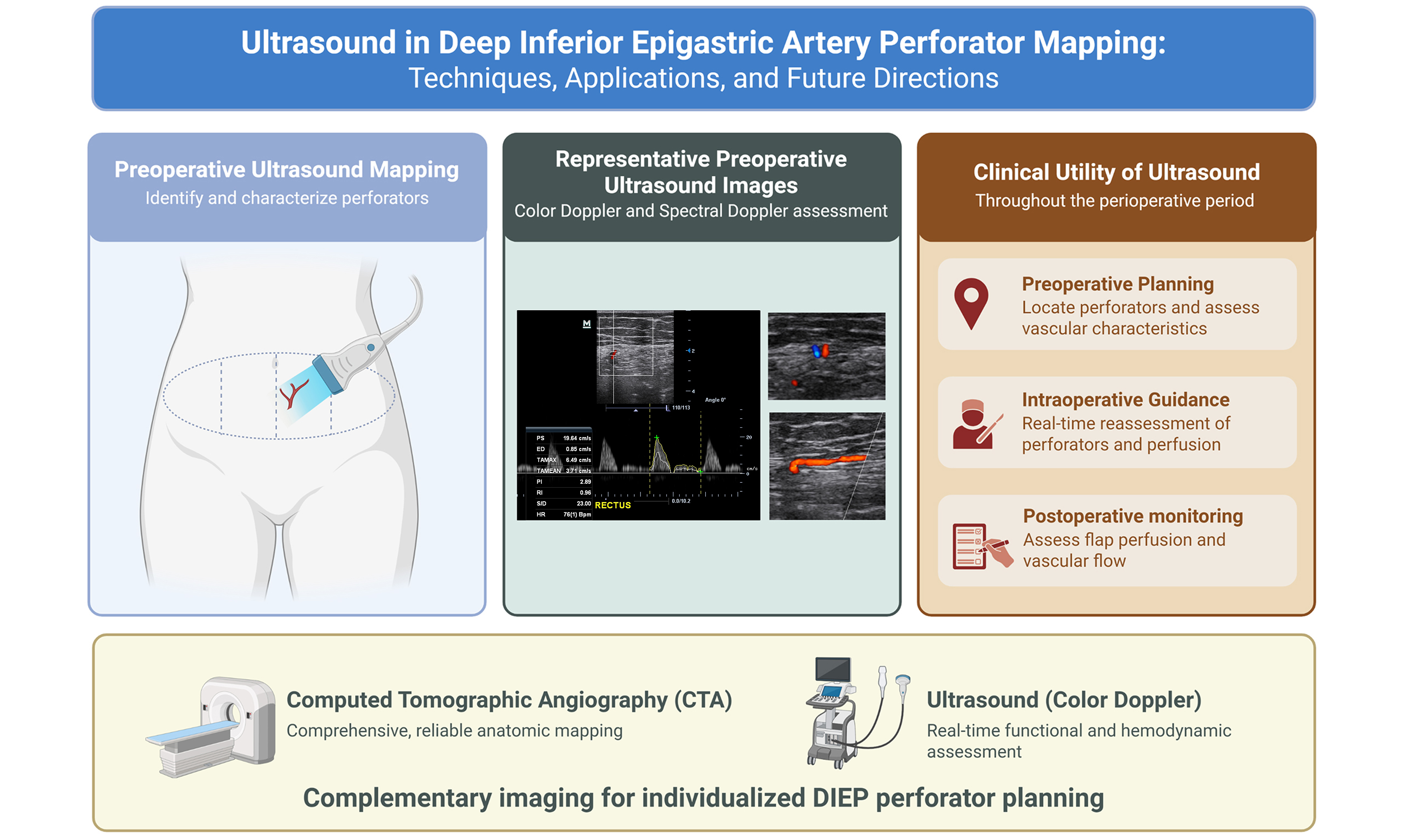
The deep inferior epigastric artery perforator (DIEP) flap is the gold standard for autologous breast reconstruction, with successful outcomes depending heavily on accurate perforator identification and selection. Computed tomographic angiography (CTA) remains the gold standard for preoperative planning; however, its limitations - including radiation exposure, contrast-related risks, and inability to provide real-time intraoperative assessment - have prompted increasing interest in ultrasound-based approaches. Ultrasound enables dynamic, real-time evaluation of both anatomical and physiological characteristics of perforator vessels, including caliber, flow velocity, and vascular resistance. Despite growing adoption, the literature describing standardized techniques and practical integration of ultrasound into plastic and reconstructive surgery workflows remains limited. This review synthesizes current evidence regarding ultrasound use in DIEP perforator mapping and presents a detailed, technique-oriented framework for its application in preoperative planning, intraoperative decision-making, and postoperative monitoring. Advantages, limitations, and emerging technologies are discussed. With appropriate training, ultrasound represents a powerful adjunct to established modalities, with the potential to enhance surgical precision, improve flap outcomes, and expand accessibility of perforator mapping and perioperative assessment in microsurgical breast reconstruction.
Keywords


 Download PDF
INTRODUCTION
The deep inferior epigastric perforator (DIEP) flap is widely considered the standard approach for autologous breast reconstruction. It offers superior aesthetic outcomes while preserving abdominal wall integrity. However, flap planning can be technically demanding due to variations in patient anatomy. As such, careful preoperative mapping is a key step in reducing complications and improving surgical efficiency[1,2].
Over the past two decades, imaging options for DIEP planning have advanced considerably. Computed tomographic angiography (CTA) has been the most commonly used modality, as it offers comprehensive three-dimensional (3D) anatomical detail including perforator location, branching patterns, intramuscular course, and superficial venous communications[2]. The 2022 American College of Radiology (ACR) criteria classify CTA with intravenous contrast and contrast-enhanced magnetic resonance angiography (MRA) as usually appropriate for initial flap planning, while color Doppler ultrasound (CDU) is classified as usually not appropriate in the initial imaging planning[2]. Subsequent clinical studies have explored surgeon-performed ultrasound as a complementary or selected alternative approach[3-5].
Prior to the adoption of CTA as the gold standard for DIEP planning, CDU was another imaging modality, although it had lost popularity due to interobserver variability. There are, however, low but non-negligible radiation and contrast risks associated with CTA. These concerns, together with the increased adoption of ultrasound in many procedural practices, have contributed to the renewed interest in CDU[6]. While CTA can provide more information on the overall vascular anatomy in a static manner, ultrasound examination can offer dynamic and real-time functional assessment of blood flow and vessel pulsatility. In a 45-patient comparative study, CTA more accurately characterized intramuscular course (95% vs. 84%) and superficial venous communications (97% vs. 80%) compared to CDU[7]. Vessel caliber measurements differed significantly between CTA and intraoperative findings (P < 0.001), whereas a difference was not detected between CDU and surgery (P = 0.33)[7]. In a 2023 single-center randomized trial with 49 patients comparing CDU alone with CTA supplemented by CDU, the average deviation from intraoperative location was 0.6 cm vs. 1.0 cm (P < 0.001)[3].
Taken together, these findings support a more integrated approach. CTA provides a broad anatomical “roadmap” that is valuable for preoperative planning, while CDU may complement cross-sectional anatomical imaging and avoid additional radiation or contrast exposure when reassessment is warranted. This combination may be particularly useful in younger patients or in cases where repeat imaging is anticipated, where minimizing radiation exposure becomes an important consideration. Although these findings are encouraging, the available evidence remains heterogeneous. Much of the comparative literature consists of retrospective single-center studies with varying ultrasound expertise, making direct comparisons between modalities challenging. Consequently, imaging decisions should be individualized based on patient characteristics, institutional resources, and operator experience. Despite the advantages of CDU, ultrasound in DIEP flap planning remains underutilized, in part due to variability in operator experience and lack of standardized protocols. This review aims to (1) summarize current literature on ultrasound in perforator mapping; (2) compare ultrasound with established imaging modalities; and (3) propose a structured, technique-oriented framework for its integration into clinical practice.
METHODS
This narrative review encompasses ultrasound-based DIEP perforator mapping, relevant vascular anatomy, complementary imaging modalities, and perioperative applications. A PubMed search was conducted for publications from January 1980 through October 2026. The most recent search was performed on October 15, 2026. The search retrieved 584 records; the complete search string is provided in Supplementary Materials. No specific language or study-design filters were applied to the search query. OpenEvidence was used as a supplementary tool for additional literature searches, though the exact searches were not recorded. Eligible publications addressed DIEP anatomy, imaging, perforator selection, venous drainage, or perioperative assessment. Non-DIEP studies were included in the emerging technologies section. The first author led literature screening, and coauthors contributed to selection and interpretation. No substantive disagreements arose. Selection followed a targeted narrative process without independent duplicate screening or exhaustive assessment of all retrieved records. References were checked using bibliographic records, abstracts, and accessible full texts. The final review cited 46 articles. No formal risk-of-bias assessment or new meta-analysis was conducted.
MAIN TEXT
Anatomical considerations in DIEP perforator selection
Successful DIEP flap breast reconstruction requires a clear understanding of the anterior abdominal wall vascular anatomy. As a branch of the external iliac artery, the deep inferior epigastric artery (DIEA) gives rise to perforators that exhibit substantial interpatient anatomic variability. These differences can meaningfully influence operative strategy and outcomes[2,8]. Representative ultrasound anatomy of the external iliac vessels and the DIEA is shown in Figure 1.
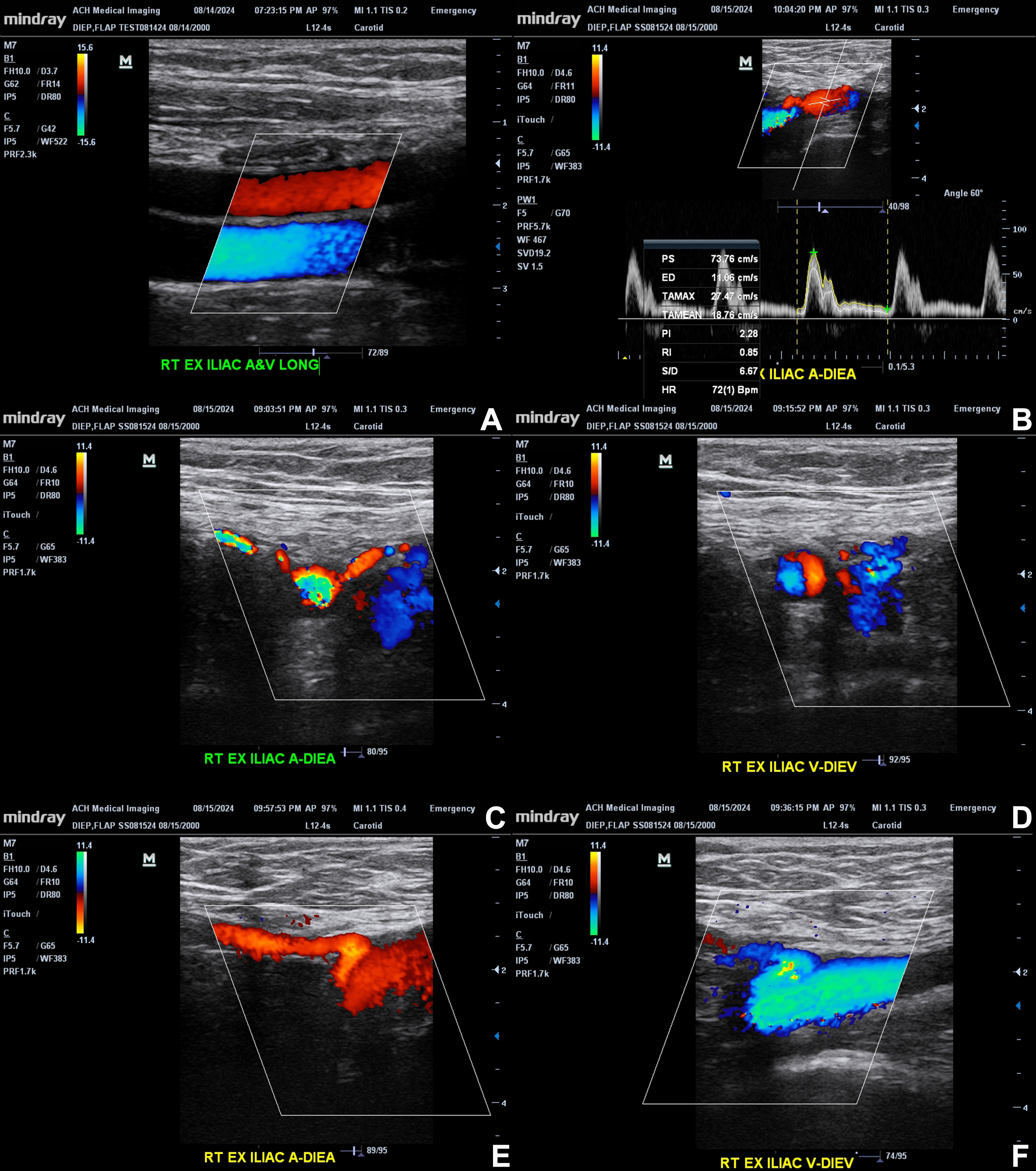
Figure 1. Ultrasound anatomy of the external iliac vessels and DIEA. Representative longitudinal and transverse color Doppler images demonstrate the right external iliac artery and vein, as well as the origin and course of the DIEA and DIEV. The top panels (A and B) show the external iliac vessels with clear arterial-venous differentiation based on flow direction. Spectral Doppler (B) confirms arterial waveforms within the DIEA, including peak systolic and end-diastolic velocities. Middle panels (C and D) illustrate the branching of the DIEA from the external iliac artery and accompanying venous structures. Bottom panels (E and F) further depict color flow within the DIEA and DIEV, highlighting vessel caliber and flow characteristics. These images demonstrate the ability of CDU to identify vessel origin, assess flow dynamics, and distinguish arterial from venous structures in preoperative DIEP flap planning. The original figure was created by the authors. Images have not been previously published. Figure 1 contains illustrative ultrasound images acquired by the first author during self-scanning; no patient images are included. A: Artery; A&V: artery and vein; CDU: color Doppler ultrasound; DIEA: deep inferior epigastric artery; DIEP: deep inferior epigastric perforator; DIEV: deep inferior epigastric vein; ED: end-diastolic velocity; EX: external; G: gain; HR: heart rate; LONG: longitudinal; PI: pulsatility index; PS: peak systolic velocity; PW: pulsed-wave Doppler; RI: resistive index; S/D: systolicto-diastolic ratio; TAMAX: time-averaged maximum velocity; TAMEAN: time-averaged mean velocity; RT: right; V: vein; bpm: beats per minute.
DIEA origin and course
After branching from the external iliac artery, the DIEA travels superiorly within the rectus sheath. In a retrospective CTA study of 184 patients, the DIEA most commonly originated medially, descended briefly, and then turned cephalad. The average descending segment measured 11.29 mm[9]. Prior abdominal surgery was associated with a longer descending course (12.22 mm vs. 9.86 mm, P < 0.01), which may complicate pedicle dissection[9]. The vessel’s branching pattern - Type I (single trunk), Type II (bifurcating), or Type III (trifurcating) - also has practical implications. Trifurcating patterns, for example, are linked to greater transverse spread (P = 0.0002) in a cadaveric study and may require more extensive rectus muscle dissection[10]. These anatomical observations, however, do not establish differences in clinical morbidity.
Medial vs. lateral row perforators
Perforators are typically grouped into medial and lateral rows, each with distinct perfusion characteristics[11]. Medial row perforators tend to provide more central perfusion and more reliably supply zone IV, whereas lateral row perforators primarily perfuse ipsilateral tissue and less frequently cross the midline[12,13]. Correspondingly, medial row perforators support a larger vascular territory on average (296 cm2 vs.
Perforators are typically grouped into medial and lateral rows, each with distinct perfusion characteristics[11]. Medial row perforators tend to provide more central perfusion and more reliably supply zone IV, whereas lateral row perforators primarily perfuse ipsilateral tissue and less frequently cross the midline[12,13]. Correspondingly, medial row perforators support a larger vascular territory on average (296 cm2 vs.
Periumbilical dominance
DIEP flap perforators commonly cluster in the periumbilical region[11,16]. In a CTA study of 180 patients, each hemiabdomen had an average of 2.9 ± 1.6 perforators, with the majority (80.5%) located below the umbilicus, and larger-caliber vessels were closer to the umbilicus[16]. This distribution makes the periumbilical area a key focus during perforator selection.
Intramuscular vs. septocutaneous course
Most DIEP perforators are musculocutaneous (33%-100%), and in many cases, they have a relatively short intramuscular course (< 4 cm) (61%-80%), though longer courses are not uncommon (9%-26%)[8]. Septocutaneous variants, such as paramedian or tendinous perforators, are generally preferred when present, given more straightforward dissection[8]. The length and complexity of the intramuscular course are closely linked to operative time. Longer or stepwise paths can significantly prolong dissection, whereas paramedian septocutaneous perforators often allow more efficient elevation[17,18].
Venous anatomy
Venous drainage occurs through both the deep venous system (venae comitantes) and the superficial inferior epigastric vein (SIEV), though the latter has historically been underemphasized[12,19]. In a retrospective MRA study of 59 patients, communication between the superficial and deep venous systems was identified in 117/118 hemiabdomens; mean diameters of the SIEV and dominant venous perforator were 3.8 and 2.8 mm, respectively[20].
Incorporating both venous systems can potentially improve outcomes. A retrospective series comparing 130 flaps with deep-system-only drainage and 114 with combined superficial and deep drainage showed lower flap loss in the dual-system group (2.5% in deep-system-only vs. 0% in dual-system; P = 0.031)[21]. A meta-analysis of 13 comparative studies demonstrated that venous supercharging with the superficial system reduces venous congestion [risk ratio (RR) of 0.12, 95% confidence interval (CI): 0.04-0.34], partial flap necrosis (RR 0.50, 95% CI: 0.30-0.84), total flap necrosis (RR 0.31, 95% CI: 0.11-0.85), and return to surgery for perfusion-related complications (RR 0.45, 95% CI: 0.21-0.99)[22]. Although these findings suggest a benefit, a subsequent systematic review rated the evidence of reducing flap complications with prophylactic venous supercharging as low certainty without clinical signs of venous congestion[23]. Evidence supporting preoperative imaging or measurements to identify patients who would benefit from augmentation was of very low certainty[23]. Future studies may be needed to clarify the evidence. Nevertheless, preoperative imaging can help identify patients who may benefit from planned venous augmentation[22].
Clinical implications
These anatomical factors, such as perforator location, intramuscular course, proximity to the umbilicus, and venous anatomy, directly impact pedicle length, ease of dissection, and flap perfusion reliability. Thoughtful perforator selection can improve efficiency while minimizing donor-site morbidity[15-17]. In this context, CTA-based planning has demonstrated high accuracy for perforator localization, with reported sensitivity approaching 99% and mean localization error within 1 cm[24].
Of note, most anatomical data summarized in this section are derived from cadaveric studies or retrospective imaging analyses. While these investigations have substantially improved understanding of perforator anatomy, relatively few studies directly correlate individual anatomic characteristics with clinically meaningful reconstructive outcomes.
Imaging modalities for DIEP planning: strengths and limitations
CTA
CTA is the most commonly used imaging modality for DIEP flap planning, as it provides 3D visualization of vascular anatomy[2,24]. A 2025 systematic review and meta-analysis found that CTA-guided reconstruction is associated with shorter operative times (by 65 min), lower flap loss (RR 0.37; 95% CI: 0.18-0.78), and fewer reoperations (RR 0.55; 95% CI: 0.33-0.89) compared to Doppler ultrasound[1]. CTA also demonstrates high accuracy for perforator localization, with 99% sensitivity and a mean error ≤ 1 cm[24].
CTA is particularly effective for assessing intramuscular perforator course (95% accuracy) and identifying superficial venous connections (97% accuracy), the absence of which has been associated with increased flap morbidity (P = 0.009)[7]. It also clearly delineates DIEA branching patterns and is especially useful in patients with prior abdominal surgery, where anatomy may be altered[2].
Limitations include exposure to ionizing radiation, the need for iodinated contrast, and a lack of real-time hemodynamic assessment[6]. CTA is also less accurate for perforator caliber measurement compared to intraoperative findings (P < 0.0001), which may affect perforator selection[7].
CDU
CDU has gained renewed interest due to the general increased adoption of ultrasound in a wide array of procedural and non-procedural practices, as well as concerns regarding radiation exposure[6]. It enables real-time, dynamic assessment without radiation or contrast and allows for the evaluation of flow characteristics and pulsatility[4]. CDU can also be performed by the operating surgeon given appropriate training and expertise[4]. In a comparative study, CDU caliber measurements did not differ significantly from intraoperative measurements (P = 0.33), whereas CTA measurements showed a statistically significant difference (P < 0.0001)[7]. A randomized trial demonstrated that CDU is non-inferior to CTA+CDU for perforator localization, with smaller deviation from intraoperative position (0.6 cm vs. 1.0 cm, P < 0.001)[3]. Cohort data also show a stronger correlation with intraoperative perforator selection for CDU (r = 0.9987) compared to CTA (r = 0.01)[6]. Limitations include operator dependence, lower accuracy in assessing intramuscular course (84% vs. 95% for CTA) and superficial venous connections (80% vs. 97%), and lack of a comprehensive anatomical overview[7]. While observational studies tend to favor CTA, randomized data show no clear difference, suggesting that CTA’s perceived advantage may be influenced by confounding factors[1].
MRA
MRA provides high-resolution, cross-sectional imaging without radiation. Meta-analyses show the strongest agreement with intraoperative findings (mean 97%; 95% CI: 86%-100%) among available modalities[25]. It offers excellent soft tissue contrast and can evaluate both arterial and venous anatomy[2]. Its drawbacks, however, include longer scan times, higher cost, limited availability, and contraindications in patients with certain implants. Although MRA demonstrates excellent diagnostic performance, the current literature is relatively limited compared with CTA; its higher cost and lower availability continue to restrict widespread clinical adoption.
Handheld doppler
Handheld Doppler is the most accessible option but is less sensitive than CTA (69% vs. 94%) and identifies fewer perforators per patient (10.4 vs. 14.8)[26]. It is best suited for intraoperative and postoperative use rather than detailed preoperative planning.
Dynamic infrared thermography (DIRT)
DIRT is an emerging modality that detects cutaneous hot spots corresponding to perforators while providing angiosome-level perfusion information. Sensitivity (92%) is comparable to CTA (94%) and higher than handheld Doppler (69%), though it cannot assess intramuscular course and remains limited by a smaller evidence base[2,26].
Extended reality technologies (XR)
XR technologies - including virtual reality (VR), augmented reality (AR), and mixed reality (MR) - enhance visualization of existing imaging rather than generating new data. VR enables immersive 3D exploration with a strong correlation to CTA (R = 0.96)[27]. AR allows intraoperative projection of perforator anatomy, reducing flap harvest time[28]. MR platforms, such as Microsoft HoloLens, enable interactive evaluation of intramuscular anatomy[29]. Its limitations include spatial registration error due to soft tissue deformation, with displacement greater than 3 mm in 30% of cases[30]. Most studies evaluating XR technologies are early feasibility investigations with small sample sizes and limited clinical validation. At present, these technologies should be considered exploratory adjuncts rather than established components of routine DIEP planning. Although several AR, VR, and MR studies have been performed specifically in DIEP reconstruction, much of the broader literature remains limited to technical feasibility studies and other microsurgical applications.
Complementary roles and practical approach
No single modality fulfills all requirements for DIEP planning. CTA remains the primary modality with the strongest outcome data. MRA serves as an alternative when CTA is contraindicated. CDU provides real-time hemodynamic information and complements cross-sectional imaging. DIRT offers a radiation-free method for perforator localization, while XR technologies enhance spatial understanding and intraoperative navigation but depend on underlying imaging and technological quality. Optimal use is multimodal and tailored to institutional resources, expertise, and patient factors [Table 1]. Future comparative studies evaluating standardized imaging protocols and patient-centered outcomes will be important to better define the incremental benefit of combining multiple imaging modalities.
Key imaging modalities for DIEP perforator mapping
| Modality | Strengths | Limitations | Key metrics |
| CTA[1,2,31] | Gold standard; rapid, reproducible; comprehensive assessment of perforator size, location, DIEA branching, and intramuscular course; strongest outcomes evidence | Ionizing radiation; iodinated contrast required; no hemodynamic flow data; static images | Partial flap loss OR 0.26; total flap loss OR 0.30 vs. no CTA; RR 0.37 for flap loss vs. CDU; -65 min operative time vs. Doppler ultrasound |
| MRA[2,25] | No ionizing radiation; equivalent ACR rating to CTA; highest agreement with intraoperative findings; pPCA technique avoids CTA vessel overestimation; image quality improves with higher BMI | Longer acquisition time; higher cost; reduced availability; gadolinium contrast required; motion artifact susceptibility | 97% mean agreement with intraoperative findings (95% CI: 86%-100%) |
| CDU[3,4,25] | Real-time hemodynamic assessment; no radiation or contrast; bedside/surgeon-performed; non-inferior to CTA+CDU for localization in RCT | Operator-dependent; lowest agreement with intraoperative findings; limited intramuscular course detail | 74% mean agreement with intraoperative findings (95% CI: 67%-81%); 0.6 cm mean localization deviation; learning curve ~7 patients |
| Handheld doppler[26] | Widely available; inexpensive; simple to use | Low sensitivity; no anatomic detail on intramuscular course, branching, or caliber | 69% sensitivity vs. 94% CTA and 92% DIRT |
| DIRT[26] | No radiation or contrast; provides angiosome hemodynamic data; sensitivity comparable to CTA | Cannot assess intramuscular course or branching; limited evidence base; requires cold challenge protocol | 92% sensitivity; comparable to CTA (94%) |
| VR[27,32] | Immersive 3D perforator exploration; intuitive spatial understanding; high correlation with CTA | Requires CTA/MRA data; no new imaging data generated; hardware cost; setup time | R = 0.96 correlation with CTA (unilateral); 0.23 cm mean localization error |
| AR[28,30,33] | Intraoperative projection of anatomy onto patient; reduces flap harvest time; usable in sterile field (Meta Quest 3) | Dependent on CTA/MRA quality; spatial registration error from skin deformation; early-stage evidence | Registration error exceeds 3 mm in 30% of cases |
| MR[29,34,35] | Interactive holographic dissection; real-time cinematic rendering; anchored to real anatomy | Registration error (median 9-13.5 mm displacement); observer/angle-dependent accuracy; limited clinical validation | Median translational displacement 9-13.5 mm depending on vantage point |
Ultrasound principles relevant to perforator mapping
Ultrasound-based perforator mapping relies on the Doppler effect, which is the frequency shift of sound waves reflected from moving red blood cells to identify, localize, and characterize DIEA perforators[2]. Understanding probe selection, Doppler modalities, and hemodynamic interpretation is essential for accurate and reproducible assessment. Representative color and spectral Doppler findings of the DIEA and deep inferior epigastric vein (DIEV) are shown in Figure 2.
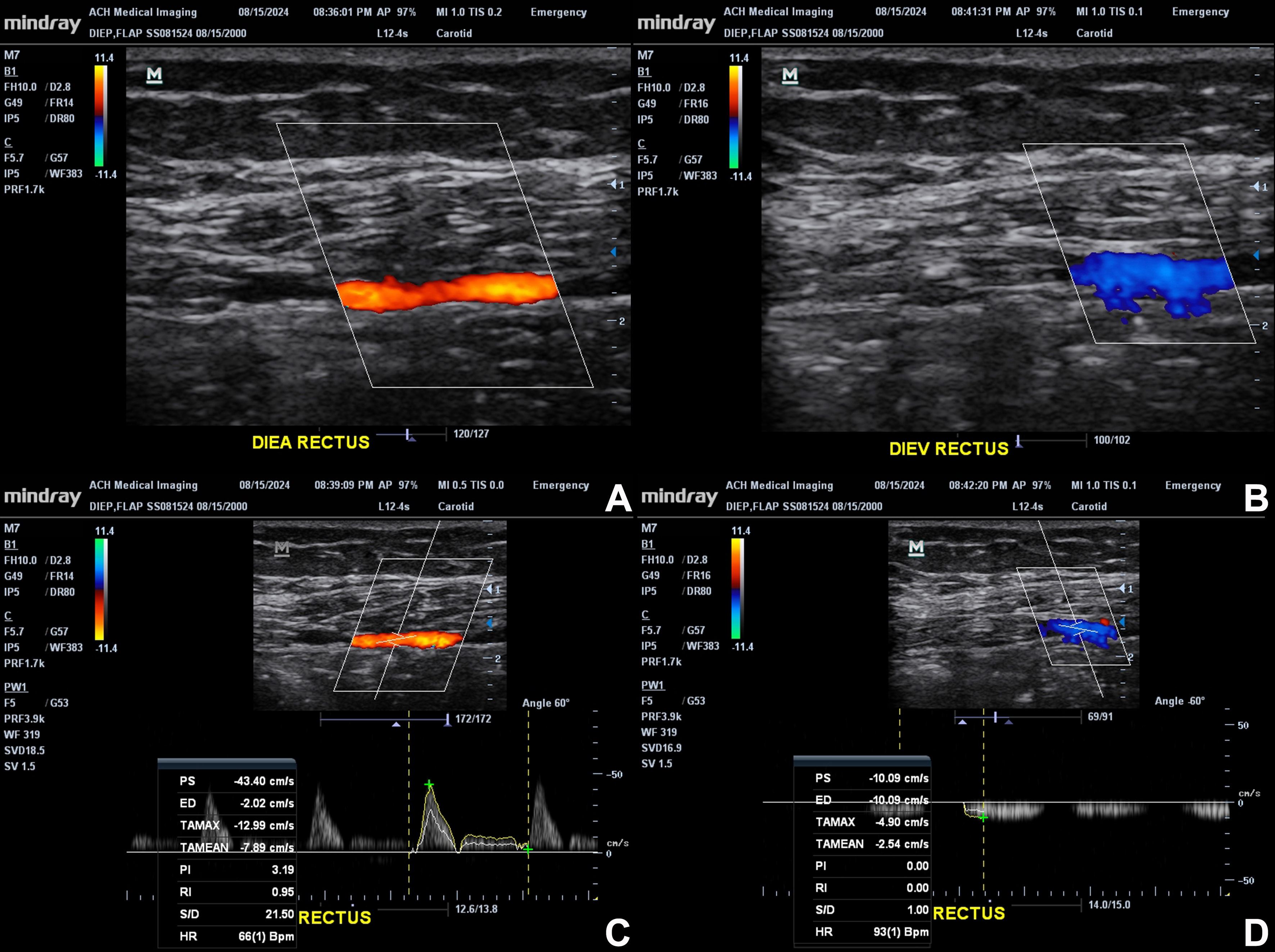
Figure 2. Color and spectral Doppler assessment of the intramuscular DIEA and DIEV. Representative images demonstrate the DIEA and DIEV as they course within the rectus muscle. Top panels (A and B) show color Doppler identification of the DIEA (left) and DIEV (right), with clear differentiation of arterial and venous flow based on direction and color encoding. Bottom panels (C and D) include corresponding spectral Doppler waveforms: the DIEA demonstrates a pulsatile arterial waveform with defined systolic peaks, while the DIEV shows low-velocity, non-pulsatile venous flow. These images highlight the ability of CDU to characterize intramuscular vessel course, confirm vessel identity, and assess flow dynamics during DIEP flap planning. Original figure created by the authors. Images have not been previously published. Figure 2 contains illustrative ultrasound images acquired by the first author during self-scanning; no patient images are included. CDU: Color Doppler ultrasound; DIEA: deep inferior epigastric artery; DIEP: deep inferior epigastric perforator; DIEV: deep inferior epigastric vein; ED: end-diastolic velocity; G: gain; HR: heart rate; PI: pulsatility index; PS: peak systolic velocity; PW: pulsed-wave Doppler; RI: resistive index; S/D: systolic-to-diastolic ratio; SV: sample volume size; SVD: sample volume depth; TAMAX: time-averaged maximum velocity; TAMEAN: time-averaged mean velocity; bpm: beats per minute.
Probe selection and device settings
High-frequency linear-array probes (typically 5-15 MHz) are standard for perforator mapping[1,31]. Higher frequencies (≥ 10 MHz) provide improved spatial resolution for superficial structures, enabling detection of vessels < 0.5 mm at the fascial level[2]. However, increased frequency reduces penetration depth. In patients with thicker subcutaneous tissue, lower frequencies (5-8 MHz) may be required to visualize the intramuscular course of perforators within the rectus abdominis[31]. Optimal settings for low-flow microvascular imaging include shallow depth (2-5 cm), low pulse repetition frequency (0.5-1.5 kHz or 3-10 cm/s), high color gain, and a low or disabled wall filter (~50 Hz)[36]. These parameters improve the detection of small, slow-flow vessels while reducing motion artifact.
Doppler modalities
Multiple Doppler modes are used in perforator mapping. Color Doppler overlays directional flow information onto B-mode imaging, allowing real-time identification of perforator location and course[2,3]. Power Doppler is more sensitive to low-velocity flow but does not provide directional information and may overestimate vessel diameter[31]. In direct comparison, color-coded duplex sonography (CCDS) produced more accurate diameter measurements (mean 1.65 ± 0.45 mm) than power Doppler (1.31 ± 0.24 mm) when correlated with intraoperative findings[37]. Spectral (pulsed-wave) Doppler enables quantitative waveform analysis, providing key hemodynamic data for perforator evaluation[36,37]. Duplex ultrasound combines structural imaging with spectral Doppler, allowing simultaneous assessment of vessel anatomy and flow. Handheld continuous-wave Doppler detects flow but lacks anatomic localization and quantitative capability, with lower sensitivity (69%) compared to CDU[26].
Hemodynamic parameters
Spectral Doppler provides quantitative parameters relevant to perforator selection. Peak systolic velocity (PSV) reflects maximal flow and serves as an indicator of arterial inflow to the perforator. In perforator flap studies, mean PSV at the fascial level is approximately 17 cm/s, with a corresponding end-diastolic velocity (EDV) of approximately 5 cm/s and a resistive index (RI) of approximately 0.7, although reported values vary depending on flap type and vessel selection[37]. A PSV threshold of ≥ 20 cm/s has been associated with successful perforator dissection in non-DIEP perforator flap studies, although DIEP-specific thresholds remain to be established[38]. The RI, calculated as (PSV - EDV)/PSV, measures distal vascular impedance, with reported mean values around 0.7[31]. The Pulsatility Index (PI) reflects resistance to flow and has demonstrated utility in postoperative monitoring, with elevated values associated with venous congestion and flap compromise[39].
Emerging microvascular flow imaging
Microvascular flow imaging (MVFI) techniques improve the detection of slow-flow, small-caliber vessels beyond conventional Doppler[40]. Technologies such as Superb Microvascular Imaging (SMI) and Micro Flow Imaging (MFI) use advanced clutter suppression to isolate low-velocity blood flow from tissue motion, enabling visualization of microvasculature without contrast[40]. SMI has been used to assess vascularity in a small cohort of buried latissimus dorsi flaps after reconstruction[40].
Contrast-enhanced ultrasound (CEUS) uses intravenously administered microbubbles that strongly reflect the sound waves to create bright, real-time images of blood vessels and perfusion. Together with 3D image rendering, it successfully identified all selected perforators in a retrospective series of 32 patients who underwent perforator flap reconstruction[41]. However, most of these published studies evaluating MVFI have not been performed specifically in DIEP reconstruction, and their application to DIEP perforator mapping is currently inferred from broader microsurgical and vascular imaging experience. B-flow imaging has also demonstrated high sensitivity for perforator detection, although DIEP-specific validation remains limited[42].
Standard ultrasound technique for DIEP perforator mapping
The examination begins by identifying the rectus abdominis muscles, usually within 5 cm lateral to the umbilicus. The umbilicus serves as a central reference point, as perforators are most commonly concentrated in the periumbilical region. Medial row perforators are typically located 1-3 cm lateral to the umbilicus, while lateral row perforators lie further laterally relative to the rectus muscle, approximately 4-6 cm from the midline[11]. A stepwise example of ultrasound-based perforator identification and hemodynamic assessment is illustrated in Figure 3.
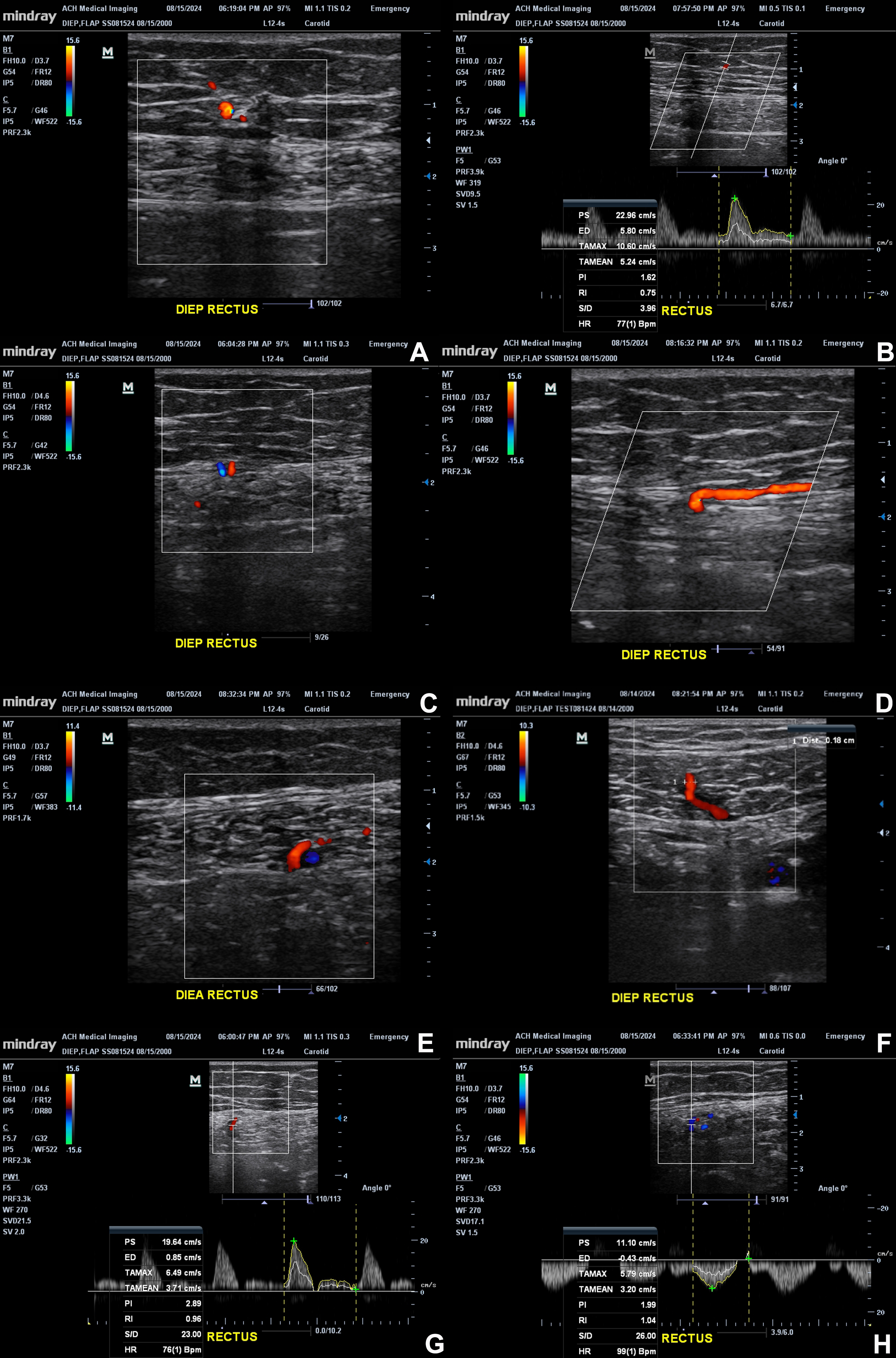
Figure 3. Representative color Doppler images demonstrate perforators arising from the DIEA and traversing the rectus muscle toward the anterior fascia and subcutaneous tissue. (A) Color Doppler visualization of a perforator within the subcutaneous tissue; (B) Pulsed-wave spectral Doppler assessment of the subcutaneous perforator segment; (C) Cross-sectional view of the perforator at its passage through the anterior rectus sheath, connecting the subcutaneous and intramuscular segments; (D) Longitudinal view showing the perforator coursing within the subcutaneous tissue along the anterior rectus sheath and traversing the sheath; (E) A deeper vascular segment within the rectus; (F) A longitudinal/oblique view of a perforator changing direction, with a caliber of 0.18 cm; (G) Spectral Doppler sampling of an arterial segment, with a sharp systolic peak and little forward end-diastolic flow; (H) Spectral sampling of another vessel segment. These images highlight the integration of anatomic and functional evaluation during perforator mapping. The original figure was created by the authors. Images have not been previously published. Figure 3 contains illustrative ultrasound images acquired by the first author during self-scanning; no patient images are included. A: Artery; A&V: artery and vein; CDU: color Doppler ultrasound; DIEA: deep inferior epigastric artery; DIEP: deep inferior epigastric perforator; DIEV: deep inferior epigastric vein; ED: end-diastolic velocity; EX: external; G: gain; HR: heart rate; LONG: longitudinal; PI: pulsatility index; PS: peak systolic velocity; PW: pulsed-wave Doppler; RI: resistive index; S/D: systolicto-diastolic ratio; TAMAX: time-averaged maximum velocity; TAMEAN: time-averaged mean velocity; RT: right; V: vein; bpm: beats per minute.
Because perforators follow a 3D and non-linear course, CDU can be used to trace vessels in real time. True perforators originate from the deep inferior epigastric system, pass through the rectus muscle and anterior rectus sheath, and enter the subcutaneous tissue. Vessels that remain intramuscular without penetrating the fascia should not be considered suitable. High-resolution CDU probes can detect vessels < 0.5 mm at the fascial level, allowing direct visualization of perforator branching[43].
After identifying a candidate perforator near the deep surface of the rectus muscle, the image is frozen, and vessel diameter is measured. The perforator is then followed cranially using CDU, with continuous adjustments in probe angle and pressure to maintain visualization. As the perforator approaches and penetrates the rectus sheath, it is reassessed at the fascial and subcutaneous levels.
At this point, vessel diameter is remeasured, and spectral (pulsed-wave) Doppler is used to evaluate flow. Accurate hemodynamic assessment requires identifying a segment where the perforator ascends vertically into the subcutaneous tissue, minimizing angle-related error. Stable probe positioning is essential to ensure reliable measurements of flow velocity and resistance.
Evaluation should also include the superficial venous system. Absence of communication between the SIEV and the deep venous system via perforator venae comitantes has been associated with increased risk of postoperative venous congestion (P < 0.0001)[44]. Although CTA is more accurate than CDU for detecting superficial venous connections (97% vs. 80%), limited visualization on ultrasound may warrant additional cross-sectional imaging[7].
Identified perforators are then marked on the skin surface to guide flap design and intraoperative dissection. In a randomized trial, surgeon-performed CDU achieved a mean localization error of 0.6 cm compared to intraoperative findings, which was more accurate than CTA-based mapping (1.0 cm, P < 0.001)[3]. Recently, D’Orsi et al. described a standardized, microsurgeon-performed duplex ultrasound protocol for DIEP perforator mapping in a prospective series of 62 flaps. The dominant perforator identified preoperatively corresponded to the intraoperative selection in approximately 81% of flaps, supporting the feasibility and reproducibility of a structured surgeon-performed ultrasound workflow[5]. However, this represents Level IV evidence from a single institution, and broader validation is warranted before widespread adoption.
Integration of ultrasound into DIEP reconstruction: a practical framework
Role across the perioperative flow
Ultrasound is applicable throughout all phases of DIEP flap reconstruction. Preoperatively, CDU enables identification, localization, caliber measurement, and hemodynamic assessment of perforators[2,43]. Intraoperatively, it can confirm perforator patency and guide dissection when findings differ from preoperative imaging[45]. Postoperatively, portable CDU allows serial monitoring of pedicle flow using the PI, which typically decreases over the first six postoperative days and provides an objective measure of perfusion, with elevated values associated with venous insufficiency (P = 0.03)[39].
Proposed imaging strategy
A tiered, patient-specific imaging approach leverages the strengths of each modality. In straightforward primary reconstructions, surgeon-performed CDU can be used as the initial mapping tool alongside established CTA or contrast-enhanced MRA modalities. In more complex cases - such as prior abdominal surgery, scarred tissue, or planned bipedicled flaps - CTA remains the preferred first-line modality due to its ability to define DIEA branching patterns, intramuscular course, and superficial venous anatomy with higher accuracy (95%-97%) than CDU (80%-84%)[7]. In this framework, CDU serves as a complementary tool, providing real-time hemodynamic assessment and more accurate caliber measurement, while CTA supplies the broader anatomic overview[6,7]. A practical summary of proposed imaging selection is provided in Table 2. Please note that this is not a validated algorithm or guideline.
Authors’ proposed practical framework for individualized DIEP imaging selection
| Clinical scenario | Preferred imaging | Rationale |
| Routine primary DIEP reconstruction | CTA or contrast-enhanced MRA; selected expert-performed CDU | CTA and MRA have an established initial-imaging role. While CDU offers dynamic assessment, the evidence does not support treating it as a universal initial default |
| Previous abdominal surgery or complex anatomy | CTA or appropriate MRA, with selective CDU | Superior evaluation of branching pattern, intramuscular course, and venous anatomy with targeted CDU to clarify specific findings |
| Multiple candidate perforators | Cross-sectional mapping with selective CDU | Combines anatomy and functional information |
| CTA contraindicated (contrast allergy, renal dysfunction) | Appropriate MRA technique or selected expert-performed CDU | Avoids iodinated contrast while maintaining vascular assessment |
| Intraoperative discrepancy between imaging and operative findings | CDU | Real-time reassessment of perforator characteristics |
When ultrasound changes management
CDU most directly impacts surgical decision-making in several scenarios. When CTA identifies multiple perforators of similar size, CDU-derived hemodynamic parameters, particularly PSV, can help identify the dominant vessel[36]. CDU also provides more accurate caliber measurements than CTA, which tends to overestimate vessel diameter, thereby helping to avoid the selection of suboptimal perforators[7].
In patients for whom CTA is contraindicated - such as those with contrast allergy, renal impairment, or concerns about radiation exposure - CDU may be a safe alternative with expertise[7]. Additionally, when discrepancies arise between preoperative imaging and intraoperative findings - situations associated with increased flap loss - CDU enables immediate reassessment to guide perforator selection[6].
Barriers to adoption
Adoption of CDU is limited by operator dependence, a learning curve of approximately seven patients to achieve CTA-equivalent accuracy, lack of standardized training pathways, and limited large-scale randomized data[1,4]. Although meta-analyses favor CTA in terms of flap loss and operative time, these findings are largely based on observational studies, while randomized data show no significant differences, suggesting that differences in operator experience and institutional expertise may contribute to the observed differences in outcomes[1]. Beyond the learning curve itself, inter-operator variability remains an important limitation. Image acquisition, probe positioning, machine settings, and interpretation may differ substantially among operators and across institutions, depending on examiner experience, equipment, and scanning technique. Standardized scanning protocols and formal training pathways may improve reproducibility across institutions.
Future directions
Artificial intelligence (AI)-assisted ultrasound analysis is an evolving area. Although early studies demonstrate encouraging proof-of-concept results, clinical validation in DIEP reconstruction remains limited, and prospective studies are needed before AI-assisted ultrasound can be routinely incorporated into DIEP reconstruction[46]. Integration of ultrasound with AR platforms may eventually allow real-time projection of perforator anatomy onto the surgical field[45]. However, most available studies remain technical feasibility reports rather than clinical outcome studies, and further prospective validation is required before widespread clinical adoption. Future research should prioritize prospective multicenter studies evaluating clinically meaningful outcomes rather than imaging accuracy alone, as improvements in perforator visualization do not necessarily translate into improved reconstructive outcomes.
CONCLUSION
Ultrasound can be a valuable adjunct in DIEP flap perforator mapping, offering real-time assessment of both vascular anatomy and hemodynamics without radiation or contrast exposure. While CTA remains the current standard due to its comprehensive anatomical visualization and strong outcomes data, ultrasound provides complementary strengths, particularly in perforator characterization, intraoperative flexibility, and postoperative monitoring. A multimodal approach that integrates CTA for global anatomical mapping with CDU for dynamic, point-of-care evaluation may optimize perforator selection and surgical decision-making. With growing evidence supporting its accuracy and utility, the broader adoption of ultrasound will depend on standardized techniques, dedicated training, and further prospective validation.
DECLARATIONS
Acknowledgments
The Graphical Abstract was created with BioRender [Song, S. (2026) https://BioRender.com/odglxin].
Authors’ contributions
Made substantial contributions to the conception and design of the study and performed data analysis and interpretation: Song SS, Schalet BJ, Lee ZH
Performed data acquisition and provided administrative, technical, and material support: Song SS, Schalet BJ, Lee ZH
Availability of data and materials
Not applicable.
AI and AI-assisted tools statement
During the preparation of this manuscript, AI tools ChatGPT (OpenAI; version 6 Sol, released 2026-09-22) were used solely for language editing and editorial assistance. These tools did not influence the study design, data collection, analysis, interpretation, or the scientific content of the work. All authors take full responsibility for the accuracy, integrity, and final content of the manuscript.
Financial support and sponsorship
None.
Conflicts of interest
All authors declared that there are no conflicts of interest.
Ethical approval and consent to participate
Not applicable.
Consent for publication
Not applicable.
Copyright
© The Author(s) 2026.
Supplementary Materials
REFERENCES
1. Collaco BG, Nogueira R, Magalhães PLM, et al. Doppler ultrasound versus computed tomography angiography prior to deep inferior epigastric perforator flap breast reconstruction: an updated systematic review and meta-analysis. J Plast Reconstr Aesthet Surg. 2026;113:532-43.
2. Singh N, Aghayev A, Ahmad S, et al. ; Expert Panel on Vascular Imaging. ACR appropriateness criteria® imaging of deep inferior epigastric arteries for surgical planning (breast reconstruction surgery): 2022 update. J Am Coll Radiol. 2022;19:S357-63.
3. Bajus A, Streit L, Kubek T, et al. Color Doppler ultrasound versus CT angiography for DIEP flap planning: a randomized controlled trial. J Plast Reconstr Aesthet Surg. 2023;86:48-57.
4. Bajus A, Kubek T, Dražan L, et al. Surgeon-conducted color Doppler ultrasound deep inferior epigastric artery perforator mapping: a cohort study and learning curve assessment. J Plast Reconstr Aesthet Surg. 2023;76:105-12.
5. D’Orsi G, Longo B, Farcomeni A, Giacalone M, La Padula S, Cervelli V. Duplex ultrasound-based step-by-step perforator mapping by a microsurgeon for DIEP flap planning: a prospective case series. J Plast Reconstr Aesthet Surg. 2026;117:94-103.
6. Mijuskovic B, Tremp M, Heimer MM, et al. Color Doppler ultrasound and computed tomographic angiography for perforator mapping in DIEP flap breast reconstruction revisited: a cohort study. J Plast Reconstr Aesthet Surg. 2019;72:1632-9.
7. Cina A, Salgarello M, Barone-Adesi L, Rinaldi P, Bonomo L. Planning breast reconstruction with deep inferior epigastric artery perforating vessels: multidetector CT angiography versus color Doppler US. Radiology. 2010;255:979-87.
8. Ireton JE, Lakhiani C, Saint-Cyr M. Vascular anatomy of the deep inferior epigastric artery perforator flap: a systematic review. Plast Reconstr Surg. 2014;134:810e-21.
9. Myung Y, Choi B, Yim SJ, et al. The originating pattern of deep inferior epigastric artery: anatomical study and surgical considerations. Surg Radiol Anat. 2018;40:873-9.
10. Rozen WM, Palmer KP, Suami H, et al. The DIEA branching pattern and its relationship to perforators: the importance of preoperative computed tomographic angiography for DIEA perforator flaps. Plast Reconstr Surg. 2008;121:367-73.
11. Cui H, Ding M, Mao Y, et al. Three-dimensional visualization for extended deep inferior epigastric perforator flaps. Ann Plast Surg. 2020;85:e48-53.
12. Schaverien M, Saint-Cyr M, Arbique G, Brown SA. Arterial and venous anatomies of the deep inferior epigastric perforator and superficial inferior epigastric artery flaps. Plast Reconstr Surg. 2008;121:1909-19.
13. Wong C, Saint-Cyr M, Mojallal A, et al. Perforasomes of the DIEP flap: vascular anatomy of the lateral versus medial row perforators and clinical implications. Plast Reconstr Surg. 2010;125:772-82.
14. Munhoz AM, Ishida LH, Sturtz GP, et al. Importance of lateral row perforator vessels in deep inferior epigastric perforator flap harvesting. Plast Reconstr Surg. 2004;113:517-24.
15. Hembd A, Teotia SS, Zhu H, Haddock NT. Optimizing perforator selection: a multivariable analysis of predictors for fat necrosis and abdominal morbidity in DIEP flap breast reconstruction. Plast Reconstr Surg. 2018;142:583-92.
16. Meyerov J, Ram R, Grinsell D. Refining the abdominal wall perforasome with clinical application. J Plast Reconstr Aesthet Surg. 2025;105:283-91.
17. Tokumoto H, Akita S, Kubo Y, et al. Does the difference in vascular anatomy in the multiple perforator DIEP flap affect the flap dissection time? Ann Plast Surg 2026;96:350-4.
18. Vandevoort M, Vranckx JJ, Fabre G. Perforator topography of the deep inferior epigastric perforator flap in 100 cases of breast reconstruction. Plast Reconstr Surg. 2002;109:1912-8.
19. Rozen WM, Pan WR, Le Roux CM, Taylor GI, Ashton MW. The venous anatomy of the anterior abdominal wall: an anatomical and clinical study. Plast Reconstr Surg. 2009;124:848-53.
20. Dortch J, Forte AJ, Bolan C, Kandel P, Perdikis G. Preoperative analysis of venous anatomy before deep inferior epigastric perforator free-flap breast reconstruction using ferumoxytol-enhanced magnetic resonance angiography. Ann Plast Surg. 2023;90:343-8.
21. Jabbour S, Youn R, Kim KG, et al. An algorithmic approach to dual-system venous drainage for DIEP flap breast reconstruction. Plast Reconstr Surg. 2024;154:1e-12.
22. Pignatti M, Pinto V, Giorgini FA, et al. Meta-analysis of the effects of venous super-drainage in deep inferior epigastric artery perforator flaps for breast reconstruction. Microsurgery. 2021;41:186-95.
23. Hansson E, Ramakrishnan V, Morgan M. A systematic review of the scientific evidence of venous supercharging in autologous breast reconstruction with abdominally based flaps. World J Surg Oncol. 2023;21:379.
24. Renzulli M, Clemente A, Brocchi S, et al. Preoperative computed tomography assessment for a deep inferior epigastric perforator (DIEP) flap: a new easy technique from the Bologna experience. Acta Radiol. 2021;62:1283-9.
25. Kiely J, Kumar M, Wade RG. The accuracy of different modalities of perforator mapping for unilateral DIEP flap breast reconstruction: a systematic review and meta-analysis. J Plast Reconstr Aesthet Surg. 2021;74:945-56.
26. Sonda R, Pandis L, Bassetto F, et al. Deep inferior epigastric perforator flap preoperative planning: a comparative analysis between dynamic infrared thermography, computerized tomography angiography, and hand-held Doppler. Microsurgery. 2022;42:649-58.
27. Di Via Ioschpe A, Golijanin B, Benharush D, Berl A, Perhulov V, Egozi D. Preoperative planning using virtual reality and computed tomography angiogram in deep inferior epigastric perforator flap breast reconstruction. J Plast Reconstr Aesthet Surg. 2023;87:161-9.
28. Wolbert TT, White AE, Han J, et al. The application of augmented reality technology in free flap reconstruction: a systematic review. Microsurgery. 2025;45:e70080.
29. Necker FN, Cholok DJ, Shaheen MS, et al. Holographic deep inferior epigastric perforator exploration in mixed reality using real-time cinematic rendering. Aesthetic Plast Surg. 2026;50:183-6.
30. Timóteo R, Pinto D, Martinho M, et al. Skin deformation analysis for pre-operative planning of DIEAP flap reconstruction surgery. Med Eng Phys. 2023;119:104025.
31. Rupra RS, Ruccia F, Daneshi K, et al. A systematic review and meta-analysis on computed tomography angiography mapping for deep inferior epigastric perforator flap breast reconstruction. Front Oncol. 2025;15:1600476.
32. Gacto-Sánchez P, Sicilia-Castro D, Gómez-Cía T, et al. Use of a three-dimensional virtual reality model for preoperative imaging in DIEP flap breast reconstruction. J Surg Res. 2010;162:140-7.
33. Edgcumbe PM, Jiang T, Ho JF, Martin ML, Stein MJ, Isaac KV. Intraoperative surgical guidance for DIEP flap harvest using augmented reality. Plast Reconstr Surg. 2025;155:932-5.
34. Wesselius TS, Meulstee JW, Luijten G, Xi T, Maal TJJ, Ulrich DJO. Holographic augmented reality for DIEP flap harvest. Plast Reconstr Surg. 2021;147:25e-9.
35. Cholok DJ, Fischer MJ, Leuze CW, Januszyk M, Daniel BL, Momeni A. Spatial fidelity of microvascular perforating vessels as perceived by augmented reality virtual projections. Plast Reconstr Surg. 2024;153:524-34.
36. Kehrer A, Sachanadani NS, da Silva NPB, et al. Step-by-step guide to ultrasound-based design of alt flaps by the microsurgeon - basic and advanced applications and device settings. J Plast Reconstr Aesthet Surg. 2020;73:1081-90.
37. Kehrer A, Lonic D, Heidekrueger P, et al. Feasibility study of preoperative microvessel evaluation and characterization in perforator flaps using various modes of color-coded duplex sonography (CCDS). Microsurgery. 2020;40:750-9.
38. Yoshida S, Koshima I, Imai H, et al. Investigation of flow velocity in recipient perforator artery for a reliable indicator for the flap transfer with perforator to perforator anastomosis. Microsurgery. 2021;41:550-6.
39. Arya R, Griffiths L, Figus A, King D, Ramakrishnan V, Griffiths M. Post-operative assessment of perfusion of deep inferior epigastric perforator (DIEP) free flaps via Pulsatility Index (PI) using a portable colour Doppler sonogram device. J Plast Reconstr Aesthet Surg. 2013;66:931-6.
40. Gün Koplay T, Uysal E, Köksal H, Babalıoğlu İ. Superb microvascular imaging at evaluation of nonvisible buried de-epitelized flap vascularization in breast reconstruction. Eurasian J Med. 2023;55:213-7.
41. Su W, Lu L, Lazzeri D, et al. Contrast-enhanced ultrasound combined with three-dimensional reconstruction in preoperative perforator flap planning. Plast Reconstr Surg. 2013;131:80-93.
42. Lin JM, Huang GF, Xie QC, et al. Application of B-flow imaging and its enhanced mode in perforator mapping. Clin Radiol. 2023;78:387-93.
43. González Martínez J, Torres Pérez A, Gijón Vega M, Nuñez-Villaveiran T. Preoperative vascular planning of free flaps: comparative study of computed tomographic angiography, color doppler ultrasonography, and hand-held Doppler. Plast Reconstr Surg. 2020;146:227-37.
44. Schaverien MV, Ludman CN, Neil-Dwyer J, et al. Relationship between venous congestion and intraflap venous anatomy in DIEP flaps using contrast-enhanced magnetic resonance angiography. Plast Reconstr Surg. 2010;126:385-92.
45. Cowan R, Mann G, Salibian AA. Ultrasound in microsurgery: current applications and new frontiers. J Clin Med. 2024;13:3412.
Cite This Article
 Open Access
How to Cite
Song SS, Schalet BJ, Lee ZH. Ultrasound in deep inferior epigastric artery flap perforator mapping: techniques, applications, and future directions. Plast Aesthet Res. 2026;13:33. https://dx.doi.org/10.20517/2347-9264.2026.55
Download Citation
If you have the appropriate software installed, you can download article citation data to the citation manager of your choice. Simply select your manager software from the list below and click on download.
Export Citation File
Type of Import
Tips on Downloading Citation
Citation Manager File Format
Type of Import
Direct Import: When the Direct Import option is selected (the default state), a dialogue box will give you the option to Save or Open the downloaded citation data. Choosing Open will either launch your citation manager or give you a choice of applications with which to use the metadata. The Save option saves the file locally for later use.
Indirect Import: When the Indirect Import option is selected, the metadata is displayed and may be copied and pasted as needed.
About This Article
Copyright

Data & Comments
Data
0










Comments
Comments must be written in English. Spam, offensive content, impersonation, and private information will not be permitted. If any comment is reported and identified as inappropriate content by OAE staff, the comment will be removed without notice. If you have any queries or need any help, please contact us at [email protected].