
fig1
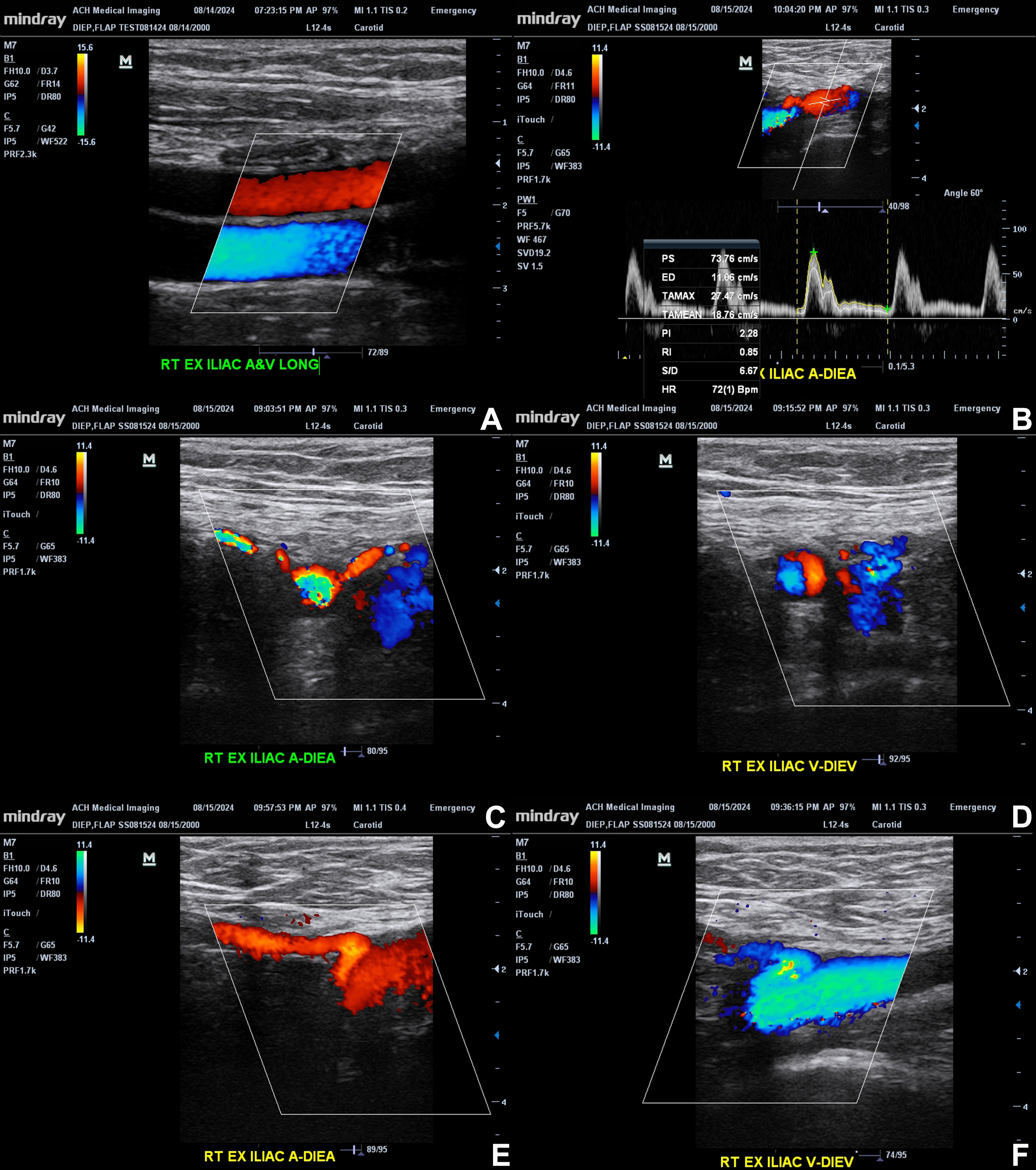
Figure 1. Ultrasound anatomy of the external iliac vessels and DIEA. Representative longitudinal and transverse color Doppler images demonstrate the right external iliac artery and vein, as well as the origin and course of the DIEA and DIEV. The top panels (A and B) show the external iliac vessels with clear arterial-venous differentiation based on flow direction. Spectral Doppler (B) confirms arterial waveforms within the DIEA, including peak systolic and end-diastolic velocities. Middle panels (C and D) illustrate the branching of the DIEA from the external iliac artery and accompanying venous structures. Bottom panels (E and F) further depict color flow within the DIEA and DIEV, highlighting vessel caliber and flow characteristics. These images demonstrate the ability of CDU to identify vessel origin, assess flow dynamics, and distinguish arterial from venous structures in preoperative DIEP flap planning. The original figure was created by the authors. Images have not been previously published. Figure 1 contains illustrative ultrasound images acquired by the first author during self-scanning; no patient images are included. A: Artery; A&V: artery and vein; CDU: color Doppler ultrasound; DIEA: deep inferior epigastric artery; DIEP: deep inferior epigastric perforator; DIEV: deep inferior epigastric vein; ED: end-diastolic velocity; EX: external; G: gain; HR: heart rate; LONG: longitudinal; PI: pulsatility index; PS: peak systolic velocity; PW: pulsed-wave Doppler; RI: resistive index; S/D: systolicto-diastolic ratio; TAMAX: time-averaged maximum velocity; TAMEAN: time-averaged mean velocity; RT: right; V: vein; bpm: beats per minute.