



Application of the bridge technique in revisional blepharoplasty in Asian patients
 0
0 Abstract
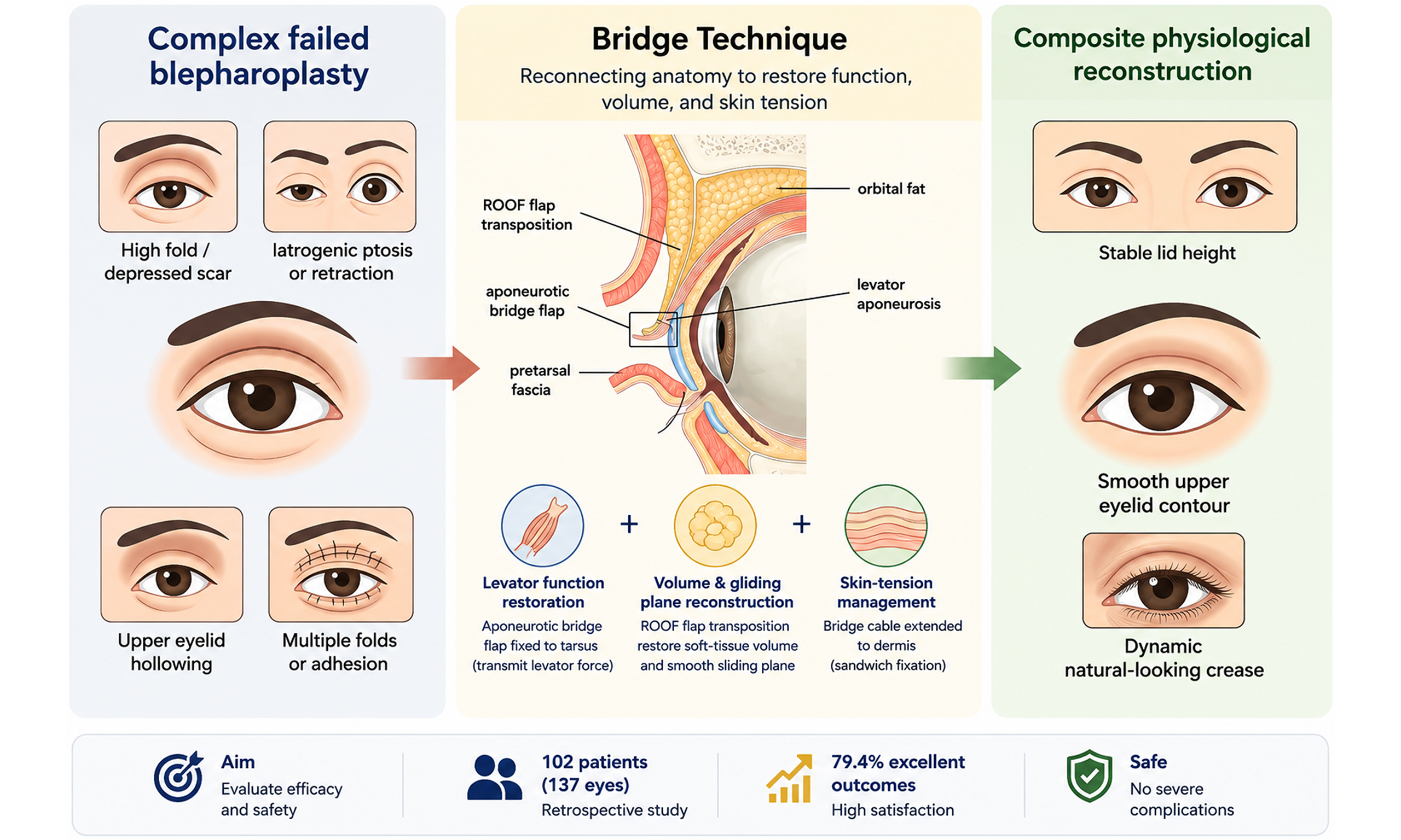
Aim: To evaluate the efficacy and safety of the Bridge Technique in revisional Asian blepharoplasty.
Methods: This retrospective study included 102 patients (137 eyes) with unsatisfactory outcomes after double-eyelid surgery between January 2024 and January 2025. Revision severity was graded using the height, shape, and function scoring system. The procedure used a levator aponeurosis-based bridge construct to adjust eyelid height and curvature, combined with local volume restoration and layered fixation to reconstruct the gliding plane. Functional outcomes were assessed using margin reflex distance 1, whereas aesthetic outcomes and complications were evaluated during follow-up.
Results: Among 137 eyes, 81 (59.1%) were classified as severe revision cases. The mean follow-up was 12.4 months. In 37 eyes with ptosis, margin reflex distance 1 improved from 1.82 ± 0.65 mm to 3.94 ± 0.42 mm (P < 0.001). In 11 eyes with upper eyelid retraction, margin reflex distance 1 decreased from 6.15 ± 0.58 mm to 4.08 ± 0.35 mm (P < 0.001). At the patient level, 81 patients (79.4%) achieved excellent, and 9 patients (8.8%) achieved good aesthetic outcomes. No severe complications occurred.
Discussion: The Bridge Technique provides a feasible and safe composite strategy for revisional Asian blepharoplasty by simultaneously addressing levator function, tissue volume, and skin tension. Further controlled studies are warranted.
Keywords
INTRODUCTION
Since Mikamo first reported double eyelid surgery in 1896, various techniques including suture methods, incisional techniques, and small-incision approaches have evolved substantially[1-3]. Anatomical studies have shown that Asian upper eyelids differ from Caucasian eyelids in the amount and distribution of pretarsal/suborbicularis fat and in the relationship between the orbital septum and levator aponeurosis, which are essential considerations for fold formation[4-6]. As one of the most popular cosmetic eye surgeries in Asia, double eyelid procedures are also influenced by trends and fashions. Some patients request high or deep folds that do not match the anatomical characteristics of Asian eyelids, and not all procedures are performed by surgeons with specialized expertise in Asian oculoplastic surgery.
This mismatch between aesthetic trends and anatomical considerations may contribute to postoperative complications, including abnormal double-eyelid depth or height, sausage-like pretarsal fullness, multiple creases, depressed incision scars, incomplete eyelid closure, upper eyelid retraction, iatrogenic ptosis, and upper eyelid depression[7,8].
Although previous studies have proposed strategies for individual problems after double eyelid surgery, there remains a lack of a comprehensive and flexible approach capable of addressing multiple coexisting deformities in a single procedure[8-15]. Modern aesthetic standards for double eyelids should emphasize a natural and dynamic appearance when the eyes are open, minimal visible scarring when the eyes are closed, and reconstruction consistent with physiological anatomy.
Based on the anatomical characteristics of the Asian upper eyelid, our team developed the Bridge Technique, a physiology-mimicking double eyelid blepharoplasty method based on relocation and reconnection of an aponeurotic flap with the tarsus[16]. The aim of the present study was to evaluate the efficacy and safety of this technique in complex revisional blepharoplasty in Asian patients.
METHODS
Patients and ethical approval
From January 2024 to January 2025, 102 Chinese patients with suboptimal double eyelids were enrolled in this retrospective study. The inclusion criteria were as follows: (1) age over 18 years; (2) at least six months since the last oculoplastic surgery; (3) no history of botulinum toxin or filler injection in the upper face within the previous 6 months; and (4) absence of neuromuscular disorders, Parkinsonism, or myasthenia gravis.
This study was approved by the Ethics Committee of Shanghai Ninth People’s Hospital, Shanghai Jiao Tong University School of Medicine (approval No. SH9H-2024-T30-1). The requirement for informed consent for participation was waived by the Ethics Committee because of the retrospective nature of this study.
Severity classification
The severity of each patient’s suboptimal double-eyelid deformity was assessed using the height, shape, and function (HSF) scoring criteria. The HSF system was adapted from Chen’s grading framework and was used as a composite difficulty classification system for revisional blepharoplasty. Unlike a simple arithmetic summation of the three dimensions, the final score represents the overall revision difficulty determined by the predominant deformity pattern[17] [Table 1].
Grading scale for revisional blepharoplasty - HSF score
| Dimension | Score | Clinical description | Evaluation criteria |
| Height | 5 | Excessively high fold with minimal residual skin | Most challenging; usually multiple prior surgeries |
| 4-1 | Low fold, shallow fold, loss of fold, or multiple folds | Scored based on residual skin volume and past surgical history | |
| Shape | 3 | Pronounced sunken scar (step-like appearance) | Caused by excessive resection of pretarsal tissue; visible when eyes are closed |
| 2 | Pronounced upper eyelid depression | Caused by excessive removal of ROOF fat and orbital septal fat | |
| 1 | Puffy eyelid, congestion of pretarsal flap, or asymmetry | Common aesthetic issues with lower surgical complexity | |
| Function | 2 | Upper eyelid retraction OR severe ptosis | Significant impairment of eyelid position or opening function |
| 1 | Mild to moderate ptosis | Evaluated via MRD1 |
To evaluate the consistency and objectivity of the assessments between two independent plastic surgeons (Jin R and Yu W), inter-rater reliability was calculated using Cohen’s kappa coefficient (κ). In instances where there was a discrepancy in HSF grading, a third senior surgeon (Yang J) reviewed the clinical photographs, and a final grade was determined through consensus.
Surgical technique
The patient was placed in the supine position. Routine disinfection and sterile draping were performed according to standard protocols. The double-eyelid incision line was delineated based on the desired height and curvature, with the original scar tissue temporarily preserved. Local anesthesia was achieved via subcutaneous infiltration, typically with 2% lidocaine and 1:100,000 epinephrine. An incision was made along the marked line. Meticulous dissection was performed to identify and isolate the previous surgical scar. Layer-by-layer separation was performed to expose the anterior orbital septum, followed by thorough release of cicatricial adhesions using high-frequency electrocautery under direct visualization. No laser was used during dissection. The orbital septum was then opened to fully mobilize the residual septal fat.
For patients presenting with volume deficiency, particularly those with upper eyelid retraction and a sunken superior sulcus, mobilized central and lateral orbital fat was transposed medially into the pre-aponeurotic space to restore a functional sliding layer. Orbital fat transposition was performed in nearly all eyes with residual orbital fat requiring volume redistribution. Concurrently, the Pendulum Movement of Orbital Fat and Retro-Orbicularis Oculi Fat technique was employed: a superior-medial pedicled flap of the retro-orbicularis oculi fat was dissected and narrowed. This flap was then rotated laterally in a pendulum-like fashion to fill the lateral tissue defect created by medial displacement of the orbital fat[18]. In cases where local autologous tissue was insufficient, distant-site autologous fat grafting was performed for volume augmentation. The graft material consisted primarily of sheet-like autologous fat or a vascularized periumbilical fat flap, selected according to the degree of soft-tissue deficiency[13,14].
Restrictive bands and ligaments between the levator aponeurosis and the posterior orbital septum were meticulously released. The orbital septum-levator aponeurosis flap was then dissected superiorly from the superior tarsal border. At this point, a flap containing the orbital septum and underlying levator aponeurosis was formed. This construct acted as a motor transmitter and was termed the “bridge”.
For patients with blepharoptosis, the motor transmitter consisted of the levator aponeurosis and Müller’s muscle. All 37 eyes with blepharoptosis underwent levator aponeurosis advancement to restore eyelid height. In severe ptosis, dissection extended beyond the check ligament to facilitate sufficient anterior advancement and force transmission. For patients with upper eyelid retraction, the levator aponeurosis-Müller’s muscle complex was lengthened using transverse incision-longitudinal suturing. All 11 eyes with upper eyelid retraction underwent this maneuver, and 3 eyes with insufficient soft-tissue volume additionally received vascularized periumbilical fat flaps.
A portion of pretarsal tissue was preserved, and the complex was anchored to the superior one-third of the tarsal plate with four to five horizontal mattress 6-0 silk sutures. At this stage, the bridge was constructed, and the knots on the tarsus were termed piers. Eyelid height, curvature, and symmetry were assessed intraoperatively, and adjustments were made until an optimal shape was achieved without entropion or angular deformity.
A multi-layered closure was performed using 7-0 nylon sutures to approximate the orbicularis oculi muscle, levator aponeurosis, and retro-orbicularis oculi fat/orbital fat layers. This step is referred to as the sandwich suturing technique, as the complex is secured between the superior and inferior segments of the orbicularis oculi muscle. Particular emphasis is placed on re-anchoring orbital fat and retro-orbicularis oculi fat superior to the intended eyelid crease to reconstruct the physiological gliding plane and prevent postoperative superior sulcus deformity.
After confirming sufficient skin redundancy, redundant skin and original scar tissue were excised. If skin laxity was inadequate, the original scar was preserved to avoid lagophthalmos. Finally, the skin incision was integrated with the bridge cable to create the double-eyelid fold. During this step, levator aponeurosis fibers were incorporated between the superior and inferior dermal edges in an interrupted fashion, simulating the physiological anatomy of a natural crease in which terminal levator aponeurosis fibers extend to the dermis. This approach was intended to maximize the long-term stability and natural dynamic appearance of the postoperative fold (Figure 1; see Supplementary Video 1 for a detailed demonstration). A diagram of the entire operation procedure is presented in Figure 2.
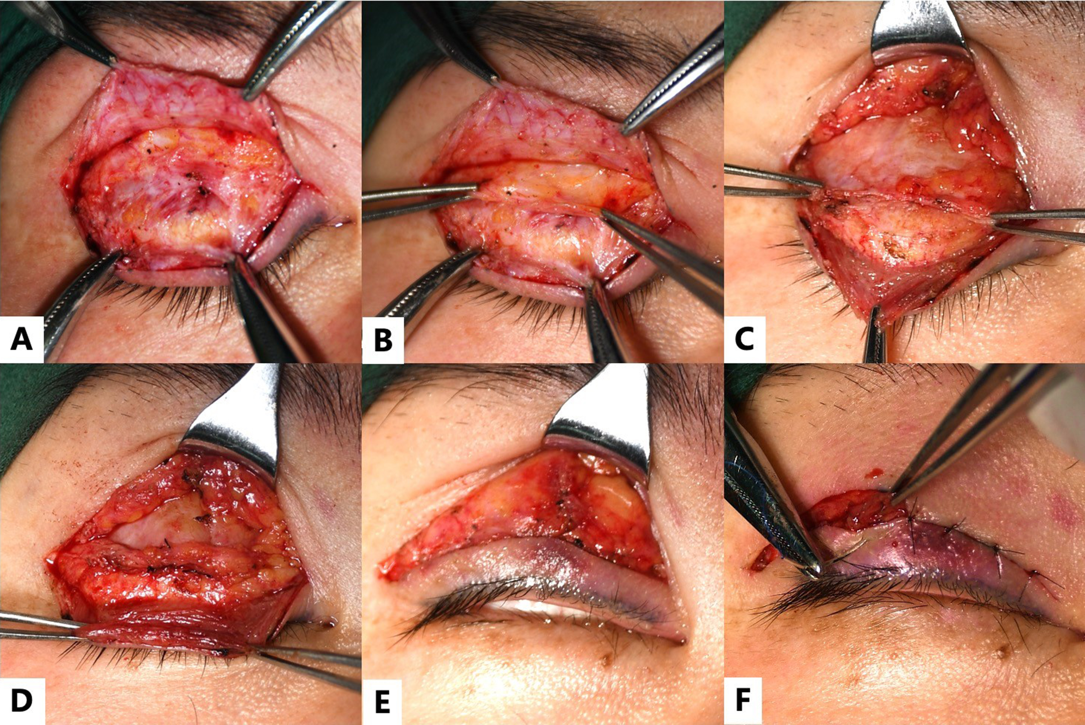
Figure 1. Application of the bridge technique in a 38-year-old female diagnosed with suboptimal double eyelids and mild ptosis on the left side. (A) After redesigning the incision, the skin and orbicularis oculi muscle are incised, and the scar tissue is fully released; (B) The ROOF and orbital fat are dissected superiorly and preserved; (C) The lower flap is dissected to expose the orbicularis oculi muscle and pretarsal fascia. The levator aponeurosis is then dissected superiorly from the upper border of the tarsal plate; (D) First-layer fixation (building the bridge): the levator aponeurosis-Muller’s muscle flap is advanced and re-anchored to the upper one-third of the tarsal plate using four to five sutures. The excessive portion of the flap is trimmed to 1-2 mm; (E) Second-layer fixation: the orbicularis oculi muscle of the lower flap, the orbital levator aponeurosis- Muller’s muscle flap, and the orbicularis oculi muscle of the upper flap are secured together, restoring the continuity of the orbicularis oculi muscle; (F) Third-layer fixation: Skin closure incorporating the “bridge cable”, enabling the levator muscle fibers to terminate at the skin. ROOF: Retro-orbicularis oculi fat.
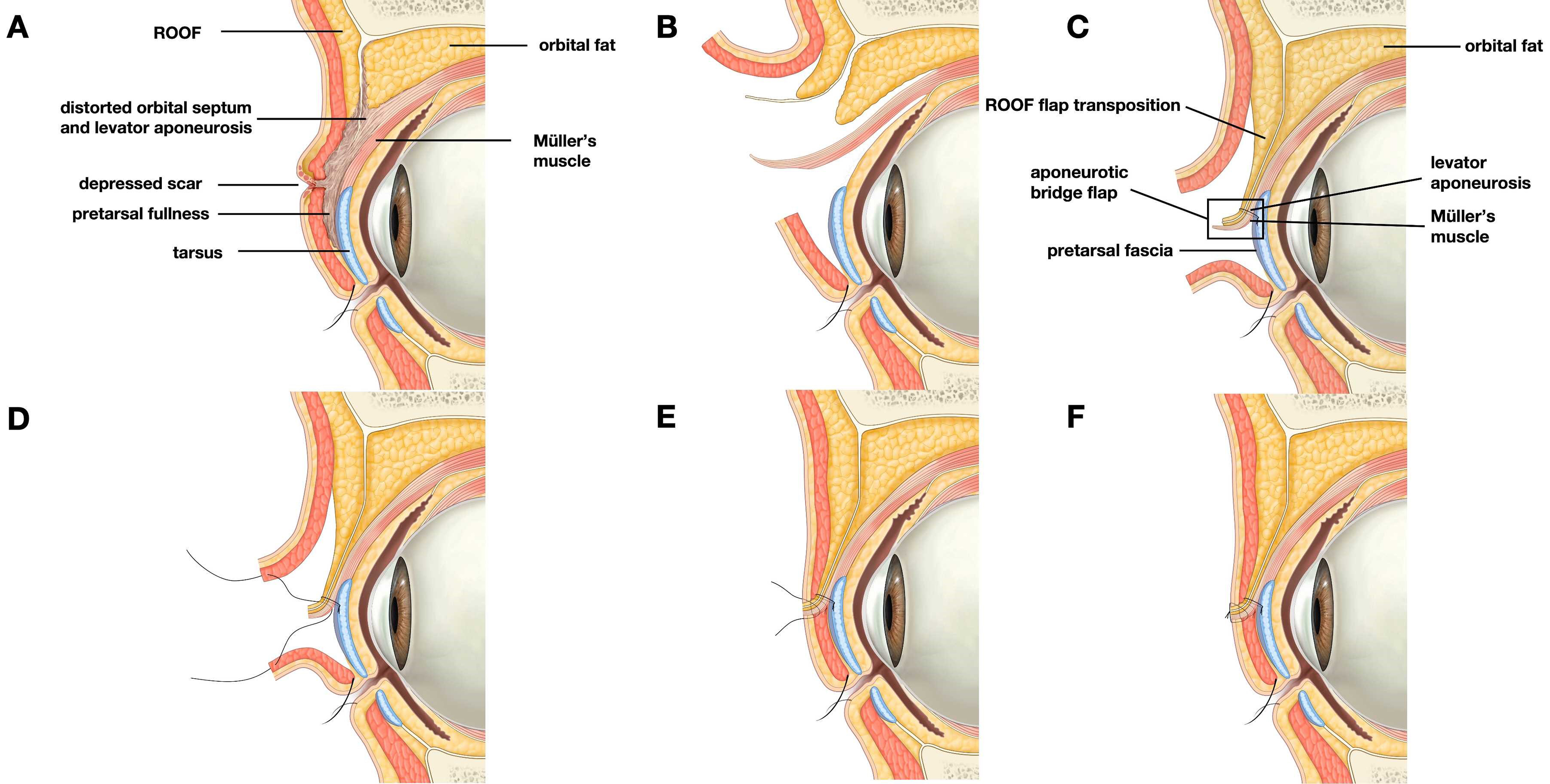
Figure 2. Schematic illustration of the Bridge Technique for secondary double-eyelid revision. The figure was illustrated by Liu Min, a professional medical illustrator. (A) Abnormal anatomy after failed double-eyelid surgery, showing a depressed scar, pretarsal fullness, scar adhesion, and distortion of the orbital septum-levator aponeurosis complex; (B) The previous scar and adhesions are released through the redesigned supratarsal incision to restore the surgical planes; (C) Orbital fat and/or ROOF flap is transposed to restore upper-eyelid volume and reconstruct the gliding plane. An aponeurotic bridge flap is prepared from the septum-levator aponeurosis complex; (D) The aponeurotic bridge flap is advanced and anchored to the pretarsal fascia/tarsal region; (E) The bridge flap is fixed to the superior tarsus, forming stable bridge piers for levator force transmission; (F) Layered sandwich fixation integrates the skin-orbicularis complex, interposed soft tissue, bridge flap, and dermis, creating a smooth and dynamic double-eyelid fold. ROOF: Retro-orbicularis oculi fat.
Antibiotic ointment was applied to the incision, followed by a light gauze dressing and intermittent cold compresses to minimize postoperative edema. Cold compresses were applied for approximately 30 min per session during the first 24 h postoperative period.
Outcome assessment
Outcomes were evaluated across three domains: aesthetic refinement, functional restoration, and surgical complications. To avoid ambiguity in data presentation, eye-level denominators were used for HSF severity classification and functional outcomes, whereas patient-level denominators were used for overall aesthetic grading and satisfaction-related outcomes.
Aesthetic outcomes were assessed by two independent plastic surgeons (Jin R and Yu W) at the final follow-up, 11-14 months postoperatively. The assessment included a retrospective review of standardized digital photographs and clinical records. Patients were seated in a neutral position with the head in the Frankfort horizontal plane. High-resolution images were captured in primary gaze, with the eyes open and closed, to evaluate fold dynamics and scar quality. The detailed aesthetic evaluation criteria are presented in Table 2.
Aesthetic assessment grading scale
| Grade | Criteria for evaluation |
| Excellent | Symmetric fold height and shape; natural-appearing contour; minimal or invisible scarring; no visible pretarsal congestion |
| Good | Slight asymmetry (< 1 mm) in fold height; satisfactory contour; mild but acceptable scar visibility; patient is highly satisfied |
| Fair | Noticeable asymmetry; mild fold irregularities or “step-like” appearance; visible scarring or mild sunken appearance |
| Poor | Severe asymmetry; recurrence of multiple folds or fold loss; significant scarring or deformity; patient requires secondary revision |
Functional efficacy was prioritized for patients presenting with preoperative upper eyelid retraction or blepharoptosis. The primary functional outcome measure was margin reflex distance 1 (MRD1). MRD1 was measured on standardized frontal photographs using ImageJ software (National Institutes of Health, Bethesda, MD, USA). Quantitative changes between preoperative and postoperative values were analyzed using paired t-tests. All statistical analyses were performed using IBM SPSS Statistics version 26.0 (IBM Corp., Armonk, NY, USA), with P < 0.05 considered statistically significant.
Adverse events and surgical morbidities were documented during follow-up. The assessment focused on recurrence of the initial deformity, undercorrection, incomplete eyelid closure, exposed sutures, and the formation of postoperative upper-eyelid fat nodules or granulomas.
RESULTS
General characteristics
A total of 102 patients (137 eyes) were included in this study. The study population consisted of 5 males (4.9%) and 97 females (95.1%), with a mean age of 31.4 years (range, 18-48 years). Based on the HSF classification, preoperative severity was categorized at the eye level as follows: 17 eyes (12.4%) had an HSF score of 1-3, 39 eyes (28.5%) scored 4-7, and 81 eyes (59.1%) scored 8-10. The mean follow-up period was 12.4 months (range, 11-14 months). Baseline demographic information and the distribution of the 137 treated eyes are summarized in Table 3 and Figure 3.
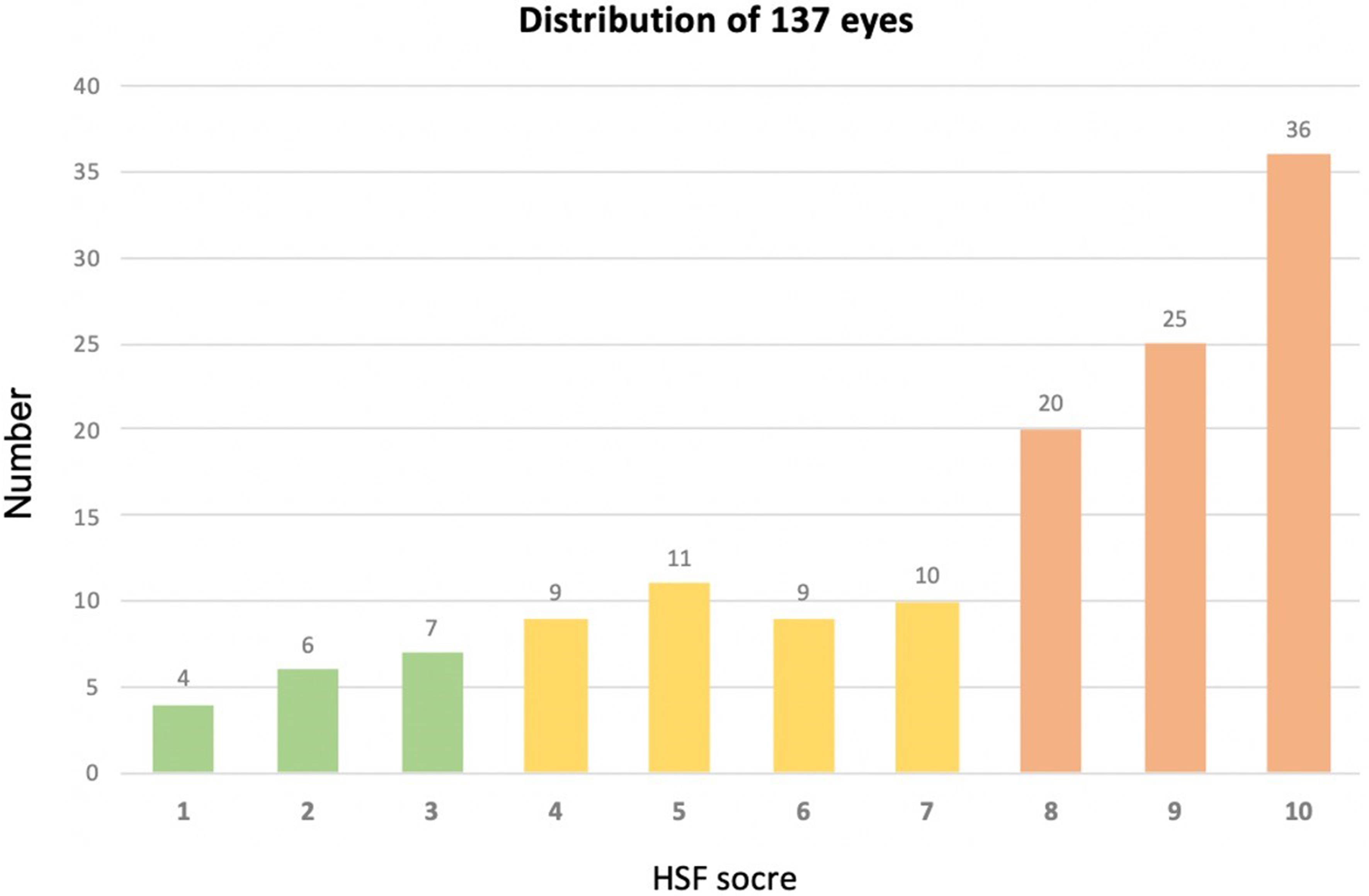
Figure 3. HSF score distribution in 137 eyes: score 1-3, relatively easy revision; score 4-7, moderate revision difficulty; score 8-10, high revision difficulty. HSF: Height, shape and function.
Demographic characteristics of patients
| Characteristic | Value |
| No. of patients/eyes | 102/137 |
| Average age (years) | 31.4 (18-48) |
| Male/female | 5/97 |
| HSF score (eyes), n (%) | |
| 1-3 | 17 (12.4%) |
| 4-7 | 39 (28.5%) |
| 8-10 | 81 (59.1%) |
The Pendulum Movement of Orbital Fat and Retro-Orbicularis Oculi Fat technique was applied in 95 eyes (approximately 70% of treated eyes). Orbital fat transposition was performed in nearly all eyes with residual orbital fat requiring volume redistribution. All 37 eyes with blepharoptosis underwent levator aponeurosis advancement. All 11 eyes with upper eyelid retraction underwent transverse incision-longitudinal suturing, and 3 of these eyes additionally received vascularized periumbilical fat flaps because of insufficient soft-tissue volume.
Inter-rater reliability for preoperative HSF severity grading was almost perfect, with a Cohen’s κ value of 0.85 (P < 0.001).
Functional outcomes: correction of ptosis and retraction
Among the cohort, 37 eyes presented with blepharoptosis, comprising 29 eyes with mild-to-moderate ptosis and 8 eyes with severe ptosis. The mean MRD1 improved significantly from 1.82 ± 0.65 mm preoperatively to 3.94 ± 0.42 mm postoperatively (P < 0.001). Additionally, 11 eyes exhibited upper eyelid retraction before surgery. After intervention, the mean MRD1 decreased from 6.15 ± 0.58 mm to 4.08 ± 0.35 mm (P < 0.001), indicating significant improvement in eyelid position [Figures 4 and 5].
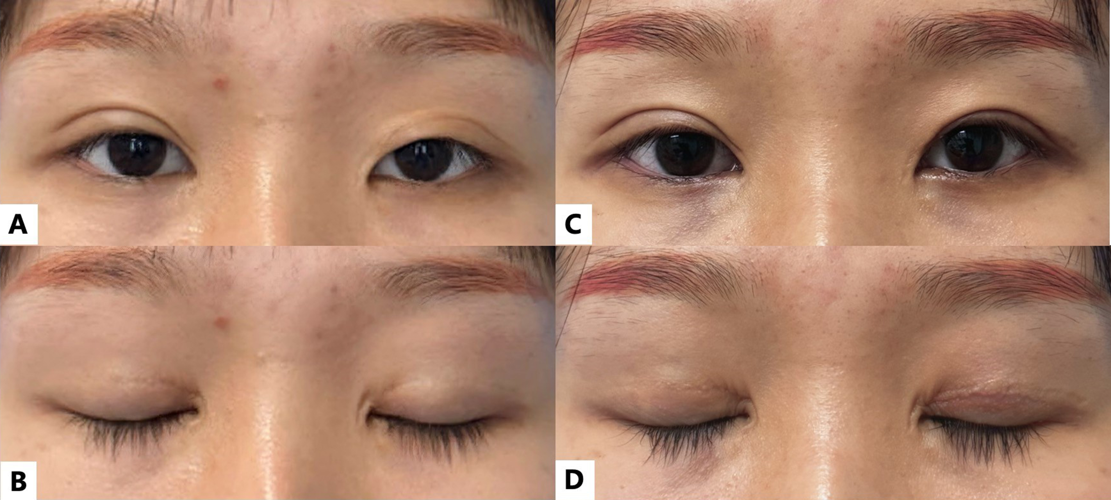
Figure 4. Case of a 26-year-old female patient with an unfavorable double-eyelid appearance and mild blepharoptosis on the left side. Preoperative views show the patient with (A) eyes open and (B) eyes closed. Postoperative outcomes at the final follow-up, 11-14 months postoperatively, after repair using the Bridge Technique are shown with (C) eyes open and (D) eyes closed.
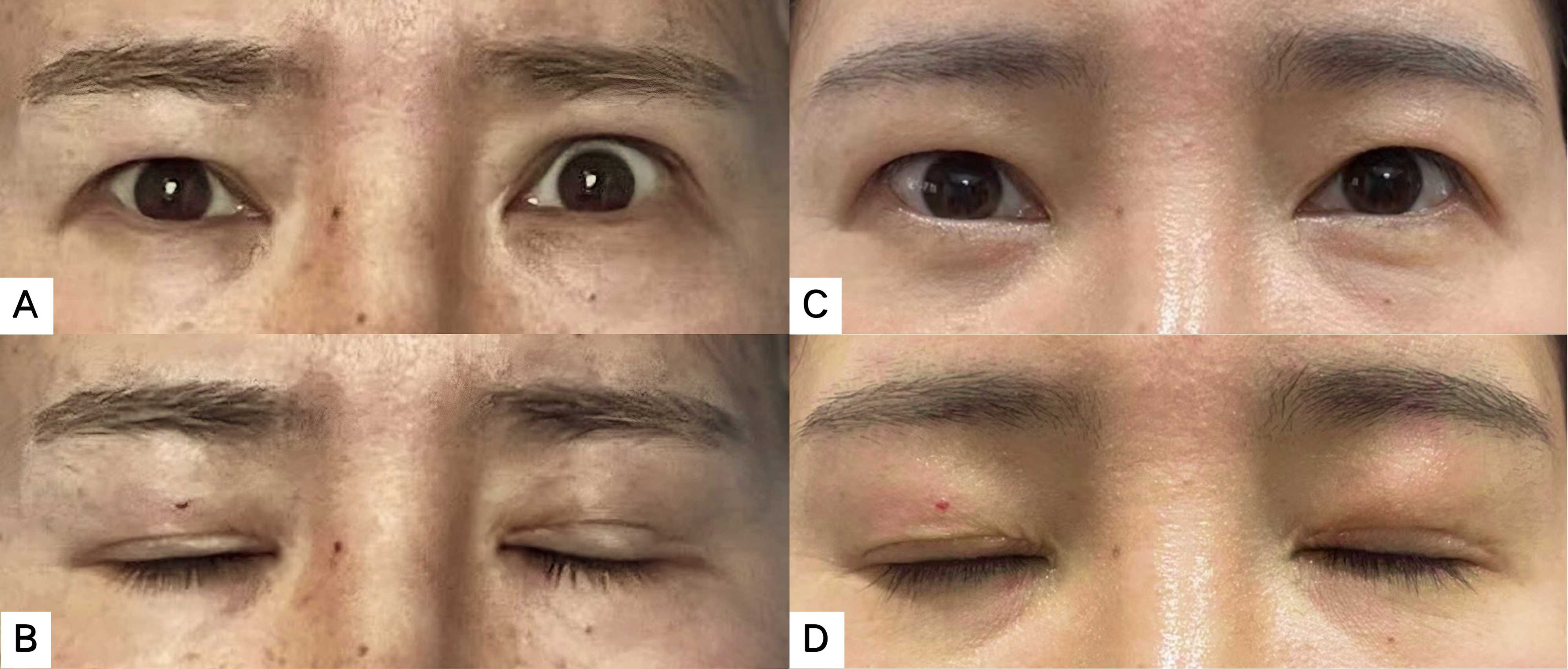
Figure 5. A 38-year-old female patient with unilateral (left) upper eyelid retraction. The left column shows the preoperative views, with (A) eyes open and (B) eyes closed. The right column shows postoperative outcomes at the final follow-up, 11-14 months postoperatively, with (C) eyes open and (D) eyes closed, demonstrating restored eyelid symmetry and a natural contour.
Aesthetic assessment
Postoperative aesthetic outcomes were independently evaluated by two raters. After minor discrepancies were resolved by consensus, 81 of 102 patients (79.4%) were graded as excellent [Figure 6]. Outcomes were graded as good in 9 patients (8.8%), primarily because of mild asymmetry. Seven patients (6.9%) received a fair grading; among these, 2 had significant asymmetry, 3 showed mild residual upper eyelid hollowing, and 2 presented with noticeable scarring. Five patients (4.9%) were graded as poor, including 4 cases of severe asymmetry and 1 case of recurrent multiple folds.
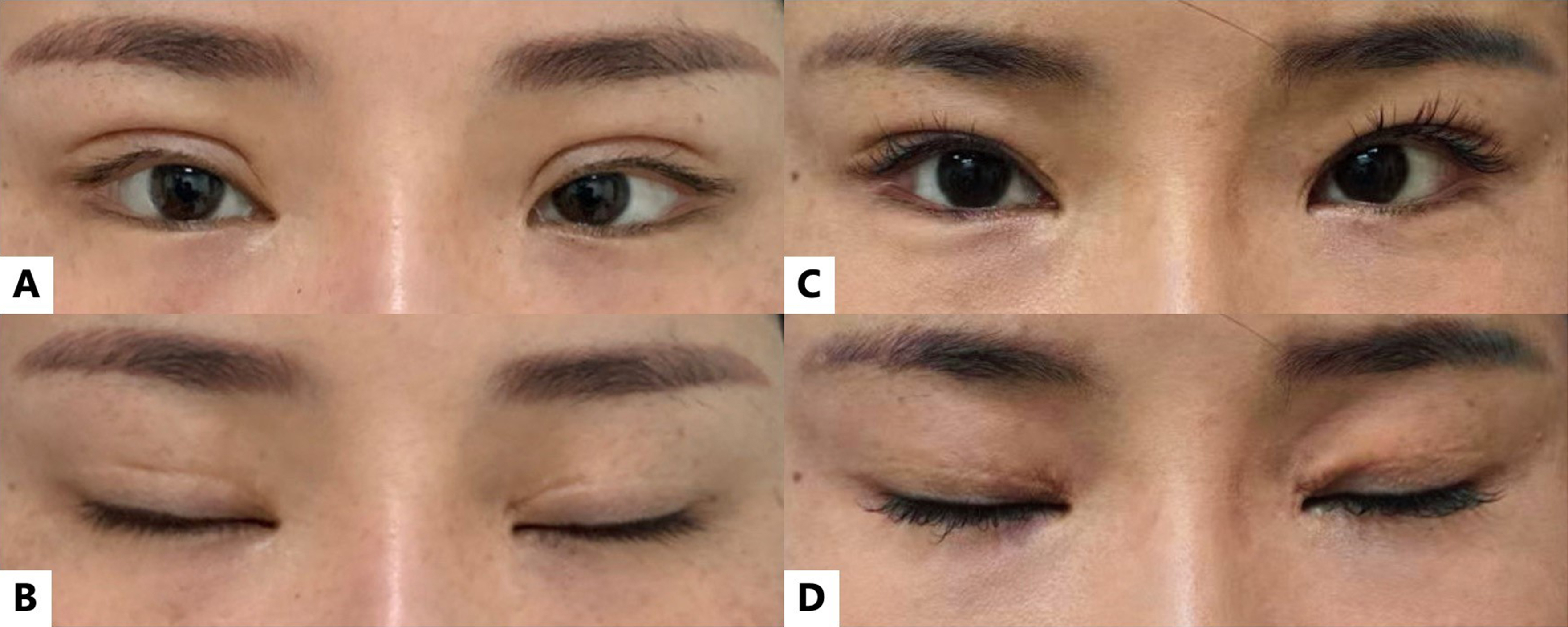
Figure 6. Case of a 34-year-old female patient with bilateral unfavorable double eyelids and prominent pretarsal fullness. Preoperative views show the patient with (A) eyes open and (B) eyes closed. Postoperative outcomes at the final follow-up, 11-14 months postoperatively, after repair using the Bridge Technique to convert the wide crease to a narrower one are shown with (C) eyes open and (D) eyes closed, demonstrating bilateral symmetry and a natural double-eyelid crease.
Complications and recurrence
The overall complication rate was low, and no severe adverse events such as retrobulbar hemorrhage, infection, or corneal injury occurred. The primary postoperative issue was partial recurrence of functional deformities in severe cases. Among the 37 eyes with preoperative ptosis, mild recurrence was observed in 2 eyes (5.4%), and 1 eye (2.7%) regressed to the preoperative level. Among the 11 eyes with retraction, 1 eye (9.1%) exhibited mild recurrence. These cases were managed conservatively or scheduled for secondary adjustment after 6 months. No cases of lagophthalmos, exposed sutures, or granuloma formation were noted during follow-up.
A detailed review of recurrent cases suggested that recurrence was associated with severe preoperative scarring, insufficient residual skin or soft tissue reserve, and reduced elasticity of the levator aponeurosis-Müller’s muscle complex after multiple prior operations. In the ptosis cases that recurred, intraoperative adjustment initially achieved acceptable eyelid height, but postoperative scar contraction and weakening of force transmission likely contributed to partial loss of correction. In the retraction case, marked preoperative fibrosis and shortage of anterior lamellar tissue may have limited stable downward repositioning despite levator complex lengthening. These observations emphasize the importance of careful preoperative grading, conservative patient counseling, and staged secondary adjustment when tissue conditions are unfavorable.
DISCUSSION
The growth in blepharoplasty procedures has inevitably led to a rise in iatrogenic complications. Secondary revision surgery is inherently complex because of scar tissue, anatomical distortion, and tissue deficiency. In recent years, various targeted strategies have emerged to address specific deformities. For instance, the beveled approach has been proposed to ameliorate step-like incision lines by preserving subcutaneous tissue[10]. Techniques focusing on adhesion release, lowering of high fixation points, and management of high-fold deformity have shown efficacy in selected cases, while fat repositioning, orbital fat fascia flap transfer, or fat grafting has become important for addressing sunken eyelids and preventing re-adhesion[11-15].
However, traditional revisional methods often focus on isolated deformities. In clinical practice, revision patients frequently present with a syndrome of coexisting complications, including iatrogenic ptosis, pretarsal fullness, incisional depression, multiple folds, skin shortage, and upper eyelid depression. In this study, 81 eyes (59.1%) were classified with HSF scores of 8-10, representing the most challenging category of revision cases. The primary etiology in these high-grade failures was often aggressive excision of physiological structures, including skin, orbicularis muscle, and orbital fat, during prior operations. This hollowed-out status leaves a paucity of local tissue available for reconstruction and limits the effectiveness of simple revision methods.
The Bridge Technique differs from conventional isolated maneuvers by integrating three repair targets into one composite reconstructive framework: levator force transmission, volume restoration, and skin-tension management. Rather than simply lowering a fixation point, releasing adhesions, or adding fat, the technique reconstructs a stable yet adjustable aponeurotic-tarsal linkage and restores a dynamic gliding plane. This may explain why the method was useful in a cohort dominated by severe revision cases.
Based on our experience in oculoplastic revision surgery, successful revision depends on meticulous management of three critical anatomical factors.
Levator management and timing
Indiscriminate manipulation of the levator complex during primary surgery may lead to iatrogenic functional disorders. In this study, 37 eyes developed iatrogenic ptosis and 11 eyes had upper eyelid retraction. The Bridge Technique restored MRD1 to the normal range in most patients by adjusting levator force transmission. Nevertheless, the recurrence observed in severe cases underscores that neuromuscular recovery in heavily scarred tissues remains variable.
The timing of intervention for iatrogenic ptosis is also critical. Patients are often anxious and request immediate correction, but premature surgery may exacerbate inflammation and increase the risk of secondary complications. We advocate a waiting period of 3 to 6 months to allow edema to subside and tissues to soften; some ptosis-like symptoms may spontaneously improve during this period. Preoperative assessment should also consider Hering’s law of equal innervation, and surgeons should anticipate the contralateral eyelid response to unilateral correction to improve postoperative symmetry[19].
Volume restoration and gliding-plane reconstruction
Soft-tissue deficiency is a major hurdle in revisional surgery. Rather than relying heavily on free fat grafts, which have unpredictable resorption, we prioritize vascularized local tissue. The Pendulum Movement of Orbital Fat and Retro-Orbicularis Oculi Fat technique and orbital fat transposition are used to correct upper eyelid hollowing. Crucially, these autologous tissues are fixed above the newly formed supratarsal crease. This placement reconstructs the physiological gliding plane between the levator aponeurosis and the overlying skin-muscle flap, creating a physical barrier against recurrent adhesion. This step is essential for preventing postoperative incisional depression and recurrence of multiple folds.
Skin and suture management
Aesthetic refinement focuses on eliminating the artificial appearance. To resolve pretarsal fullness and step-like depression, the incision line is redesigned to lower excessively high fixation points to an optimal height. Pretarsal skin of the lower flap is tightened superiorly, while continuity of the orbicularis oculi muscle is restored to reduce incisional depression. Skin excision requires extreme caution. We adopt a preservation-first strategy in which the original scar is initially retained, and excision is performed only after internal fixation is complete and skin redundancy can be accurately assessed.
For severe cases with HSF scores of 8-10, particularly high-fold patients with significant skin shortage, a preoperative tissue expansion strategy may be considered. Patients are instructed to perform manual traction exercises on the upper eyelid for 3 to 6 months before surgery. This regimen may increase skin laxity and recruit a degree of tissue surplus, providing a safer margin for the revision procedure.
Revision surgery demands a high level of anatomical expertise because normal landmarks are often destroyed or obscured by fibrosis. To navigate this, we employ a touch-bottom, or retrograde dissection, strategy. The surgeon first identifies the deep, healthy, unscarred anatomical planes and then dissects layer by layer toward the scarred superficial tissues. This reverse-engineering approach maximizes preservation of remaining normal tissue and minimizes collateral damage. Given the uncertainty and great difficulty of these procedures, thorough preoperative communication regarding realistic expectations is as important as the surgery itself.
The Bridge Technique offers a biomimetic solution to revisional blepharoplasty. The bridge pier provides a stable anchor to the tarsus and allows precise adjustment of crease height, depth, and levator force. The sandwich technique incorporates the orbicularis oculi muscle, retro-orbicularis oculi fat, and orbital fat into internal fixation to prevent adhesion, depression, and rigidity. The bridge cable mimics the natural terminal fibers of the levator aponeurosis extending to the skin. By modulating suture depth between the bridge cable and the muscle, the technique fine-tunes eyelid contour and creates a floating pretarsal segment rather than a stiff static scar.
Limitations
This study has several limitations. First, it was a retrospective case series without a control group, which limits the ability to draw definitive comparative conclusions between the Bridge Technique and other revisional methods. Second, although the total number of treated eyes was relatively large for complex revision blepharoplasty, several important subgroups, particularly upper eyelid retraction and severe recurrent ptosis, contained limited numbers of eyes. This restricts subgroup analysis and may underestimate recurrence in the most severe categories. Third, aesthetic evaluation was based on standardized photographs and independent surgeon assessment; however, validated patient-reported outcome instruments, such as the Facial Assessment and Cosmetic Enhancement Quality of Life Questionnaire (FACE-Q) scale for double-eyelid blepharoplasty, were not prospectively collected[20]. Fourth, because the study population consisted of Asian patients seeking revision after double-eyelid surgery, the findings may not be directly generalizable to non-Asian eyelid anatomy or to revision strategies commonly used in Western patients. Finally, the technique requires familiarity with scarred eyelid anatomy, levator complex manipulation, and local tissue transposition; therefore, outcomes may vary according to surgeon experience. Future prospective studies with longer follow-up, larger subgroup samples, standardized patient-reported measures, and comparative control groups are needed to further validate the clinical advantages and boundaries of this technique.
Conclusion
The Bridge Technique represents a composite physiological reconstruction strategy for revisional Asian blepharoplasty, moving beyond simple static fixation. By addressing the triad of levator function, volume restoration, and skin tension, this method addresses multifactorial revision challenges, including iatrogenic ptosis, pretarsal fullness, upper eyelid hollowing, multiple folds, and extensive scarring. The technique appears feasible, safe, and reproducible in experienced hands, with favorable functional and aesthetic outcomes in this retrospective cohort. Nevertheless, prospective controlled studies with larger sample sizes, validated patient-reported outcomes, and longer follow-up are needed to further validate its comparative efficacy.
DECLARATIONS
Acknowledgments
The authors thank Liu Min for the professional medical illustration of Figure 2 and the Graphical Abstract.
Authors’ contributions
Contributed equally to data acquisition, data analysis, and manuscript drafting: Shen Y, Yu S
Contributed to clinical evaluation, data interpretation, and critical revision of the manuscript: Jin R, Lu L
Conceived and supervised the study, performed senior surgical assessment, and critically revised the manuscript for important intellectual content: Yang J, Yu W
All authors read and approved the final manuscript.
Availability of data and materials
The datasets generated and analyzed during the current study are available from the corresponding author upon reasonable request and in accordance with institutional data-sharing policies.
AI and AI-assisted tools statement
During the preparation of this manuscript, the AI tool ChatGPT (GPT-5.5, released 2026-04-23) was used solely for language editing. The tool did not influence the study design, data collection, analysis, interpretation, or the scientific content of the work. All authors take full responsibility for the accuracy, integrity, and final content of the manuscript.
Financial support and sponsorship
This research was financially supported by Wuxi Taihu Lake Talent Plan, Supports for Leading Talents in Medical and Health Profession (No. 453210902THDJ), and the Project of Biobank (No. YBK202502) from Shanghai Ninth People’s Hospital, Shanghai Jiao Tong University School of Medicine.
Conflicts of interest
Yang J is an Editorial Board Member of the journal Plastic and Aesthetic Research. Yang J was not involved in any steps of editorial processing, notably including reviewers’ selection, manuscript handling and decision making. The other authors declare that there are no conflicts of interest.
Ethical approval and consent to participate
This study was approved by the Ethics Committee of Shanghai Ninth People’s Hospital, Shanghai Jiao Tong University School of Medicine (approval No. SH9H-2024-T30-1). The requirement for informed consent for participation was waived by the Ethics Committee because of the retrospective nature of this study.
Consent for publication
Written informed consent for publication was obtained from all patients whose clinical images are included in this manuscript.
Copyright
© The Author(s) 2026.
Supplementary Materials
REFERENCES
1. Mikamo M. Mikamo’s double-eyelid operation: the advent of Japanese aesthetic surgery. 1896. Plast Reconstr Surg. 1997;99:664-9.
2. Wang C, Pu LLQ. Asian upper blepharoplasty: a comprehensive approach. Clin Plast Surg. 2023;50:101-9.
3. Yu P, Chen S, Gu T, Zhao M, Teng L, Lu J. Small-incisional techniques for double-eyelid blepharoplasty: a systematic review. Aesthetic Plast Surg. 2023;47:1067-75.
4. Jeong S, Lemke BN, Dortzbach RK, Park YG, Kang HK. The Asian upper eyelid: an anatomical study with comparison to the Caucasian eyelid. Arch Ophthalmol. 1999;117:907-12.
5. Kiranantawat K, Suhk JH, Nguyen AH. The Asian eyelid: relevant anatomy. Semin Plast Surg. 2015;29:158-64.
7. Liu J, Song B. Review of complications in double eyelid surgery. Indian J Ophthalmol. 2022;70:1460-5.
10. Chen WP. Beveled approach for revisional surgery in Asian blepharoplasty. Plast Reconstr Surg. 2007;120:545-52.
11. Liu C, Zhao Q, Zhu S, Zhang Z, Li J, Chen H. Correction of high eyelid folds using three-plane dissection and fat repositioning/grafting in revisional blepharoplasty. Aesthetic Plast Surg. 2025;49:4597-606.
12. Huang J, Li Z, Chi Y, et al. Individualized high double eyelid fold correction in secondary blepharoplasty: a free-style design. Aesthetic Plast Surg. 2023;47:1843-50.
13. Gao S, Yi M, Li C, Gao W. Correction of sunken upper eyelid in Asian blepharoplasty based on anatomical and histological study of orbital fat fascia flap. Aesthetic Plast Surg. 2024;48:134-40.
14. Ramil ME. Fat grafting in hollow upper eyelids and volumetric upper blepharoplasty. Plast Reconstr Surg. 2017;140:889-97.
15. Kim BG, Youn DY. Management of adhesion using a pretarsal fibromuscular flap or graft in secondary blepharoplasty. Plast Reconstr Surg. 2006;117:782-9.
16. Jin R, Shen Y, Yu W, et al. Tarsal-fixation with aponeurotic flap linkage in blepharoplasty: bridge technique. Aesthet Surg J. 2020;40:NP648-54.
17. Chen WP. The eyelid crease height, depth, and shape: a scoring system for revisional Asian blepharoplasty. Plast Reconstr Surg Glob Open. 2020;8:e2802.
18. Wang X, Li R, Qiu CS, et al. The pendulum movement of orbital fat and retro-orbicularis oculi fat: a new strategy for correction of sunken eyelid deformity in revision upper blepharoplasty for Asian patients. Aesthetic Plast Surg. 2024;48:1104-10.
19. Chen AD, Lai YW, Lai HT, et al. The impact of hering’s law in blepharoptosis: literature review. Ann Plast Surg. 2016;76:S96-100.
Cite This Article
 Open Access
How to Cite
Download Citation
Export Citation File:
Type of Import
Tips on Downloading Citation
Citation Manager File Format
Type of Import
Direct Import: When the Direct Import option is selected (the default state), a dialogue box will give you the option to Save or Open the downloaded citation data. Choosing Open will either launch your citation manager or give you a choice of applications with which to use the metadata. The Save option saves the file locally for later use.
Indirect Import: When the Indirect Import option is selected, the metadata is displayed and may be copied and pasted as needed.
About This Article
Copyright

Data & Comments
Data
0









Comments
Comments must be written in English. Spam, offensive content, impersonation, and private information will not be permitted. If any comment is reported and identified as inappropriate content by OAE staff, the comment will be removed without notice. If you have any queries or need any help, please contact us at [email protected].