
fig1
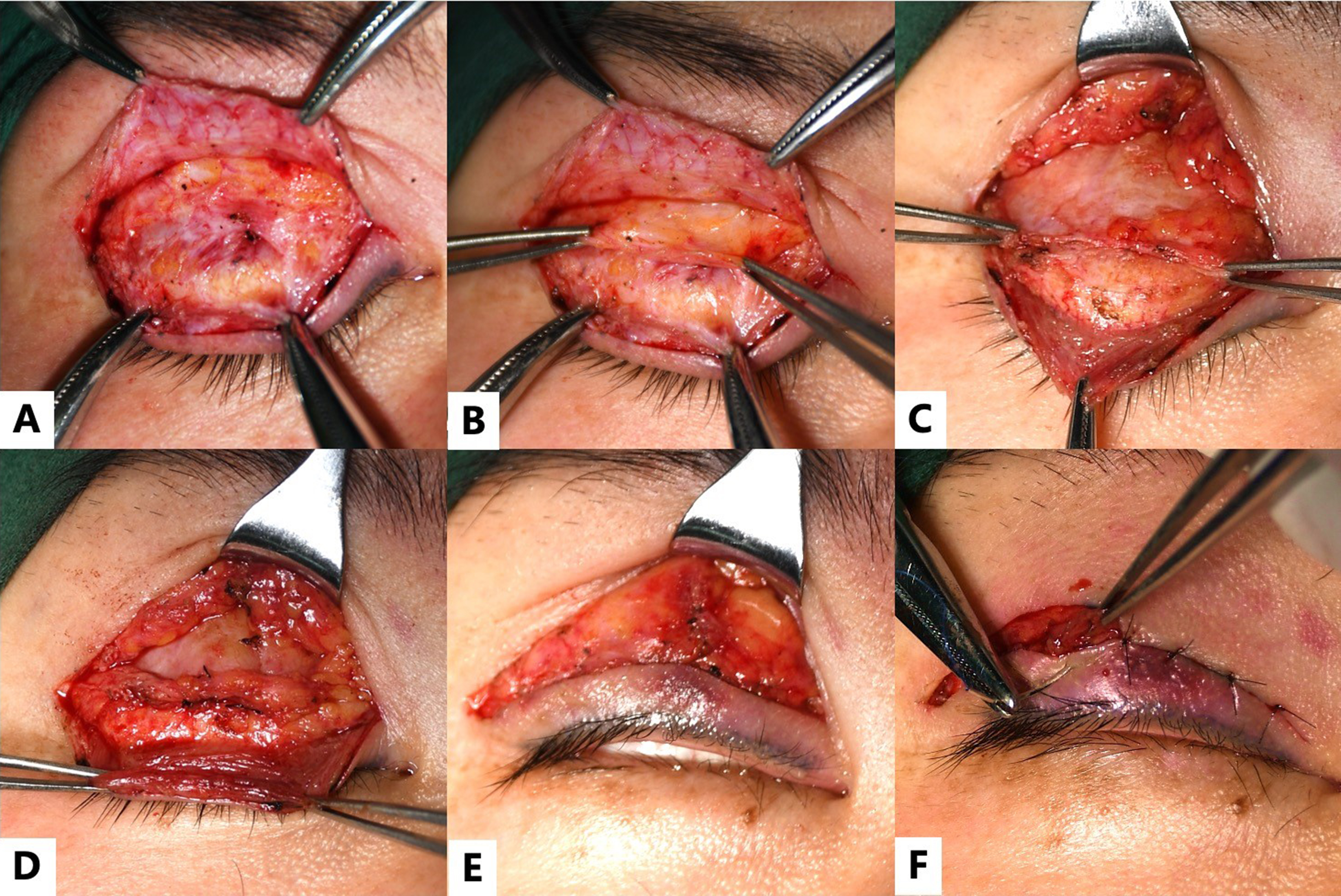
Figure 1. Application of the bridge technique in a 38-year-old female diagnosed with suboptimal double eyelids and mild ptosis on the left side. (A) After redesigning the incision, the skin and orbicularis oculi muscle are incised, and the scar tissue is fully released; (B) The ROOF and orbital fat are dissected superiorly and preserved; (C) The lower flap is dissected to expose the orbicularis oculi muscle and pretarsal fascia. The levator aponeurosis is then dissected superiorly from the upper border of the tarsal plate; (D) First-layer fixation (building the bridge): the levator aponeurosis-Muller’s muscle flap is advanced and re-anchored to the upper one-third of the tarsal plate using four to five sutures. The excessive portion of the flap is trimmed to 1-2 mm; (E) Second-layer fixation: the orbicularis oculi muscle of the lower flap, the orbital levator aponeurosis- Muller’s muscle flap, and the orbicularis oculi muscle of the upper flap are secured together, restoring the continuity of the orbicularis oculi muscle; (F) Third-layer fixation: Skin closure incorporating the “bridge cable”, enabling the levator muscle fibers to terminate at the skin. ROOF: Retro-orbicularis oculi fat.