
fig2
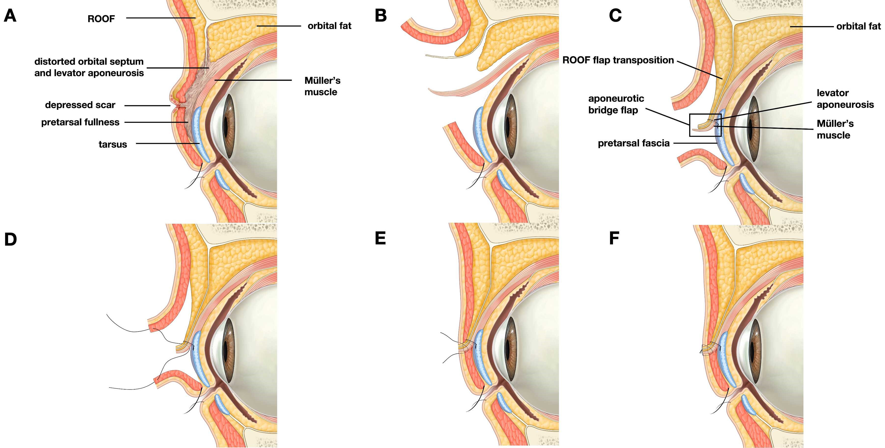
Figure 2. Schematic illustration of the Bridge Technique for secondary double-eyelid revision. The figure was illustrated by Liu Min, a professional medical illustrator. (A) Abnormal anatomy after failed double-eyelid surgery, showing a depressed scar, pretarsal fullness, scar adhesion, and distortion of the orbital septum-levator aponeurosis complex; (B) The previous scar and adhesions are released through the redesigned supratarsal incision to restore the surgical planes; (C) Orbital fat and/or ROOF flap is transposed to restore upper-eyelid volume and reconstruct the gliding plane. An aponeurotic bridge flap is prepared from the septum-levator aponeurosis complex; (D) The aponeurotic bridge flap is advanced and anchored to the pretarsal fascia/tarsal region; (E) The bridge flap is fixed to the superior tarsus, forming stable bridge piers for levator force transmission; (F) Layered sandwich fixation integrates the skin-orbicularis complex, interposed soft tissue, bridge flap, and dermis, creating a smooth and dynamic double-eyelid fold. ROOF: Retro-orbicularis oculi fat.