
Volume 6 (2019) – 16 articles
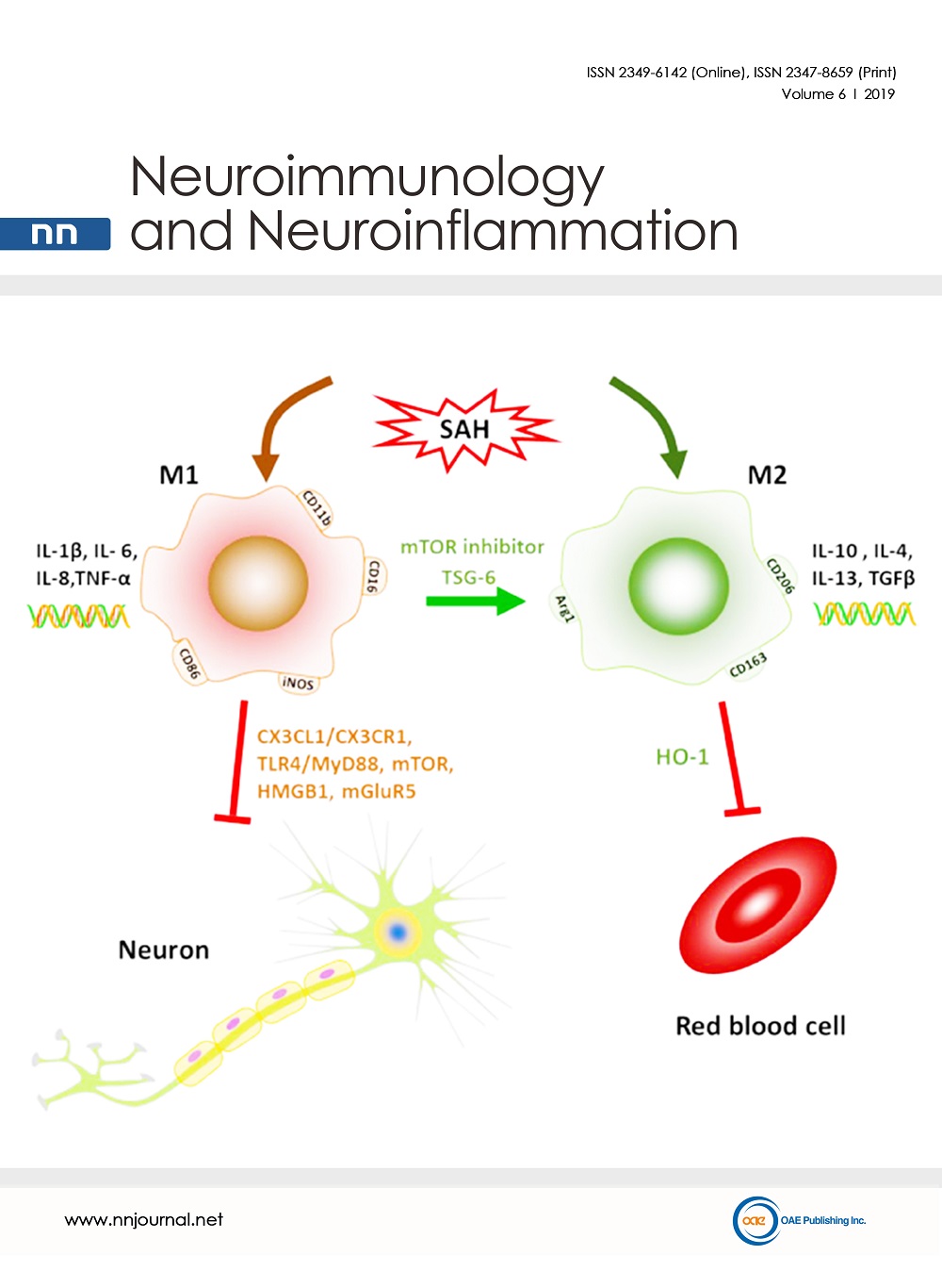
Cover Picture: Schematic description of microglial polarization after SAH and the interaction of microglia with other cell types in central nervous system. Microglia are activated and polarized to M1 or M2 direction after SAH. M1 phenotype is characterized by the cell surface marker CD11b, CD16, CD32, CD86. M2 phenotype is characterized by the cell surface markers CD206, CD163, Arg1. M1 microglia exhibit the pro-inflammatory responses with the expression of IL-1β, IL-6, IL-8 and TNF-α. M2 exhibit the pro-inflammatory responses with the expression of IL-10, IL-4, IL-13 and TGF-β. The M1 1 microglial activation has an unfavorable effect on the neurons through the interaction of CX3CL1 and CX3CR1. The neuronal apoptosis is modulated by microglia-dependent TLR4-MyD88, mTOR, HMGB1 and mGluR5. The extracellular ATP released from the surrounding astrocytes trigger the rapid chemotactic response of microglia towards injury. This response can be inhibited by blocking G protein-coupled purinergic receptors and connexin channels, which are highly expressed in astrocytes. The expression of HO-1 in microglia helps to clear the extravasated red blood cells and attenuated neuronal apoptosis.
view this paper
Read Online Viewed:
Download This Volume Viewed:


