



Beyond copper retention: a dynamic flux-compartment-response model of Wilson disease
 0
0 
A SUCCESSFUL MODEL THAT HAS BECOME TOO NARROW
Wilson disease (WD) has traditionally been understood through a straightforward causal sequence: pathogenic ATP7B variants impair the incorporation of copper into ceruloplasmin and, more importantly, reduce biliary copper excretion; copper then accumulates in the liver and subsequently in other organs, producing hepatic, neurological, psychiatric, ophthalmological, and systemic manifestations[1-3]. This model has enormous explanatory and therapeutic value. It established the molecular basis of WD, justified lifelong copper-lowering therapy, and remains the foundation of current clinical guidelines[1,2].
Its success, however, may have encouraged an overly static interpretation of the disease. In the classical model, ATP7B loss is the initiating event, copper is treated largely as a cumulative burden, and tissue injury is positioned as a downstream consequence. That sequence is necessary, but it is no longer sufficient. We argue that ATP7B deficiency creates an unstable copper-handling state in which copper entry, intracellular distribution, adaptive buffering, organelle vulnerability, intercellular signaling, and systemic modifiers continuously reshape disease expression. WD is therefore better conceptualized as a dynamic copper systems disorder than as a passive copper storage disease.
Several long-recognized observations expose the limits of a burden-centered model. First, WD is monogenic but phenotypically diverse. Patients with similar or identical ATP7B variants may differ markedly in age at onset, dominant organ involvement, rate of progression, and treatment response. In a large international cohort, age and sex influenced clinical presentation more effectively than ATP7B genotype[4]. This does not diminish the causal role of ATP7B; rather, it indicates that genotype establishes susceptibility while additional biological processes shape phenotype.
Second, total copper content does not consistently predict the timing or severity of injury. In ATP7B-deficient mice, liver pathology was related more closely to the duration of copper exposure than to the absolute hepatic copper concentration[5]. At the single-cell level, temporal and spatial copper distribution was more informative than total copper alone, with compartment-specific copper relocation accompanied by distinct metabolic responses[6]. Thus, where copper is located, in what chemical form, and for how long it remains there may matter as much as how much copper is present.
Third, ATP7B deficiency does not produce a uniform cellular response. Comparisons of different Atp7b-deficient mouse models have shown that the magnitude and character of hepatic injury can vary despite copper overload, and that proteomic changes may correlate more closely with buffering capacity, including metallothionein abundance, than with total liver copper[7]. Copper burden is therefore not synonymous with copper toxicity. Toxicity emerges when copper flux exceeds the capacity of sequestration, trafficking, metabolic adaptation, immune control, and tissue repair.
OUR PERSPECTIVE: A FLUX-COMPARTMENT-RESPONSE FRAMEWORK
We propose a flux-compartment-response framework for WD [Figure 1]. The framework does not replace the classical ATP7B model; it expands it. ATP7B deficiency remains the initiating lesion, but the disease state at any point in time reflects the interaction of three dimensions: copper flux, intracellular compartmentalization, and cellular/tissue response. These dimensions are modified by age, sex, diet, extrahepatic ATP7B deficiency, other metals, the intestine and microbiome, immune-cell interactions, treatment, and time [Table 1].
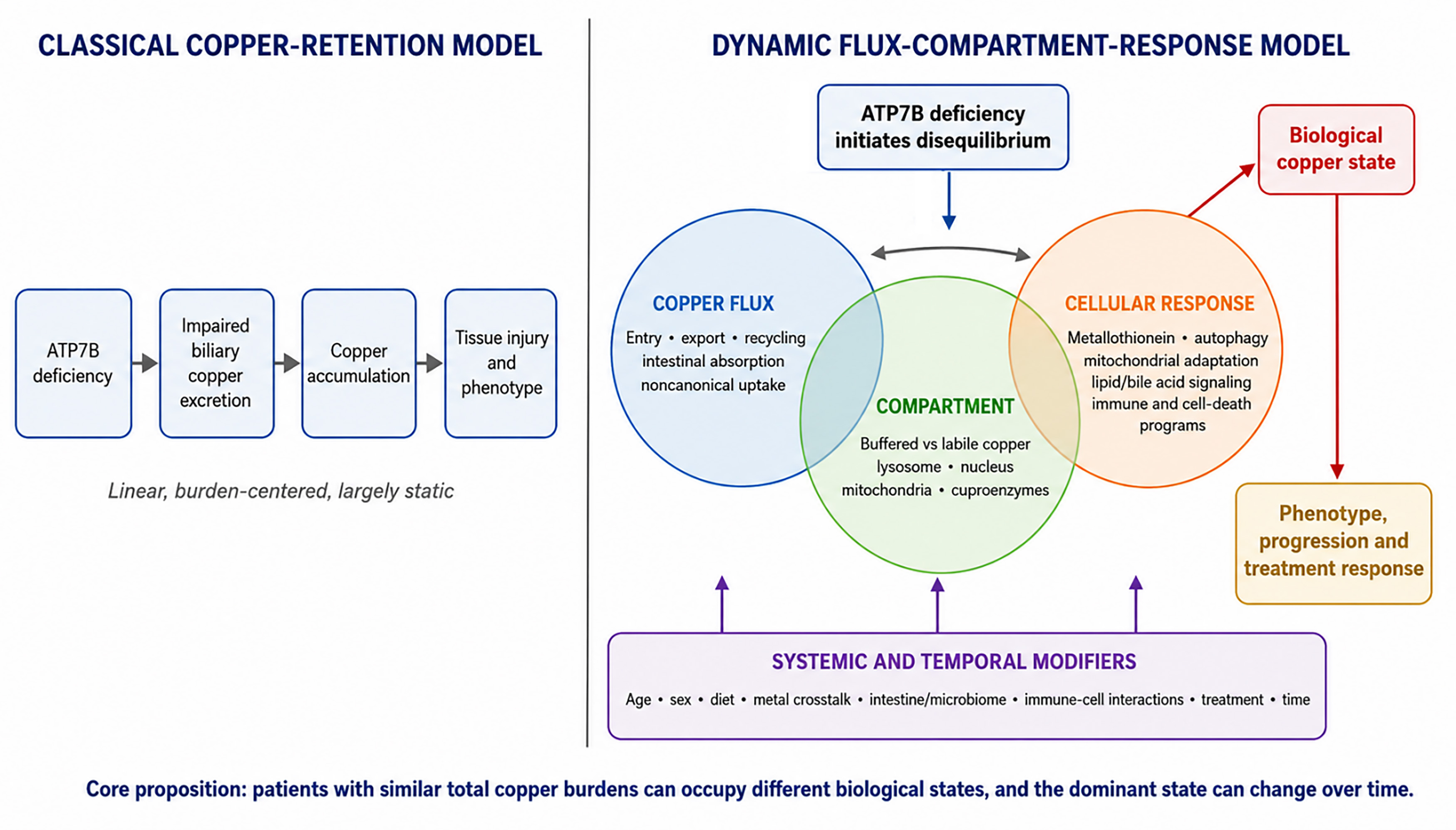
Figure 1. Classical copper-retention model versus the dynamic flux-compartment-response framework. The classical model links ATP7B deficiency linearly to impaired biliary excretion, copper accumulation, and injury. The proposed framework retains this initiating sequence but adds three interacting, time-dependent dimensions - copper flux, intracellular compartmentalization, and cellular/tissue response - together with systemic and temporal modifiers. Their interaction defines a biological copper state that may better explain phenotype, progression, and treatment response.
From the classical copper-retention model to a dynamic flux-compartment-response perspective
| Dimension | Classical copper-retention model | Dynamic interpretation and evidence | What needs to be done |
| Causal logic | ATP7B deficiency causes impaired biliary excretion, copper accumulation, and tissue injury[1-3] | ATP7B deficiency initiates disequilibrium; phenotype emerges from interactions among flux, compartmentalization, response, systemic context, and time[4-7] | Test whether state variables improve prediction of onset, progression, and treatment response beyond total copper burden |
| Meaning of copper burden | Total tissue or circulating copper is treated as the principal indicator of toxicity | Toxicity depends on bioavailability, exposure duration, and delivery to vulnerable organelles[5,6,10,11] | Develop and validate markers of labile, exchangeable, and organelle-directed copper; relate them longitudinally to outcomes |
| Cellular behavior | Cells are largely passive recipients of retained copper | ATP7B-deficient cells reprogram transport, antioxidant, autophagic, lipid/sterol, and cell-death pathways; responses may protect or become maladaptive[9,14,15] | Map response states by disease stage; test which adaptive programs should be enhanced and which maladaptive programs should be inhibited |
| Phenotypic heterogeneity | Variation is attributed mainly to residual ATP7B function and overall copper burden | Age, sex, buffering capacity, mitochondrial sensitivity, immune state, diet, metal crosstalk, and extrahepatic ATP7B deficiency modify phenotype[4,7,10,16,20,21] | Build standardized longitudinal cohorts; integrate genotype, clinical phenotype, treatment exposure, omics, and imaging with external validation |
| Systemic context | The liver is the principal site of copper retention and injury | Intestine, microbiome, immune cells, and stromal cells modify metabolic and inflammatory consequences of ATP7B deficiency[16,19-23] | Use tissue-specific models and human multi-omics to establish directionality and identify clinically actionable gut-liver or immune pathways |
| Monitoring | Ceruloplasmin, serum copper, urinary copper, and liver copper estimate disease and treatment status[1,2] | Conventional indices are combined with exchangeable/accurately measured non-ceruloplasmin copper and response-specific biomarkers[24-27] | Validate early-warning and treatment-response markers in presymptomatic, untreated, and longitudinally followed cohorts |
| Therapy | Remove copper, reduce intestinal absorption, restore export, or transplant the liver[1-3] | Copper lowering remains foundational; adjuncts are matched to dominant mechanisms, and ATP7B restoration may correct the initiating defect[9,11,23,28] | Evaluate combination therapies with mechanistic endpoints; develop AAV safety/durability data and international repositories for stratified trials |
The conceptual contribution is not the individual components, many of which have been described separately. The added value is to treat flux, compartmentalization, and response as coupled, time-dependent state variables rather than as a linear list of downstream events. This yields three testable propositions. First, patients with similar total copper burdens may have different risks because they occupy different biological copper states. Second, the dominant mechanism may shift during disease progression or after treatment. Third, biomarkers and therapeutic combinations should be selected to identify and modify the mechanism active at a particular time, rather than relying on a single cumulative copper value. In this sense, the framework converts mechanistic heterogeneity into a falsifiable strategy for longitudinal phenotyping and treatment selection.
Copper flux: defective export is only one side of the balance
Copper homeostasis is a balance of entry, intracellular transfer, storage, recycling, and export. The classical model emphasizes the export defect, but net accumulation is determined by both influx and efflux. ATP7B itself is not simply a fixed canalicular pump; under high-copper conditions, it traffics through intracellular compartments, including lysosomes, and lysosomal exocytosis contributes to copper clearance[8]. This behavior illustrates that copper excretion is a dynamic trafficking process rather than a single membrane-transport event.
The same logic applies to copper entry. Experimental work by Petruzzelli et al. showed that ATP7B loss induced expression of cellular prion protein (PrP), which promoted endocytic copper uptake; PRNP suppression reduced copper accumulation and injury in experimental models[9]. This study provides a useful proof of principle that maladaptive influx may amplify an export defect. However, the finding is currently preclinical; its relevance across human phenotypes and treatment states is unknown, and PrP should not be considered a universal mechanism or an established therapeutic target. Other canonical and noncanonical uptake pathways may be equally or more important. The broader implication is that ATP7B-deficient cells can reprogram copper flux, and the balance between entry and exit may vary by disease stage.
Compartmentalization: total copper is not the same as toxic copper
Cells do not experience copper as a single homogeneous pool. Copper may be bound to metallothioneins, incorporated into cuproenzymes, stored in lysosomal compartments, redistributed to the nucleus, or delivered to mitochondria and other vulnerable organelles. These pools differ in bioavailability and toxicity. A large buffered pool may coexist with limited injury, whereas a smaller labile or organelle-directed pool may trigger profound dysfunction.
This distinction is particularly important for mitochondria. A striking comparison can be made between Long-Evans Cinnamon (LEC) rats and Atp7b-/- mice. Both accumulate hepatic copper, yet LEC rats develop severe hepatitis and frequently die from liver failure at a young age, whereas Atp7b-/- mice generally survive much longer. LEC rats show extensive mitochondrial membrane crosslinking and near-complete mitochondrial destruction, while Atp7b-/- mice display structural and functional mitochondrial abnormalities without the same degree of organelle loss[10]. Moreover, rapid removal of mitochondrial copper with methanobactin restored liver function and survival in LEC rats[11]. These observations do not prove that mitochondrial sensitivity alone determines fulminant versus chronic disease, but they strongly support organelle-specific copper as a determinant of injury severity.
Copper-dependent cell death can involve binding to lipoylated tricarboxylic acid cycle proteins, aggregation of lipoylated proteins, and loss of iron-sulfur-cluster proteins[12,13]. Cuproptosis is therefore a useful mechanistic concept from copper-toxicity research, but its quantitative contribution to human WD has not been established, and it should not be presented as a dominant or universal pathway. The clinically relevant principle is broader: copper toxicity depends on molecular targets, chemical availability, exposure duration, and subcellular destination.
Cellular and tissue response: adaptation may protect, fail, or become maladaptive
Copper overload activates protective programs, including metallothionein induction, antioxidant responses, autophagy, organelle remodeling, and changes in intermediary metabolism. These responses can delay injury, but their capacity is finite and may differ according to ATP7B variant, cell type, developmental stage, nutritional environment, and prior treatment. In ATP7B R778L mutant hepatocytes, for example, autophagy activation and necroptosis inhibition modified susceptibility to copper toxicity[14]. Such findings help explain why similar copper exposure may produce divergent outcomes.
Adaptation may also create secondary metabolic injury. Dev et al. showed that the early antioxidant response in Atp7b-deficient liver was accompanied by induction of Sult1e1, enhanced sulfation of oxysterols, reduced liver X receptor activity, and disruption of lipid homeostasis[15]. This links copper stress to lipid and sterol signaling rather than to nonspecific oxidative damage alone. Altered nuclear receptor activity, lipid handling, and bile acid-related pathways may therefore influence steatosis, inflammation, regeneration, and disease progression. These pathways broaden the model’s response component beyond PrP and regulated cell death.
The response state is also intercellular. Copper-stressed hepatocytes interact with Kupffer cells, neutrophils, sinusoidal endothelial cells, and hepatic stellate cells through cytokines, metabolites, damage-associated signals, and extracellular matrix remodeling. In experimental WD, N2-polarized neutrophils promoted hepatic fibrogenesis through transforming growth factor-beta (TGF-β) signaling[16]. Immune activation may thus be both a consequence of copper-induced injury and an amplifier of chronic inflammation and fibrosis. Future studies should distinguish cell-autonomous copper toxicity from injury propagated by immune and stromal cells.
Systemic context: intestine, microbiome, metals, and time
The disease state extends beyond the hepatocyte. Copper and iron metabolism intersect in the intestine and liver, influencing metal distribution, redox balance, and mitochondrial function[17,18]. Copper overload can impair intestinal barrier integrity[19], while intestine-specific loss of ATP7B alters intestinal and systemic metabolic responses, lipid profiles, and cell-growth programs even when hepatic copper accumulation is not the primary driver[20,21]. These findings provide a more explicit explanation of how extrahepatic copper handling can modify the metabolic status of the liver.
The microbiome may contribute through several nonexclusive routes: altered intestinal barrier function, changes in bile-acid transformation, microbial competition for metals, and production of metabolites that affect hepatic immunity and metabolism. Multi-omics studies in patients with WD have identified alterations in microbial composition and circulating or fecal metabolites, although causality and the effects of diet and treatment remain difficult to separate[22]. In ATP7B-deficient mice, reduced Akkermansia muciniphila (A. muciniphila) and propionate were linked to increased hepatic TGF-β1 production, N2-neutrophil polarization, and fibrosis; microbiota transfer, A. muciniphila, or propionate attenuated these changes[23]. This emerging evidence is mechanistically provocative but remains insufficient to support microbiome-directed treatment in routine clinical practice.
Time is a central modifier. Early disease may be dominated by sequestration and metabolic compensation, whereas progression may involve copper redistribution, mitochondrial failure, inflammatory amplification, and tissue remodeling. Treatment changes flux and can mobilize stored copper, creating another transition in biological state. The framework therefore predicts that the same biomarker or therapeutic target may have different meanings at different stages of disease.
FROM COPPER BURDEN TO BIOLOGICAL COPPER STATE
A dynamic model has immediate implications for biomarkers. Current diagnosis and monitoring rely on combinations of ceruloplasmin, serum copper, 24-h urinary copper, hepatic copper, clinical findings, and genetic testing[1,2]. These tools are indispensable, but most are indirect or static. Calculated non-ceruloplasmin-bound copper is vulnerable to analytical error, and total copper measurements do not distinguish buffered from biologically active pools. Exchangeable copper and relative exchangeable copper were developed to estimate a labile circulating fraction[24], and accurate measurement of non-ceruloplasmin-bound copper by high-performance liquid chromatography (HPLC) coupled to inductively coupled plasma tandem mass spectrometry (ICP-MS/MS) may improve assessment and monitoring[25].
Metabolic biomarkers may add information about the state of response. Serum sphingolipid profiling has identified candidate markers that distinguish patients with WD from healthy controls[26], while untargeted metabolomics has demonstrated a distinct metabolic signature involving lipid, amino-acid, and energy pathways[27]. Such signals could potentially identify metabolic stress before irreversible organ injury, but most available studies are cross-sectional and include patients with established disease. Demonstration of an early-warning role will require longitudinal studies in presymptomatic genetically diagnosed individuals, newly diagnosed untreated patients, and patients undergoing decoppering therapy.
The next step is therefore to define a biological copper state rather than a single copper value. This state would integrate burden with bioavailability, organ distribution, mitochondrial and metabolic injury, inflammatory activity, and adaptive capacity. Research could combine exchangeable copper, organ-specific imaging, liver and brain injury markers, lipidomic or metabolomic profiles, inflammatory signatures, and spatial or single-cell methods. The objective is not to create an impractically large panel, but to identify the smallest set of measures that predicts disease onset, progression, or therapeutic response better than conventional copper indices alone.
Hypothetical retention-dominant, uptake-amplified, mitochondrial-injury, immunometabolic, or mixed states are testable rather than definitive categories. Their value will depend on reproducibility, longitudinal stability, and clinical utility. They may ultimately prove more therapeutically informative than a purely hepatic-versus-neurological classification, but this remains a research hypothesis.
THERAPEUTIC IMPLICATIONS: MECHANISM-INFORMED CARE BUILT ON CURRENT PRACTICE
Current therapies reduce intestinal copper absorption, increase copper excretion, or replace the failing organ in advanced disease[1-3]. Their effectiveness confirms the central importance of copper lowering. The proposed framework does not weaken this rationale. Instead, it explains why clinical response may vary despite apparently acceptable conventional copper indices and why adjunctive therapies should be evaluated against a defined biological state rather than added empirically.
Restoration of ATP7B function is the most direct way to correct the initiating defect. In preclinical models, adeno-associated virus (AAV)-mediated delivery of a shortened but functional ATP7B construct produced long-term hepatic expression, normalized copper handling, and prevented liver injury[28]. These results establish proof of concept for gene replacement, but AAV therapy for WD remains investigational. Vector dose, pre-existing immunity, durability in a growing liver, transgene expression, redosing, and long-term safety must be resolved before claims of complete or permanent correction can be made in patients.
Other mechanism-informed strategies could reduce pathological copper entry, redirect intracellular copper away from vulnerable organelles, strengthen adaptive buffering, or limit mitochondrial, immunometabolic, and fibrotic injury. PrP-directed intervention is one experimental example, but human pathway activation, tissue-selective delivery, physiological functions of PrP, and interactions with chelators or zinc would require careful evaluation[9]. Likewise, microbiome, immune, or metabolic interventions should be regarded as adjunctive research directions rather than alternatives to established decoppering therapy.
Translation will also require better clinical infrastructure. Comprehensive phenotyping should be collected with standardized definitions, sampling schedules, copper assays, treatment-exposure data, neurological and hepatic outcomes, and biobanking protocols. International, well-curated repositories would allow data sharing across centers and provide sufficiently large datasets for externally validated machine-learning models. Such models may assist diagnosis, identify clinically meaningful subgroups, forecast treatment response, and detect nonadherence or early deterioration. They should complement, not replace, mechanistic reasoning and must be evaluated for calibration, interpretability, transportability, and bias.
A RESEARCH AGENDA FOR THE DYNAMIC MODEL
Human studies should determine whether noncanonical uptake pathways, lipid and bile-acid responses, immune-cell states, and microbiome-derived metabolites are activated in specific subgroups and how these responses change with treatment. Spatial methods should relate cellular and organelle copper distribution to mitochondrial function, histology, and phenotype. Longitudinal cohorts should track the transition from presymptomatic disease to hepatic or neurological manifestations and should test whether metabolic or inflammatory signatures predict onset before conventional organ injury markers become abnormal.
Experimental models should be interpreted as representations of different phases or components of WD rather than ranked simply as accurate or misleading. Liver-specific Atp7b knockout models, for example, are valuable because they can reveal early hepatocyte compensation before systemic manifestations emerge. Systemic knockout mice, LEC rats, cell models, and organoids provide complementary information about chronic adaptation, mitochondrial failure, immune amplification, and tissue-specific vulnerability. Therapeutic testing should therefore define which response state a model reproduces and match that state to the proposed mechanism of action.
The framework must remain falsifiable. It will be useful only if measures of flux, compartmentalization, or response predict phenotype, progression, or therapeutic response better than total copper burden alone, or identify clinically actionable subgroups. The aim is not to replace a simple model with untestable complexity, but to identify the minimum additional biology needed to explain meaningful variation and improve care.
CONCLUSION
The classical copper-retention model of WD remains correct, indispensable, and incomplete. ATP7B deficiency initiates copper accumulation, but it does not by itself explain when, where, and how injury occurs. A dynamic flux-compartment-response framework offers a broader interpretation: disease expression reflects the balance between copper entry and export, the intracellular destination and chemical availability of copper, and the capacity of cells, immune and stromal partners, and extrahepatic tissues to adapt to or amplify stress.
Future research should move beyond total copper burden toward biological copper states that can be measured longitudinally and linked to outcomes. Copper lowering remains the therapeutic foundation; the opportunity is to combine it, when supported by evidence, with interventions targeting pathological flux, organelle vulnerability, immunometabolic injury, or restoration of ATP7B. The value of the framework will ultimately be judged by whether it improves prediction, patient stratification, and treatment response.
DECLARATIONS
Authors’ contributions
Conceptualization, literature review, evidence synthesis, visualization, and writing - original draft: Hou W
Conceptualization, supervision, and writing - review and editing: Zheng S
Both authors read and approved the final manuscript and agree to be accountable for the work.
Availability of data and materials
Not applicable.
AI and AI-assisted tools statement
During the preparation of this manuscript, the AI tool ChatGPT (version GPT 5.6, released 2026-07-09) was used for English-language editing, organization, and readability improvement. The tool did not influence the study design, data collection, analysis, interpretation, or the scientific content of the work. Both authors take full responsibility for the accuracy, integrity, and final content of the manuscript.
Financial support and sponsorship
This work was supported by the Beijing Hospitals Authority’s Ascent Plan (DFL20241701).
Conflicts of interest
Both authors declared that there are no conflicts of interest.
Ethical approval and consent to participate
Not applicable.
Consent for publication
Not applicable.
Copyright
© The Author(s) 2026.
REFERENCES
1. Schilsky ML, Roberts EA, Bronstein JM, et al. A multidisciplinary approach to the diagnosis and management of Wilson disease: 2022 Practice Guidance on Wilson disease from the American Association for the Study of Liver Diseases. Hepatology. 2025;82:E41-90.
2. European Association for the Study of the Liver. EASL-ERN Clinical Practice Guidelines on Wilson’s disease. J Hepatol. 2025;82:690-728.
3. Roberts EA, Schilsky ML. Current and emerging issues in Wilson’s disease. N Engl J Med. 2023;389:922-38.
4. Ferenci P, Stremmel W, Członkowska A, et al. Age and sex but not ATP7B genotype effectively influence the clinical phenotype of Wilson disease. Hepatology. 2019;69:1464-76.
5. Huster D, Finegold MJ, Morgan CT, et al. Consequences of copper accumulation in the livers of the Atp7b-/- (Wilson disease gene) knockout mice. Am J Pathol. 2006;168:423-34.
6. Ralle M, Huster D, Vogt S, et al. Wilson disease at a single cell level: intracellular copper trafficking activates compartment-specific responses in hepatocytes. J Biol Chem. 2010;285:30875-83.
7. Muchenditsi A, Talbot CC, Gottlieb A, et al. Systemic deletion of Atp7b modifies the hepatocytes’ response to copper overload in the mouse models of Wilson disease. Sci Rep. 2021;11:5659.
8. Polishchuk EV, Concilli M, Iacobacci S, et al. Wilson disease protein ATP7B utilizes lysosomal exocytosis to maintain copper homeostasis. Dev Cell. 2014;29:686-700.
9. Petruzzelli R, Catalano F, Crispino R, et al. Prion protein promotes copper toxicity in Wilson disease. Nat Commun. 2025;16:1468.
10. Zischka H, Lichtmannegger J, Schmitt S, et al. Liver mitochondrial membrane crosslinking and destruction in a rat model of Wilson disease. J Clin Investig. 2011;121:1508-18.
11. Lichtmannegger J, Leitzinger C, Wimmer R, et al. Methanobactin reverses acute liver failure in a rat model of Wilson disease. J Clin Invest. 2016;126:2721-35.
12. Tsvetkov P, Coy S, Petrova B, et al. Copper induces cell death by targeting lipoylated TCA cycle proteins. Science. 2022;375:1254-61.
14. Tang S, Liang C, Hou W, et al. ATP7B R778L mutant hepatocytes resist copper toxicity by activating autophagy and inhibiting necroptosis. Cell Death Discov. 2023;9:344.
15. Dev S, Muchenditsi A, Gottlieb A, et al. Oxysterol misbalance critically contributes to Wilson disease pathogenesis. Sci Adv. 2022;8:eadc9022.
16. Mi X, Song Y, Deng C, et al. Stimulation of liver fibrosis by N2 neutrophils in Wilson’s disease. Cell Mol Gastroenterol Hepatol. 2023;16:657-84.
17. Doguer C, Ha JH, Collins JF. Intersection of iron and copper metabolism in the mammalian intestine and liver. Compr Physiol. 2018;8:1433-61.
18. Pak K, Ordway S, Sadowski B, Canevari M, Torres D. Wilson’s disease and iron overload: pathophysiology and therapeutic implications. Clin Liver Dis. 2021;17:61-6.
19. Fontes A, Pierson H, Bierła JB, et al. Copper impairs the intestinal barrier integrity in Wilson disease. Metabolism. 2024;158:155973.
20. Sarode GV, Mazi TA, Neier K, et al. The role of the intestine in metabolic dysregulation in murine Wilson disease. Hepatology Communications. 2023;7:e0247.
21. Caceres A, Shibata NM, Davalos-Gutierrez CD, et al. Inactivation of Atp7b copper transporter in intestinal epithelial cells is associated with altered lipid processing and cell growth machinery independent from hepatic copper accumulation and severity of liver histology. Am J Pathol. 2026;196:407-27.
22. Cai X, Dai J, Xie Y, Xu S, Liu M. Multi-omics study unravels gut microbiota and metabolites alteration in patients with Wilson’s disease. Sci Rep. 2024;14:21025.
23. Mi X, Liu R, Jiang Z, et al. Gut microbiota-derived propionate governs hepatic N2 neutrophils in Wilson’s disease. Cell Mol Gastroenterol Hepatol. 2026;20:101770.
24. Woimant F, Djebrani-Oussedik N, Poujois A. New tools for Wilson’s disease diagnosis: exchangeable copper fraction. Ann Transl Med. 2019;7:S70.
25. Harrington CF, Carpenter G, Coverdale JPC, et al. Accurate non-ceruloplasmin bound copper: a new biomarker for the assessment and monitoring of Wilson disease patients using HPLC coupled to ICP-MS/MS. Clin Chem Lab Med. 2024;63:320-8.
26. Tang S, Liang C, Yu H, et al. The potential serum sphingolipid biomarkers for distinguishing Wilson disease. Clin Chim Acta. 2024;553:117740.
27. Sarode GV, Kim K, Kieffer DA, et al. Metabolomics profiles of patients with Wilson disease reveal a distinct metabolic signature. Metabolomics. 2019;15:43.
Cite This Article
 Open Access
How to Cite
Download Citation
Export Citation File:
Type of Import
Tips on Downloading Citation
Citation Manager File Format
Type of Import
Direct Import: When the Direct Import option is selected (the default state), a dialogue box will give you the option to Save or Open the downloaded citation data. Choosing Open will either launch your citation manager or give you a choice of applications with which to use the metadata. The Save option saves the file locally for later use.
Indirect Import: When the Indirect Import option is selected, the metadata is displayed and may be copied and pasted as needed.
About This Article
Copyright

Data & Comments
Data
0










Comments
Comments must be written in English. Spam, offensive content, impersonation, and private information will not be permitted. If any comment is reported and identified as inappropriate content by OAE staff, the comment will be removed without notice. If you have any queries or need any help, please contact us at [email protected].