
fig6
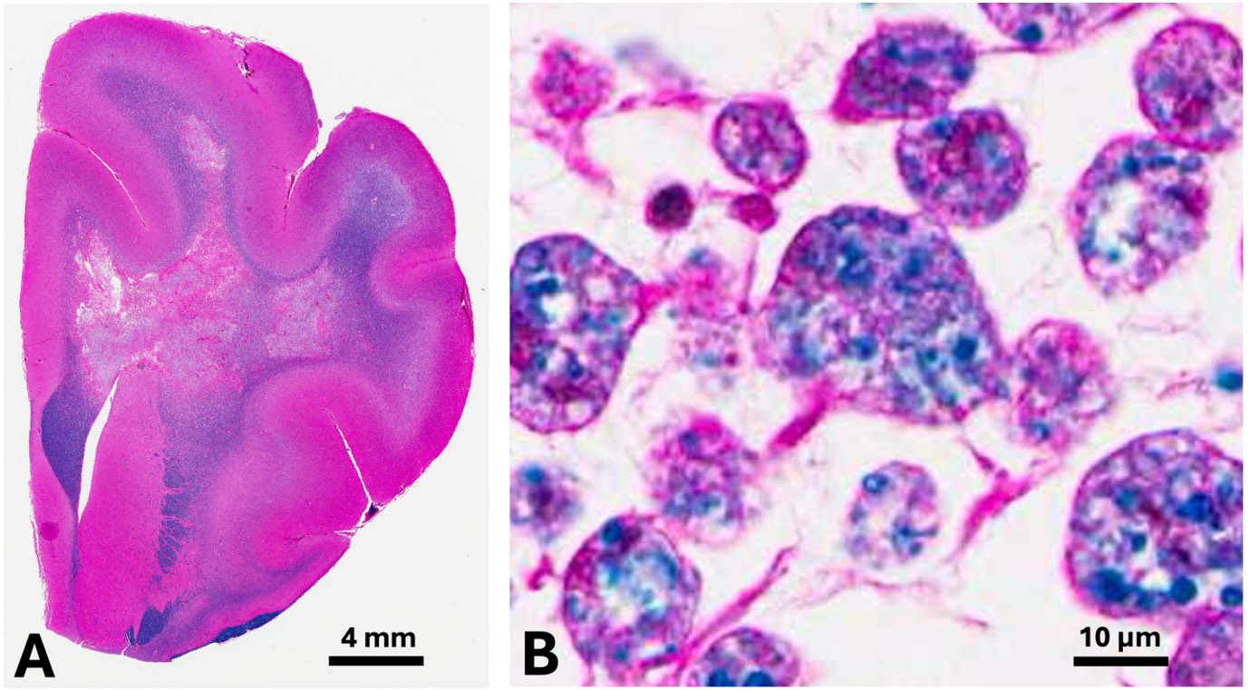
Figure 6. Histopathologic lesions of globoid cell leukodystrophy in a 6-month-old West Highland white terrier dog. Note the loss of blue-stained myelin within the white matter of the corona radiata (A) and characteristic large macrophages (globoid cells) containing phagocytized myelin lipids (B). Hematoxylin and eosin with a Luxol fast blue counterstain (H&E-LFB). Bar = 4 mm in (A) and 10 µm in (B).