
fig4
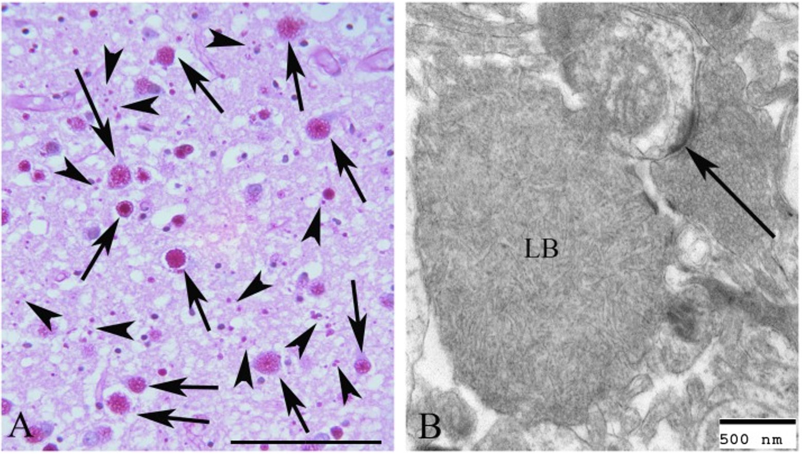
Figure 4. Lafora bodies in the frontal cortex of a dachshund dog seen with PAS diastase staining (A) and electron microscopy (B). Classical larger (arrows) and smaller (arrowheads) inclusions are evident. Bar = 50 µm in (A) and 500 nm in (B). The arrow in (B) identifies a synaptic density. The figure is reprinted with permission from[153]. Adhering to the CC BY 4.0 agreement.