
fig1
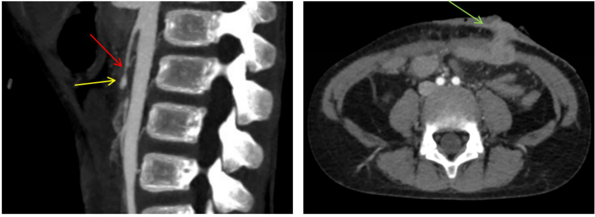
Figure 1. Preoperative CTA scan and CT showing the mesenteric lesion and post-jejunostomy changes. Left: Sagittal view showing the superior mesenteric artery trunk with luminal narrowing (red arrow) and a distal cystic protrusion (yellow arrow). Right: Axial view showing anterior abdominal wall post-jejunostomy changes (green arrow). CTA: Computed tomography angiography; CT: computed tomography.