



Advances in the application of deep learning in computed tomography imaging analysis of rare lung diseases
 0
0 
Abstract
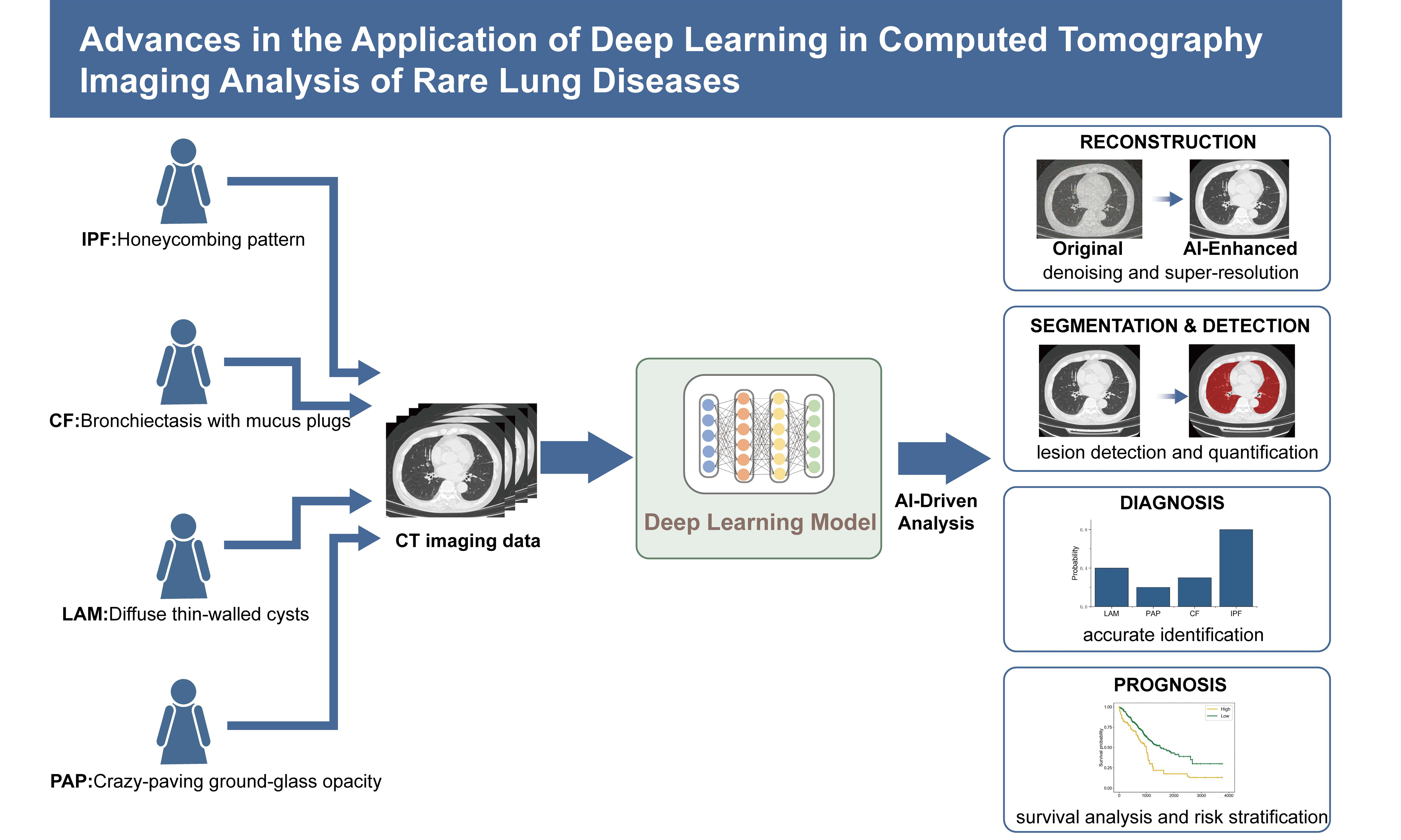
Diagnosis and management of rare lung diseases remain challenging owing to their low incidence, heterogeneous manifestations, and limited therapeutic options. High-resolution computed tomography (CT) is central to imaging assessment, yet conventional visual interpretation is subjective and lacks good reproducibility. Recent advances in artificial intelligence, especially deep learning, provide new approaches for automated, quantitative and objective chest computed tomography image analysis. This review summarizes deep learning applications across core stages of CT imaging analysis for rare respiratory diseases: image reconstruction and generation, lesion segmentation and detection, disease classification and diagnosis, and treatment response and prognosis prediction. With typical cases of rare lung diseases, we illustrate that deep learning models enable accurate quantification of imaging biomarkers, elevated diagnostic accuracy and optimized outcome stratification. Despite notable progress, key challenges remain in model generalization, interpretability, and clinical validation.
Keywords
INTRODUCTION
Rare lung diseases represent a category of conditions characterized by low incidence rates and multifaceted etiologies, encompassing a diverse array of disorders that affect a substantial number of patients. These diseases typically manifest with varied clinical symptoms, exhibit heterogeneous disease progression, and present limited therapeutic options, thereby imposing significant economic burdens on healthcare systems and adversely affecting patients’ quality of life[1,2].
Medical imaging is instrumental in the clinical diagnosis and research of rare respiratory diseases. Imaging modalities, particularly high-resolution computed tomography (HRCT), non-invasively visualize subtle alterations in structures such as the lung parenchyma, airways, blood vessels, and pleura. Such imaging serves as a fundamental basis for confirming diagnoses, classifying disease subtypes, and evaluating treatment efficacy across numerous rare respiratory conditions. However, challenges persist in disease imaging, including the phenomena of “the same disease exhibiting diverse manifestations” and “different diseases presenting with similar manifestations”. Traditional interpretations of medical images are heavily contingent upon the subjective expertise of radiologists, and the human perception often struggles to accurately detect early-stage or subtle structural changes. These limitations impede progress in achieving precise diagnoses, conducting quantitative research, and implementing personalized treatment strategies for rare lung diseases.
In recent years, rapid advancements in artificial intelligence, particularly in deep learning, which is a pivotal branch of machine learning have yielded groundbreaking developments in the analysis of medical images. By emulating the hierarchical neural network architecture of the human brain, deep learning algorithms can autonomously learn high-dimensional features and perform pattern recognition on extensive image datasets, thereby overcoming the constraints associated with conventional manual feature extraction. For rare lung diseases, which are often characterized by limited sample sizes and intricate pathologies, deep learning methodologies can automatically capture latent phenotypic information from imaging data. This capability facilitates quantitative, objective, and reproducible assessments of lesions, offering innovative technical approaches to precise diagnosis, mechanistic research, and translational applications in the realm of rare diseases.
APPLICATIONS OF DEEP LEARNING IN CHEST COMPUTED TOMOGRAPHY IMAGING
The clinical diagnosis and management of pulmonary diseases are highly reliant on imaging assessments. Chest radiography and computed tomography (CT) function as the fundamental imaging modalities. Chest X-rays are commonly utilized for preliminary screening and subsequent monitoring owing to their simplicity and minimal radiation exposure. Nevertheless, they are intrinsically limited by challenges such as tissue overlap and restricted spatial resolution. HRCT is regarded as the gold standard in pulmonary imaging evaluations. HRCT offers superior clarity in depicting the intricate structures of the pulmonary interstitium, airways, and vascular system, and serves as an essential tool in the diagnosis and assessment of rare lung diseases.
However, conventional imaging evaluation techniques predominantly depend on subjective visual assessments by clinicians, thereby imposing notable limitations. These issues encompass inconsistent diagnostic standards, challenges in obtaining accurate quantitative assessments, and low inter-observer reliability - problems that are especially evident when evaluating complex pathologies such as rare diseases.
In recent years, deep learning has made significant advancements in the analysis of chest CT scans. Its developmental trajectory demonstrates an evolutionary progression from two-dimensional to three-dimensional, from single-task to multi-task, and from fully supervised learning to self-supervised or weakly supervised learning. Through the development of multi-layer nonlinear network models, deep learning is capable of autonomously learning and extracting intricate pulmonary pathological features from extensive image datasets, facilitating intelligent identification and mapping from raw images to disease phenotypes. The subsequent sections comprehensively delineate the principal technical applications of deep learning in chest CT image analysis [Figure 1] and offer an overview and discussion of the key network architectures involved (as shown in Figure 2).
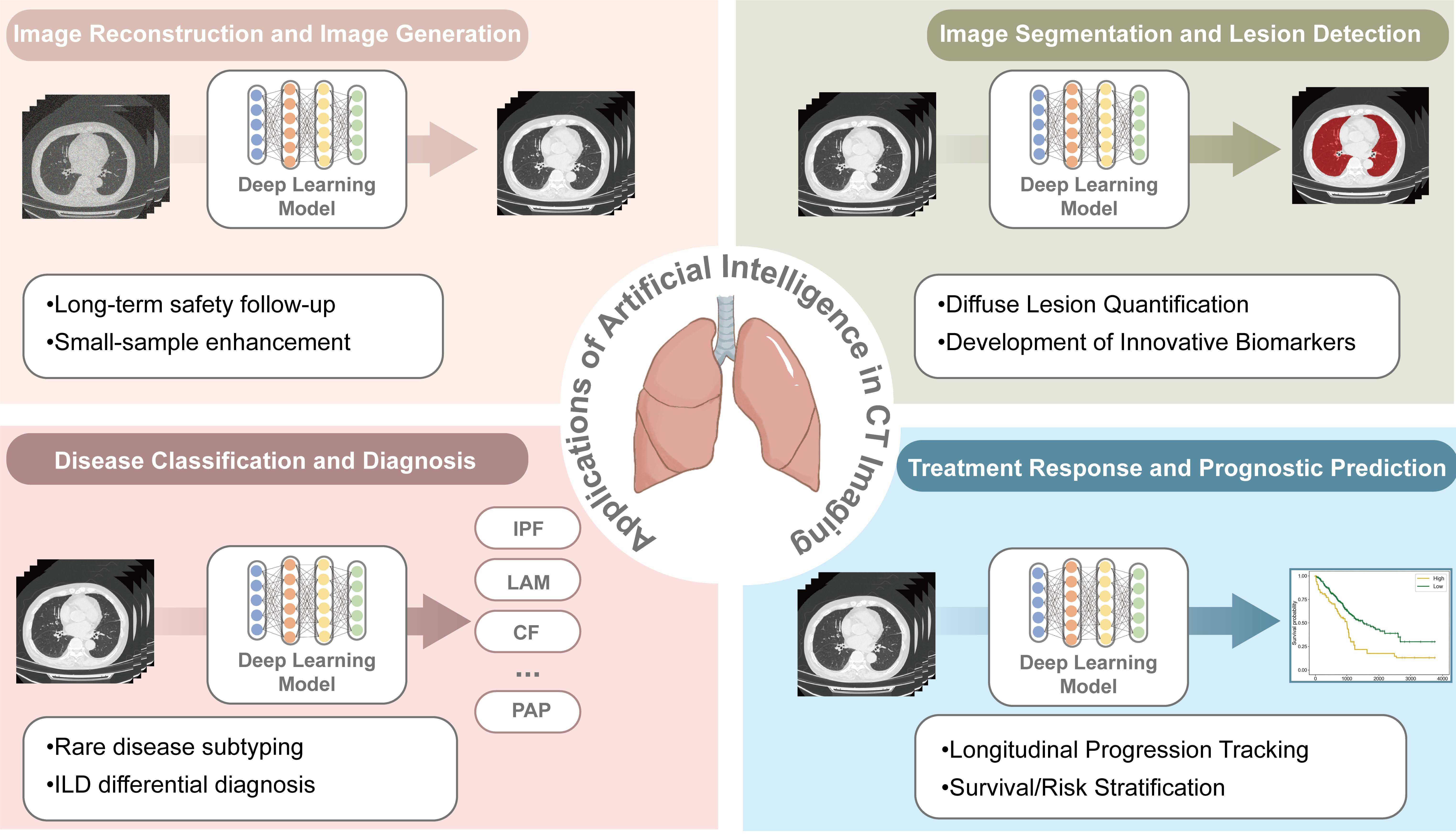
Figure 1. Applications of Deep Learning in Chest CT Imaging The framework illustrates the integration of artificial intelligence across four core clinical stages: (Top Left) Image Reconstruction and Image Generation, focusing on enhancing image quality and denoising; (Top Right) Image Segmentation and Lesion Detection, involving the automated delineation of lung structures and pathological regions; (Bottom Left) Disease Classification and Diagnosis, facilitating the differential diagnosis of various respiratory conditions such as idiopathic pulmonary fibrosis (IPF), cystic fibrosis (CF), lymphangioleiomyomatosis (LAM), and pulmonary alveolar proteinosis (PAP), and (Bottom Right) Treatment Response and Prognostic Prediction, utilizing longitudinal data for survival analysis and outcome stratification. All CT images shown are anonymized clinical data from the authors’ institution. The bar chart and survival curves are for illustration only, with no real data implication. Created in Adobe Illustrator. ILD: Interstitial lung disease; CT: computed tomography.
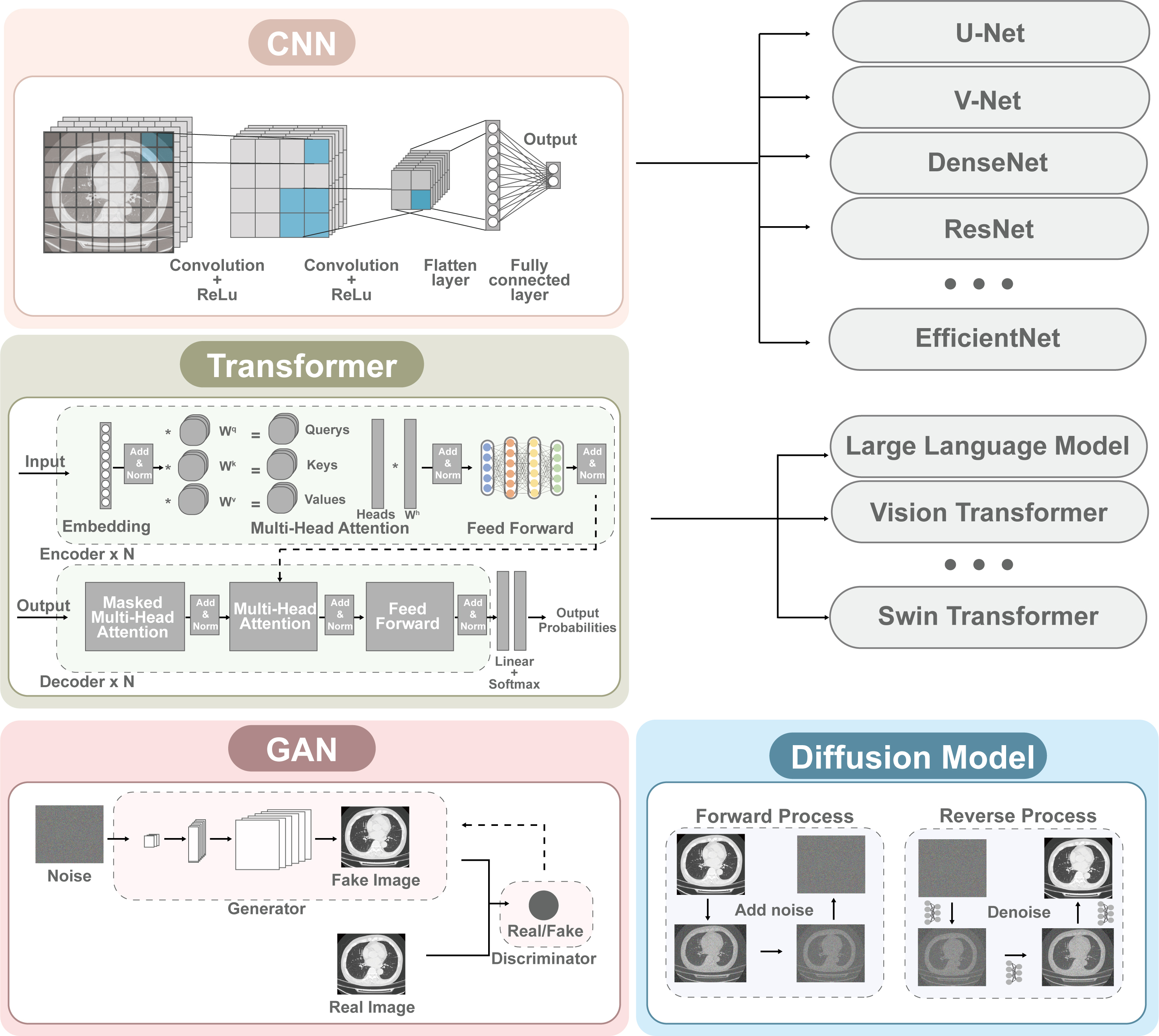
Figure 2. Overview of Commonly Used Deep Learning Network Architectures The diagram categorizes four major architectural paradigms: (Top Left) Convolutional Neural Networks (CNNs), emphasizing local feature extraction through hierarchical convolution and pooling layers, which form the basis for models like U-Net, V-Net, and ResNet; (Middle) Transformers, utilizing self-attention mechanisms and multi-head attention (MHA) to capture global contextual dependencies, represented by Vision Transformer (ViT) and Swin Transformer; (Bottom Left) Generative Adversarial Networks (GANs), consisting of a generator and a discriminator in an adversarial training loop for image synthesis; and (Bottom Right) Diffusion Models, based on a two-step stochastic process involving the progressive addition of noise (forward process) and iterative denoising (reverse process) to generate high-quality images. All CT scan samples included are anonymized clinical data from the authors’ institution and are free of copyright restrictions. Created in Adobe Illustrator. CT: Computed tomography.
The subsequent sections detail foundational deep learning architectures and their general implementation in CT image analysis. While these frameworks were primarily developed for broader radiologic applications, they constitute the essential technical basis for the disease-specific adaptations and rare-disease contexts discussed later in this review.
Image reconstruction and image generation
CT image reconstruction refers to the use of advanced algorithms to transform raw projection data into high-quality images while suppressing noise and artifacts, thereby improving image clarity and diagnostic reliability. CT image reconstruction constitutes the fundamental initial stage in the clinical imaging analysis process. The quality of image reconstruction directly influences lesion detectability and the accuracy of subsequent quantitative analysis. CT image reconstruction has historically depended on traditional approaches grounded in mathematical-physical models, including filtered back-projection (FBP) and adaptive statistical iterative reconstruction (ASIR). Among these, FBP provides benefits such as high computational efficiency and ease of implementation; however, it is highly susceptible to noise, leading to considerable deterioration in image quality under low-dose CT (LDCT) conditions. ASIR enhances noise performance to a certain extent through the integration of statistical noise modeling and regularization constraints. However, they frequently generate artifacts and excessive flattening of textures, which restricts the visualization of anatomical intricacies and undermines the accuracy of subsequent quantitative assessments[3].
With the progress of deep learning, Convolutional neural network (CNN)-based image reconstruction methods have become a prominent area of research[4,5]. CNN learns high-dimensional mapping relationships from low-dose CT to standard-dose CT using extensive paired data, effectively balancing noise reduction and the preservation of structural details. The Generative Adversarial Network (GAN) introduced by Goodfellow et al.[6]. substantially improves the structural integrity and visual clarity of low-dose images through adversarial learning techniques, representing a significant advancement in deep learning for CT image reconstruction. However, regular CNN and GAN architectures continue to encounter challenges in capturing long-range dependencies and preserving global structural coherence because of their inherently local convolutional design.
In recent years, CT reconstruction research has increasingly integrated more sophisticated networks. Transformer-based reconstruction models utilize self-attention techniques[7,8]. In contrast to convolutional operations, which are limited by local receptive fields, self-attention incorporates global contextual information within a single layer. This enables noise reduction while maintaining structural integrity and anatomical coherence.
Building upon this foundation, certain studies started efforts to extend deep learning reconstruction from single-modality to cross-modal image translation. Through the study of correspondences among various imaging modalities, researchers aim to generate CT images from X-ray data[9,10] or establish reciprocal mappings between CT and magnetic resonance imaging (MRI)[11,12]. These cross-modal reconstruction and generation techniques provide innovative imaging solutions for patients unable to tolerate conventional CT scans or necessitating multimodal integrated evaluation, thereby further broadening the scope of deep learning applications in medical image reconstruction.
In addition to CT reconstruction, the technology for medical image generation has also progressed significantly. Notably, the advancement of GAN and diffusion models has introduced innovative methods for data augmentation and low-dose imaging. Diffusion models produce images via an iterative denoising process, providing benefits such as enhanced training stability and superior output quality. Since Ho et al.[13] introduced the denoising diffusion probabilistic model in 2020, diffusion models have been effectively utilized across a range of medical image generation applications[14]. Numerous studies have utilized synthetic chest CT images produced by these models to mitigate the shortage of real data[15-17], thereby substantially improving the performance of subsequent classification or prediction models. This illustrates their considerable potential for applicability in contexts involving limited sample sizes, such as rare respiratory diseases.
Image segmentation and lesion detection
Quantitative analysis of medical images requires accurate segmentation of lung structures and lesions. Conventional segmentation techniques predominantly depend on manual or semi-automatic delineation, which is highly influenced by the operator’s level of expertise. This is particularly true for intricate structures such as the airway walls, small blood vessels, and irregularly shaped lesions, where notable inter- and intra-observer variability is present. This inconsistency not only restricts the feasibility of large-scale, multi-center studies but also introduces systematic bias in subsequent lesion assessment and image feature extraction, potentially impacting the stability and clinical interpretability of the imaging evaluation outcomes. Deep learning techniques, through end-to-end learning, offer an effective approach to resolving these challenges.
In the initial development of deep learning segmentation models, CNN-based encoder-decoder architectures established the foundational framework for medical image segmentation. Among these, the U-Net model proposed by Ronneberger et al.[18] employs a symmetric encoder-decoder architecture with cross-layer skip connections, which effectively maintains spatial localization precision while compressing semantic information, thereby substantially improving segmentation performance in scenarios with limited sample sizes. Milletari et al.’s V-Net[19] extended two-dimensional convolutions to three-dimensional space, directly modeling the spatial continuity between voxels through 3D convolution kernels, thereby further enhancing the consistency of volumetric segmentation and the integrity of anatomical structures. Although several general-purpose visual models have been introduced and demonstrated progress in lung imaging tasks, the U-Net series continues to serve as the foundational framework for most state-of-the-art approaches currently.
Classical CNN-based models have achieved notable improvements in overall segmentation accuracy; however, their capacity to accurately identify essential anatomical regions and minor structures remains constrained. Attention mechanisms, boundary-aware techniques, and Transformer architectures, such as Attention U-Net, BG-Net, and Swin-UNETR, which have shown benefits in the segmentation of irregular lesions and structures[20-22], have all been incorporated by researchers to improve the recognition of small structures and critical anatomical regions.
Simultaneously, the development and dissemination of open-source tools have expedited the clinical implementation of deep learning technologies. The nnU-Net, introduced by Isensee et al.[23], which autonomously adapts network architectures, training protocols, and preprocessing procedures, has established a robust baseline model for various medical imaging applications. Its outstanding stability and reproducibility have contributed to its extensive adoption in multi-center investigations. Building upon this, the TotalSegmentator[24] and Medical Open Network for AI (MONAI)[25] frameworks have further extended the capabilities of automatic segmentation, facilitating fully automated delineation of multi-organ and complex structures within the thorax, including pulmonary vessels and subsegmental airways. This has markedly decreased the labor expenses associated with large-scale imaging studies and has enhanced the practicality of lung image segmentation algorithms in clinical environments. The core characteristics and typical application scenarios of representative deep learning architectures applied in medical image analysis are summarized in Table 1.
Common deep learning models’ architectures and their applications in medical image segmentation
| Parent architecture | Representative network | Architectural characteristics | Typical application scenarios |
| CNN-based networks | FCN[26] | First fully convolutional network for dense prediction, replacing FC layers with convolutions | Semantic segmentation of natural and medical images, establishing the basic paradigm |
| U-Net[18] | Symmetric encoder-decoder with skip connections to combine multi-scale features | Medical image segmentation, especially with limited annotated data | |
| V-Net[19] | 3D volumetric extension of U-Net with dice loss optimization | Volumetric medical image segmentation requiring 3D context | |
| Transformer-based Networks | Vision Transformer[27] | Pure self-attention architecture treating images as sequences of patches | Medical image analysis requiring global contextual understanding |
| Swin transformer[28] | Hierarchical design with shifted windows for efficient self-attention | High-resolution medical image segmentation with multi-scale requirements | |
| Hybrid networks | TransUNet[29] | Combines CNN for local feature extraction and Transformer for global context | Medical image segmentation benefiting from both local details and global semantics |
Disease classification and diagnosis
Automated classification and differential diagnosis utilizing CT imaging hold significant clinical importance in the management of rare respiratory diseases. These diseases generally exhibit low prevalence, rare instances, and heterogeneous imaging features, often lacking high specificity, which hinders the ability to establish stable and precise differential diagnoses through qualitative assessment reliant on human expertise, thereby constraining early detection and intervention. Deep learning has the capability to autonomously extract latent high-dimensional imaging features with discriminative significance from complex chest CT, thereby facilitating accurate disease diagnosis.
Early studies predominantly employed CNN architectures including Visual Geometry Group (VGG), ResNet, DenseNet, and EfficientNet[30-33]. These networks conducted comprehensive feature learning on chest CT images, with hierarchical convolutional architectures systematically extracting features pertaining to image texture, morphology, and density distribution. The networks calibrated their parameters through the process of learning the ultimate classification labels, thereby distinguishing images of distinct diseases. Nevertheless, as CNNs predominantly depend on localized receptive fields, their capacity for discrimination typically emphasizes distinctions in local image features. Consequently, the model’s capacity for generalization and clinical stability remains constrained.
To improve the model’s capacity to identify essential anatomical regions and concealed lesion patterns, researchers incorporated network architectures featuring attention mechanisms into classification tasks[34,35]. These models are capable of incorporating global contextual information during feature learning and can adaptively prioritize regions that significantly contribute to diagnosis, which is especially beneficial for tasks involving small lesions or substantial background interference.
However, the aforementioned networks predominantly rely on static image pattern recognition, which impedes their ability to comprehensively identify the systemic structural alterations and cross-scale phenotypic characteristics associated with rare diseases. Considering the complexity of imaging manifestations and the considerable clinical heterogeneity associated with rare lung diseases, research efforts have progressively transitioned from isolated image classification towards comprehensive integrated modeling[36,37]. By integrating CT images, clinical indicators, pulmonary function parameters, and molecular or genetic data, multimodal deep learning models can comprehensively characterize disease phenotypes from structural, functional, and biological viewpoints. This method markedly enhances diagnostic consistency and clinical interpretability in respiratory disease classification endeavors. This research trajectory has facilitated the transition from image appearance-based pattern recognition to comprehensive phenotypic modeling focused on the core characteristics of the disease, offering a more clinically pertinent technological approach for the accurate diagnosis of rare lung diseases.
Treatment response and prognostic prediction
In respiratory diseases, the assessment of treatment response and prognosis prediction are vital elements of precision medicine. Conventional prognostic analysis primarily depends on manually extracted imaging biomarkers and clinical variables, employing statistical techniques such as the Cox proportional hazards model for modeling. However, these approaches frequently rely on linear assumptions, which hinder their ability to comprehensively represent the high-dimensional, nonlinear information contained within imaging data. Furthermore, their capacity to detect intricate disease phenotypes and accommodate heterogeneous evolutionary patterns remains restricted. Deep learning models are capable of directly extracting latent phenotypic features associated with outcomes from raw images, offering a novel methodological approach for developing prognostic prediction models with enhanced expressiveness and generalization capabilities.
Early studies on integrating deep learning into prognostic prediction predominantly concentrated on the combination of image features with survival analysis models. For instance, several studies employed convolutional neural networks to extract imaging features and incorporated them into the Cox proportional hazards model, enabling joint analysis of imaging features and survival outcomes[38-41]. As research advanced, it was determined that single-timepoint imaging is inadequate for accurately characterizing disease progression. Following 2020, longitudinal imaging modeling emerged as a prominent focus of research. Temporal networks utilizing 3D-CNN in conjunction with long short-term memory (LSTM), gated recurrent unit (GRU), or Transformer architectures can effectively model the temporal evolution of imaging features, thereby representing disease progression and response to treatment[42-44]. Research indicates that longitudinal modeling provides more consistent and clinically meaningful prognostic predictions across a range of pulmonary conditions.
Furthermore, the scope of research has broadened from single-task models to encompass multi-task learning[45,46]. Multi-task networks are capable of concurrently predicting multiple tasks, such as lesion volume alterations and clinical outcomes, thereby improving the model’s robustness and generalization capabilities. Deep learning is enhancing the prediction of treatment response and prognosis in respiratory diseases, progressing from conventional statistical methods to a dynamic approach that incorporates imaging, temporal data, and mechanistic insights. This establishes a novel methodological basis for precise therapeutic decision-making and sustained disease management. Simultaneously, the incorporation of multimodal data allows the model to identify more comprehensive and multivariate disease characteristics, thereby facilitating clinical decisions informed by more extensive information and supporting accurate treatment and personalized management[47-50].
ADVANCES IN DEEP LEARNING APPLICATIONS FOR CT OF RARE LUNG DISEASES
Owing to its superior capabilities in representational learning and automated feature extraction, deep learning can discern structural and textural features within complex, high-dimensional chest CT images that are challenging to detect through conventional visual evaluation. This advantage is especially important for rare respiratory diseases marked by limited sample sizes, significant clinical heterogeneity, and imaging alterations challenging to assess visually. In recent years, diverse deep learning models have facilitated multi-tiered applications - from structural quantification to functional prediction and prognostic stratification - in diffuse fibrosis, cystic lesions, and hereditary airway diseases, advancing the field of precise diagnosis and treatment of rare diseases into a new era. The subsequent portions will demonstrate the development and application of these technologies through case studies involving idiopathic pulmonary fibrosis (IPF), cystic fibrosis (CF), lymphangioleiomyomatosis (LAM) and pulmonary alveolar proteinosis (PAP).
Idiopathic pulmonary fibrosis
IPF is a chronic progressive pulmonary fibrosis disease characterized by dyspnea and the progressive deterioration of lung function[51]. Its imaging evaluation predominantly depends on HRCT to visually identify features such as honeycombing, reticular patterns, and traction bronchiectasis. However, these manifestations substantially overlap with other interstitial lung diseases, and visual evaluation remains subjective, exhibiting limited inter-observer reliability, which hampers consistent quantification of fibrosis burden and precise assessment of disease progression.
To address these limitations, deep learning models have markedly enhanced non-invasive diagnostic capabilities by autonomously recognizing characteristic imaging patterns of IPF. In 2022, Refaee et al.[52] developed an integrated radiomics and deep learning model utilizing high-resolution computed tomography data from 139 patients with IPF and 335 patients with non-IPF interstitial lung disease (ILD). The model achieved an area under the curve (AUC) of 0.917 on an external test set, markedly surpassing the performance of individual models and clinical visual evaluations. This model effectively mitigated the diagnostic challenge resulting from the imaging similarities between IPF and non-IPF ILD. Bratt et al.[53] designed a model utilizing EfficientNet-B3, attaining an AUC of 0.87 for the prediction of histologically confirmed Usual Interstitial Pneumonia (UIP) within a biopsy-validated ILD cohort. The model demonstrated markedly greater consistency than radiologists and successfully identified atypical UIP cases that had been visually misclassified. However, this study has some limitations in data collection: the training and test sets were both derived from the same three Mayo Clinic centers without an independent external validation cohort, and the inclusion of only biopsy-proven cases introduces significant selection bias toward diagnostically challenging patients. Huang et al.[54] additionally attained an AUC of 0.96 in detecting acute exacerbation of IPF on HRCT, offering prompt assistance for clinical emergency management.
Traditional HRCT evaluation of IPF encounters challenges in structural quantification, in addition to qualitative assessment, due to its dependence on manual delineation. Manual recognition requires significant time investment, and the scarcity of available data complicates longitudinal follow-up and risk modeling efforts. Deep learning facilitates the objective quantification of structural abnormalities in IPF. In 2022, Sun et al.[55] developed a segmentation model that exhibited a significant correlation between deep learning-based quantification of honeycombing changes and Diffusing Capacity for Carbon Monoxide (DLCO). Thillai et al.[56] developed an automatic segmentation model utilizing data from 446 patients to extract CT-derived lung volume, fibrosis volume, and pulmonary vascular volume. The metrics demonstrated a strong correlation with Forced Vital Capacity (FVC), and longitudinal changes were able to independently predict survival risk. Deep learning has identified novel imaging biomarkers in addition to fibrosis burden. In 2024, Maetani et al.[57] employed AI-based image analysis software (AIQCT and SYNAPSE VINCENT) to segment reticular and honeycombing regions, as well as the airway tree, identifying airway wall thickening and tracheal tortuosity as independent predictors of IPF progression. It should be noted that this study only included male patients, which may limit the generalizability of the findings to female IPF patients. Nan et al.[58] developed the SABRE AI model for biomarker recognition, utilizing a dataset of HRCT images from 460 patients with various diseases. This model quantifies airway volume to predict the mortality risk associated with fibrotic lung diseases, including idiopathic pulmonary fibrosis. The integration of DLCO resulted in an AUC of 0.852 and a C-index of 0.752 at one year, facilitating precise stratification of patients into low, medium, and high-risk categories.
Regarding disease stratification and prognosis evaluation in IPF, Humphries et al.[59] formulated a data-driven texture analysis model utilizing convolutional neural networks, which quantified the extent of fibrosis in 393 IPF patients. The fibrosis score demonstrated a significant correlation with lung function and composite physiological indices, and it persisted as an independent prognostic factor even among patients with relatively preserved pulmonary function. Further, Humphries et al.[60] introduced a multi-instance learning model that reliably detected UIP across various cohorts in 2024. In patients diagnosed with UIP, the transplant-free survival rate was markedly decreased, and the decline in FVC occurred more rapidly. The model additionally exhibited independent prognostic significance in populations with ambiguous UIP diagnoses determined through visual assessment.
Cystic fibrosis
CF is an inherited autosomal recessive disorder resulting from mutations in the CFTR gene[61]. For the evaluation of pulmonary lesions in cystic fibrosis patients, conventional chest CT continues to serve as the standard method for assessing structural alterations, such as bronchiectasis, airway wall hypertrophy, and mucus impaction. Nevertheless, its evaluation frequently depends on semi-quantitative scoring systems that do not possess adequate sensitivity to detect subtle alterations in disease progression in a timely manner. Furthermore, the risk of radiation exposure from recurrent scans restricts its feasibility as a standard monitoring method.
In 2019, Nezamabadi et al.[62] developed a CF HRCT image pattern classification system utilizing CNN to evaluate its effectiveness in categorizing three patterns: normal lung tissue, bronchiectasis, and inflammation. The results indicated that the system attained a classification accuracy of 93.64%, with an average sensitivity of 93.47% and a specificity of 96.61% on a patch dataset derived from 1,125 HRCT segments of 45 CF patients with the final patch distribution consisting of 36,889 normal, 24,820 bronchiectasis, and 2,700 inflammation patches, markedly surpassing conventional approaches. In a 2020 study, Zucker et al.[63] devised a deep convolutional neural network (DCNN) utilizing ResNet-18 architecture for automated Brasfield scoring. The model evaluated 200 test cases in 5 s and attained a correlation of 0.79-0.83 with the total Brasfield score, which was comparable to the correlation between the total Brasfield score and the scores assigned by five pediatric radiologists (0.85-0.90). This study was conducted at a single center using a single X-ray system model, and did not include an independent external validation cohort, which may limit the generalizability of the findings.
Furthermore, Dournes et al.[64] developed the NOVAA-CT system in 2022, which consists of three 2D convolutional neural networks, utilizing CT data from 184 patients. The system exhibited the capability to detect and quantify five categories of pulmonary structural abnormalities, encompassing bronchiectasis, bronchial wall thickening, and mucus blockage. The volume of aberrant structures segmented by the system exhibited a Dice coefficient of 0.71 in comparison to manual segmentation outcomes and demonstrated a significant correlation with pulmonary function tests and visual CT scoring. The system has the capability to precisely identify treatment-associated structural modifications, such as notable decreases in mucus accumulation and bronchial wall thickening volume following lumacaftor/ivacaftor therapy. The single-CT analysis was completed in just 2 min, and enhancements in specific indicators were noted in 10 patients receiving Lumacaftor/Ivacaftor treatment. In 2025, Hadj Bouzid et al.[65] further confirmed the reproducibility and efficacy of this system in 139 CF patients through the application of the NOVAA-CT model. They quantified the substantial therapeutic effects of Elexacaftor/tezacaftor/ivacaftor (ETI) therapy and corticosteroid treatment, concluding that ETI therapy could significantly reverse the extent of bronchiectasis (P < 0.001), whereas corticosteroids showed no such effect. The study emphasized the precision of artificial intelligence in comprehensive lung analysis and the continuous monitoring of treatment progress. Based on the reversibility of bronchiectasis observed in this study, Hadj Bouzid et al.[66] performed a multivariate analysis involving 106 patients treated with ETI, revealing that younger individuals, patients without chronic Pseudomonas aeruginosa colonization, and those with a lower pulmonary mucus burden were more likely to derive benefit from the therapy. Deep learning has surpassed the constraints of conventional visual scoring methods, including lengthy processing times and limited reproducibility, thereby advancing the automated and standardized quantification of CF.
Lymphangioleiomyomatosis
LAM is a rare systemic neoplastic disorder of low malignant potential, characterized by the widespread proliferation of smooth muscle-like LAM cells and primarily affecting women[67]. In clinical practice, the imaging features of LAM significantly coincide with those of other diffuse cystic lung diseases, such as Langerhans cell histiocytosis and Birt-Hogg-Dubé syndrome, all of which may manifest as diffusely distributed thin-walled cysts in both lungs. Dependence solely on morphological features for manual interpretation necessitates significant subspecialty expertise, and inter-observer agreement continues to be limited. Jonas et al.[68] devised an automated classification model utilizing a TensorFlow Inception-v3 network to differentiate LAM from other diffuse cystic lung diseases, attaining a sensitivity of 0.85 and a specificity of 0.92, comparable to expert-level performance and valuable in minimizing diagnostic delays.
Most patients with LAM are women of reproductive age, and the cumulative radiation exposure resulting from prolonged, recurrent CT surveillance poses a significant consideration in clinical management. In a 2024 study, Golbus et al.[69] employed ultra-low-dose chest CT utilizing a SilverBeam filter in conjunction with a deep learning-based reconstruction algorithm (AiCE), achieving an 85.5% reduction in radiation exposure while preserving precise cyst quantification (R2 = 0.98), thereby significantly enhancing the safety of longitudinal follow-up examinations.
Although deep learning holds significant potential for LAM imaging analysis, its implementation remains limited by the fundamental challenge of limited availability of high-quality annotated data. Chest CT of LAM is characterized by numerous thin-walled cysts with diffuse distribution; meticulous manual annotation on a slice-by-slice basis is highly time-consuming and labor-intensive, with limited inter-observer consistency, significantly restricting the advancement and applicability of supervised deep learning models. To confront this challenge, Zhang et al.[70] introduced an unsupervised recursive self-learning U-Net architecture. This method produces preliminary segmentation outcomes employing spatial K-means clustering and graph-cut algorithms, which are subsequently utilized as pseudo-labels for iterative network training. At each stage, the output of the preceding network functions as a new supervisory signal, with parameter transfer and learning rate decay strategies employed to progressively enhance segmentation accuracy. Without manual annotations, this approach attained a Dice score of 0.7587, effectively mitigating the high annotation costs and limited sample sizes associated with rare disease imaging, thereby offering a viable pathway for the application of deep learning to low-sample diseases such as LAM.
Pulmonary alveolar proteinosis
PAP is a rare interstitial lung disease characterized by the abnormal accumulation of surfactant material within the alveoli[71]. HRCT is the cornerstone imaging modality for diagnosis and therapeutic evaluation; however, clinical assessment still largely relies on subjective visual interpretation and coarse grading. This approach makes it difficult to achieve stable and reproducible quantification of disease burden and is insufficiently sensitive to longitudinal changes in disease progression and treatment response, thereby limiting precise assessment and individualized management.
To address these limitations, deep learning has markedly improved the objectivity and granularity of PAP imaging evaluation through automated segmentation and quantitative modeling. Shi et al.[72] applied a fully convolutional neural network to HRCT images from 50 PAP patients to automatically segment ground-glass opacities (GGO) and quantify attenuation in Hounsfield unit ranges. Their approach effectively differentiated disease severity, and the quantitative metrics showed significant reductions after 12 months of statin therapy, with positive correlations to improvements in PaO2 and DLCO %pred. These findings demonstrated the advantage of deep learning-derived parameters for objective assessment of disease burden and treatment response. In PAP patients without hypercholesterolemia, Shi et al.[73] further showed that deep learning-quantified GGO metrics could identify 65% of treatment responders and, when combined with baseline granulocyte-macrophage colony-stimulating factor (GM-CSF) antibody levels and the total cholesterol to high-density lipoprotein cholesterol ratio (TC/HDL) ratio, could predict therapeutic efficacy, thereby extending its clinical utility to non-traditional patient subgroups.
Given the inherent scarcity of samples in rare diseases such as PAP, Gao et al.[74] demonstrated that the LCT found foundation model pretrained on the LungCT-28M dataset could achieve robust performance with minimal annotated data. In PAP diagnosis, the model achieved an area under the receiver operating characteristic curve (AUROC) of 0.9532, and even with the training data reduced by half, performance remained high (AUROC = 0.9130), significantly outperforming mainstream pretrained models. Overall, deep learning not only overcomes the limitations of traditional imaging assessment characterized by subjectivity and insufficient sensitivity, but also provides scalable and reusable technical pathways for objective phenotyping, treatment monitoring, and individualized therapeutic decision-making in PAP, underscoring its central role in precision diagnosis and management of rare lung diseases.
Table 2 summarizes the key characteristics of studies that trained their own deep learning models for rare lung diseases, including clinical tasks, model architectures and cohort sizes.
Overview of deep learning studies with original model training for rare lung diseases
| Disease | Reference | Clinical task | Model architecture | Cohort size |
| IPF | Refaee et al.[52] | Diagnosis: IPF vs. non-IPF ILDs | DenseNet | 474 scans |
| Bratt et al.[53] | UIP vs non-UIP | EfficientNet-B3 | 1,239 scans | |
| Huang et al.[54] | AE-IPF vs stable IPF vs healthy | DeepLabV3+ + SlowFast | 306 scans | |
| Thillai et al.[56] | Segmentation:lung, airway, vessel, fibrosis | UNet | 621 scans | |
| Nan et al.[58] | Segmentation:airway and branch classification | Fuzzy attention neural network | 1,744 scans | |
| Humphries et al.[60] | Diagnosis: UIP classification | MIL with attention | 3,488 scans | |
| CF | Nezamabadi et al.[62] | Segmentation:normal, bronchiectasis, inflammation | Custom CNN | 1,125 slices (64,409 patches) |
| Zucker et al.[63] | Brasfield radiographic scoring | ResNet-18 | 2,058 X-rays | |
| Dournes et al.[64] | Segmentation: bronchiectasis, peribronchial thickening, bronchial mucus plug, bronchiolar mucus plug, consolidation | Ensemble of three 2D CNNs | 23,530 slices | |
| LAM | Jonas et al.[68] | Diagnosis: LAM vs. non-LAM | Inception-v3 | 389 slices |
| Zhang et al.[70] | Segmentation: cyst | U-Net | 183 scans | |
| PAP | Gao et al.[74] | Multi-task: diagnosis, prognosis, segmentation, image reconstruction/enhancement | Unet+crossattention+diffusion pretrain | 105,184 scans |
CONCLUSION
In recent years, deep learning has achieved significant progress in the imaging analysis of rare lung diseases. By leveraging its powerful feature extraction capabilities and techniques such as self-attention mechanisms, deep learning can efficiently process complex CT images and identify critical pathological features, providing substantial support for image-based characterization and evaluation of these conditions. Despite this promise, translating these advances into clinical practice faces several challenges, particularly in model generalization, interpretability, and real-world validation.
From a methodological perspective, standardization across study designs is notably lacking in the current literature. A particular concern is that CT acquisition parameters, such as slice thickness, reconstruction kernels, and contrast enhancement, can act as confounding factors that distort model predictions rather than capturing true disease features. This phenomenon, known as acquisition shift, occurs when models are trained on data from one protocol and applied to another, often leading to substantial performance degradation[75]. For example, Bratt et al.[53] included CT scans with slice thickness up to 20 mm alongside standard 0.5-1.5 mm slices but did not report performance stratified by slice thickness, leaving it unclear whether their model generalizes to thinner-slice scans used in routine clinical practice. Similarly, Thillai et al.[56] pooled data from five scanner manufacturers with slice thicknesses ranging from 0.5 mm to 5 mm but did not quantify performance variation across these parameters. Annotation strategies also lack consistency: while most studies relied on radiologist-annotated ground truth, others used unsupervised pseudo-labeling[70] or commercial black-box software without disclosure[72], making it impossible to compare the reliability of reference standards. We therefore recommend that future studies explicitly report acquisition parameters, quantify performance variation across different protocols, and, where possible, employ domain adaptation or data harmonization techniques to mitigate confounding effects[75,76].
From a data perspective, rare diseases are characterized by small, scattered patient populations and variability in imaging protocols across centers, resulting in limited and heterogeneously distributed datasets prone to overfitting and poor external validity. For instance, Zucker et al.[63] lacked external validation despite achieving promising internal performance. Even among studies that employed external test sets, such as Refaee et al.[52], Huang et al.[54] sample sizes remained modest, and all validations were retrospective in nature. Strategies such as transfer learning from large-scale pre-trained models, federated learning for multi-center collaboration, and generative models for data augmentation offer potential solutions to enhance model robustness in small-sample settings.
At the model level, the “black-box” nature of many deep learning systems limits interpretability and hinders their clinical adoption, especially in high-stakes diagnostic and prognostic tasks. Among the reviewed studies, interpretability appears to be addressed infrequently. Only a limited number of studies, such as Refaee et al.[52], have employed Grad-CAM for visualization, yet they also acknowledged that the method “can only highlight regions, not explain why” a region is considered important. The majority of other studies seem to concentrate primarily on performance metrics, with limited attention given to interpretability analysis. Future work should integrate explainability techniques such as saliency mapping, attention visualization, and feature attribution to link model decisions to specific anatomical or pathological findings, thereby improving transparency and facilitating clinician trust.
Finally, regarding clinical translation, there remains a lack of large-scale, multi-center prospective validation, along with standardized evaluation metrics and reporting frameworks. There is an urgent need to establish open-access, multi-center datasets with standardized annotations, develop rigorous AI assessment protocols, and foster closer collaboration between clinicians and engineers. Such efforts will be important for facilitating the transition of deep learning from experimental research into routine clinical practice.
DECLARATIONS
Acknowledgements
The Graphical Abstract was created in Adobe Illustrator.
Authors’ contributions
Design, literature review, writing: Liu B
Manuscript editing, manuscript revision: Xie M
Availability of data and materials
Not applicable.
AI and AI-assisted tools statement
During the preparation of this manuscript, the AI tool GPT (version gpt-4o, released 2024-5-14) was used solely for language editing. The tool did not influence the study design, data collection, analysis, interpretation, or the scientific content of the work. All authors take full responsibility for the accuracy, integrity, and final content of the manuscript.
Financial support and sponsorship
None.
Conflicts of interest
All authors declared that there are no conflicts of interest.
Ethical approval and consent to participate
Not applicable.
Consent for publication
Not applicable.
Copyright
© The Author(s) 2026.
REFERENCES
2. McCarthy C, Lara Gallego B, Trapnell BC, et al. Epidemiology of rare lung diseases: The challenges and opportunities to improve research and knowledge. In: Posada de la Paz M, Taruscio D, Groft SC, Editors. Rare diseases epidemiology: update and overview. Cham: Springer International Publishing; 2017. pp. 419-42.
3. Willemink MJ, Noël PB. The evolution of image reconstruction for CT-from filtered back projection to artificial intelligence. Eur Radiol. 2019;29:2185-95.
4. Chen H, Zhang Y, Zhang W, et al. Low-dose CT via convolutional neural network. Biomed Opt Express. 2017;8:679-94.
5. Chen H, Zhang Y, Kalra MK, et al. Low-dose CT with a residual encoder-decoder convolutional neural network. IEEE Trans Med Imaging. 2017;36:2524-35.
6. Goodfellow IJ, Pouget-Abadie J, Mirza M, et al. Generative adversarial networks. arXiv. 2014;arXiv:1406.2661. Available from: https://arxiv.org/abs/1406.2661 [Last accessed on 20 Jul 2026].
7. Pan J, Zhang H, Wu W, Gao Z, Wu W. Multi-domain integrative swin transformer network for sparse-view tomographic reconstruction. Patterns. 2022;3:100498.
8. Hu J, Zheng S, Wang B, Luo G, Huang W, Zhang J. Super-resolution swin transformer and attention network for medical CT imaging. Biomed Res Int. 2022;2022:4431536.
9. Tan Z, Li J, Tao H, Li S, Hu Y. XctNet: Reconstruction network of volumetric images from a single X-ray image. Comput Med Imaging Graph. 2022;98:102067.
10. Ying X, Guo H, Ma K, et al. X2CT-GAN: reconstructing CT from biplanar X-rays with generative adversarial networks. In: 2019 IEEE/CVF Conference on Computer Vision and Pattern Recognition (CVPR); 2019 Jun 15-20; Long Beach, CA, USA. IEEE; 2019. pp. 10611-20.
11. Jiang J, Hu YC, Tyagi N, et al. Cross-modality (CT-MRI) prior augmented deep learning for robust lung tumor segmentation from small MR datasets. Med Phys. 2019;46:4392-404.
12. Jue J, Jason H, Neelam T, et al. Integrating cross-modality hallucinated MRI with CT to aid mediastinal lung tumor segmentation. In: Shen D, Liu T, Peters TM, et al. Editors. Medical image computing and computer assisted intervention-MICCAI 2019. Cham: Springer International Publishing; 2019. pp. 221-9.
13. Ho J, Jain A, Abbeel P. Denoising diffusion probabilistic models. In: Proceedings of the 34th International Conference on Neural Information Processing Systems; Red Hook, NY, USA. Curran Associates Inc.; 2020. pp. 6840-51. Available from: https://dl.acm.org/doi/abs/10.5555/3495724.3496298 [Last accessed on 20 Jul 2026].
14. Kazerouni A, Aghdam EK, Heidari M, et al. Diffusion models in medical imaging: a comprehensive survey. Med Image Anal. 2023;88:102846.
15. Jiang Y, Chen H, Loew M, Ko H. COVID-19 CT image synthesis with a conditional generative adversarial network. IEEE J Biomed Health Inform. 2021;25:441-52.
16. Onishi Y, Teramoto A, Tsujimoto M, et al. Automated pulmonary nodule classification in computed tomography images using a deep convolutional neural network trained by generative adversarial networks. Biomed Res Int. 2019;2019:6051939.
17. Apostolopoulos ID, Papathanasiou ND, Panayiotakis GS. Classification of lung nodule malignancy in computed tomography imaging utilising generative adversarial networks and semi-supervised transfer learning. Biocybern Biomed Eng. 2021;41:1243-57.
18. Ronneberger O, Fischer P, Brox T. U-net: Convolutional networks for biomedical image segmentation. In: Navab N, Hornegger J, Wells WM, et al. Editors. Medical Image Computing and Computer-Assisted Intervention-MICCAI 2015. Cham: Springer International Publishing; 2015. pp. 234-41.
19. Milletari F, Navab N, Ahmadi S-A. V-net: fully convolutional neural networks for volumetric medical image segmentation. In: 2016 Fourth International Conference on 3D Vision (3DV); 2016 Oct 25-28; Stanford, CA, USA. IEEE; 2016. pp. 565-71.
20. Hu H, Li Q, Zhao Y, Zhang Y. Parallel deep learning algorithms with hybrid attention mechanism for image segmentation of lung tumors. IEEE Trans Ind Inf. 2021;17:2880-9.
21. Nan Y, Ser JD, Tang Z, et al. Fuzzy attention neural network to tackle discontinuity in airway segmentation. IEEE Trans Neural Netw Learn Syst. 2024;35:7391-404.
22. Abdalaoui Slimani F, Bentourkia M. Improving deep learning U-Net++ by discrete wavelet and attention gate mechanisms for effective pathological lung segmentation in chest X-ray imaging. Phys Eng Sci Med. 2025;48:59-73.
23. Isensee F, Jaeger PF, Kohl SAA, Petersen J, Maier-Hein KH. nnU-Net: a self-configuring method for deep learning-based biomedical image segmentation. Nat Methods. 2021;18:203-11.
24. Wasserthal J, Breit HC, Meyer MT, et al. TotalSegmentator: robust segmentation of 104 anatomic structures in CT images. Radiol Artif Intell. 2023;5:e230024.
25. Diaz-Pinto A, Alle S, Nath V, et al. MONAI label: a framework for AI-assisted interactive labeling of 3D medical images. Med Image Anal. 2024;95:103207.
26. Shelhamer E, Long J, Darrell T. Fully convolutional networks for semantic segmentation. IEEE Trans Pattern Anal Mach Intell. 2017;39:640-51.
27. Dosovitskiy A, Beyer L, Kolesnikov A, et al. An image is worth 16x16 words: transformers for image recognition at scale. arXiv. 2021;arXiv:2010.11929. Available from: http://arxiv.org/abs/2010.11929 [Last accessed on 20 Jul 2026].
28. Liu Z, Lin Y, Cao Y, et al. Swin transformer: hierarchical vision transformer using shifted windows. In: 2021 IEEE/CVF International Conference on Computer Vision (ICCV); 2021 Oct 10-17; Montreal, QC, Canada. IEEE; 2021. pp. 9992-10002.
29. Chen J, Lu Y, Yu Q, et al. TransUNet: transformers make strong encoders for medical image segmentation. arXiv. 2021;arXiv:2102.04306. Available from: http://arxiv.org/abs/2102.04306 [Last accessed on 20 Jul 2026].
30. Raza R, Zulfiqar F, Khan MO, et al. Lung-EffNet: Lung cancer classification using EfficientNet from CT-scan images. Eng Appl Artif Intell. 2023;126:106902.
31. Huang ML, Liao YC. A lightweight CNN-based network on COVID-19 detection using X-ray and CT images. Comput Biol Med. 2022;146:105604.
32. Kathamuthu ND, Subramaniam S, Le QH, et al. A deep transfer learning-based convolution neural network model for COVID-19 detection using computed tomography scan images for medical applications. Adv Eng Softw. 2023;175:103317.
33. Jiang H, Tang S, Liu W, Zhang Y. Deep learning for COVID-19 chest CT (computed tomography) image analysis: a lesson from lung cancer. Comput Struct Biotechnol J. 2021;19:1391-9.
34. Sabir M, Farhan M, Almalki NS, Alnfiai MM, Sampedro GA. FibroVit-Vision transformer-based framework for detection and classification of pulmonary fibrosis from chest CT images. Front Med. 2023;10:1282200.
35. UrRehman Z, Qiang Y, Wang L, et al. Effective lung nodule detection using deep CNN with dual attention mechanisms. Sci Rep. 2024;14:3934.
36. Sangeetha SKB, Mathivanan SK, Karthikeyan P, et al. An enhanced multimodal fusion deep learning neural network for lung cancer classification. Syst Soft Comput. 2024;6:200068.
37. Huang SC, Pareek A, Zamanian R, Banerjee I, Lungren MP. Multimodal fusion with deep neural networks for leveraging CT imaging and electronic health record: a case-study in pulmonary embolism detection. Sci Rep. 2020;10:22147.
38. Hasenstab KA, Yuan N, Retson T, et al. Automated CT staging of chronic obstructive pulmonary disease severity for predicting disease progression and mortality with a deep learning convolutional neural network. Radiol Cardiothorac Imaging. 2021;3:e200477.
39. González G, Ash SY, Vegas-Sánchez-Ferrero G, et al. Disease staging and prognosis in smokers using deep learning in chest computed tomography. Am J Respir Crit Care Med. 2018;197:193-203.
40. Mukherjee P, Zhou M, Lee E, et al. A shallow convolutional neural network predicts prognosis of lung cancer patients in multi-institutional CT-image data. Nat Mach Intell. 2020;2:274-82.
41. Katzman JL, Shaham U, Cloninger A, Bates J, Jiang T, Kluger Y. DeepSurv: personalized treatment recommender system using a Cox proportional hazards deep neural network. BMC Med Res Methodol. 2018;18:24.
42. Mei X, Liu Z, Singh A, et al. Interstitial lung disease diagnosis and prognosis using an AI system integrating longitudinal data. Nat Commun. 2023;14:2272.
43. Han J, Xiao N, Yang W, et al. MS-ResNet: disease-specific survival prediction using longitudinal CT images and clinical data. Int J Comput Assist Radiol Surg. 2022;17:1049-57.
44. Aslani S, Alluri P, Gudmundsson E, et al. Enhancing cancer prediction in challenging screen-detected incident lung nodules using time-series deep learning. Comput Med Imaging Graph. 2024;116:102399.
45. Zhang S, Liu X, Zhou L, et al. Intelligent prognosis evaluation system for stage I-III resected non-small-cell lung cancer patients on CT images: a multi-center study. EClinicalMedicine. 2023;65:102270.
46. Gong J, Fu F, Ma X, et al. Hybrid deep multi-task learning radiomics approach for predicting EGFR mutation status of non-small cell lung cancer in CT images. Phys Med Biol. 2023;68:245021.
47. Ye G, Wu G, Qi Y, et al. Non-invasive multimodal CT deep learning biomarker to predict pathological complete response of non-small cell lung cancer following neoadjuvant immunochemotherapy: a multicenter study. J Immunother Cancer. 2024;12:e009348.
48. Niu Y, Jia HB, Li XM, et al. Deep learning radiomics and mediastinal adipose tissue-based nomogram for preoperative prediction of postoperative brain metastasis risk in non-small cell lung cancer. BMC Cancer. 2025;25:1133.
49. Ortiz A, Trivedi A, Desbiens J, et al. Effective deep learning approaches for predicting COVID-19 outcomes from chest computed tomography volumes. Sci Rep. 2022;12:1716.
50. She Y, He B, Wang F, et al. Deep learning for predicting major pathological response to neoadjuvant chemoimmunotherapy in non-small cell lung cancer: a multicentre study. EBioMedicine. 2022;86:104364.
51. Raghu G, Remy-Jardin M, Richeldi L, et al. Idiopathic pulmonary fibrosis (an update) and progressive pulmonary fibrosis in adults: an official ATS/ERS/JRS/ALAT clinical practice guideline. Am J Respir Crit Care Med. 2022;205:e18-47.
52. Refaee T, Salahuddin Z, Frix AN, et al. Diagnosis of idiopathic pulmonary fibrosis in high-resolution computed tomography scans using a combination of handcrafted radiomics and deep learning. Front Med. 2022;9:915243.
53. Bratt A, Williams JM, Liu G, et al. Predicting usual interstitial pneumonia histopathology from chest CT imaging with deep learning. Chest. 2022;162:815-23.
54. Huang X, Si W, Ye X, et al. Novel 3D-based deep learning for classification of acute exacerbation of idiopathic pulmonary fibrosis using high-resolution CT. BMJ Open Respir Res. 2024;11:e002226.
55. Sun H, Liu M, Kang H, et al. Quantitative analysis of high-resolution computed tomography features of idiopathic pulmonary fibrosis: a structure-function correlation study. Quant Imaging Med Surg. 2022;12:3655-65.
56. Thillai M, Oldham JM, Ruggiero A, et al. Deep learning-based segmentation of computed tomography scans predicts disease progression and mortality in idiopathic pulmonary fibrosis. Am J Respir Crit Care Med. 2024;210:465-72.
57. Maetani T, Tanabe N, Tanizawa K, et al. Computed tomography morphological assessments of central airways in interstitial lung abnormalities and idiopathic pulmonary fibrosis. Respir Res. 2024;25:404.
58. Nan Y, Felder FN, Humphries S, et al. Prognostication in patients with idiopathic pulmonary fibrosis using quantitative airway analysis from HRCT: a retrospective study. Eur Respir J. 2025;66:2500981.
59. Humphries SM, Mackintosh JA, Jo HE, et al. Quantitative computed tomography predicts outcomes in idiopathic pulmonary fibrosis. Respirology. 2022;27:1045-53.
60. Humphries SM, Thieke D, Baraghoshi D, et al. Deep learning classification of usual interstitial pneumonia predicts outcomes. Am J Respir Crit Care Med. 2024;209:1121-31.
62. Nezamabadi K, Naseri Z, Moghaddam HA, Modarresi M, Pak N, Mahdizade M. Lung HRCT pattern classification for cystic fibrosis using convolutional neural network. SIViP. 2019;13:1225-32.
63. Zucker EJ, Barnes ZA, Lungren MP, et al. Deep learning to automate brasfield chest radiographic scoring for cystic fibrosis. J Cyst Fibros. 2020;19:131-8.
64. Dournes G, Hall CS, Willmering MM, et al. Artificial intelligence in computed tomography for quantifying lung changes in the era of CFTR modulators. Eur Respir J. 2022;59:2100844.
65. Hadj Bouzid AI, Bui S, Benlala I, et al. Artificial intelligence-driven volumetric CT outcome score in cystic fibrosis: longitudinal and multicenter validation with/without modulators treatment. Eur Radiol. 2025;35:815-27.
66. Hadj Bouzid AI, Pasche D, Benlala I, et al. Quantitative CT evaluation of bronchiectasis improvement in cystic fibrosis after CFTR-modulator therapy. Radiol Cardiothorac Imaging. 2025;7:e250116.
67. McCormack FX, Gupta N, Finlay GR, et al. Official American Thoracic Society/Japanese Respiratory Society clinical practice guidelines: lymphangioleiomyomatosis diagnosis and management. Am J Respir Crit Care Med. 2016;194:748-61.
68. Jonas A, Muelly M, Gupta N, Reicher JJ. Machine learning to distinguish lymphangioleiomyomatosis from other diffuse cystic lung diseases. Respir Investig. 2022;60:430-3.
69. Golbus AE, Steveson C, Schuzer JL, et al. Ultra-low dose chest CT with silver filter and deep learning reconstruction significantly reduces radiation dose and retains quantitative information in the investigation and monitoring of lymphangioleiomyomatosis (LAM). Eur Radiol. 2024;34:5613-20.
70. Zhang L, Gopalakrishnan V, Lu L, et al. Self-learning to detect and segment cysts in lung CT images without manual annotation. In: 2018 IEEE 15th International Symposium on Biomedical Imaging (ISBI 2018); 2018 Apr 4-7; Washington, DC, USA. IEEE; 2018. pp. 1100-3.
71. McCarthy C, Bonella F, O'Callaghan M, et al. European Respiratory Society guidelines for the diagnosis and management of pulmonary alveolar proteinosis. Eur Respir J. 2024;64:2400725.
72. Shi S, Zou R, Chen L, et al. Quantitative chest CT assessment of pulmonary alveolar proteinosis with deep learning: a real-world longitudinal study. Quant Imaging Med Surg. 2022;12:5394-403.
73. Shi S, Gui X, Ding J, et al. Assessment of statin treatment for pulmonary alveolar proteinosis without hypercholesterolemia: a 12-month prospective, longitudinal, and observational study. Biomed Res Int. 2022;2022:1589660.
74. Gao Z, Zhang G, Liang H, et al. A lung CT vision foundation model facilitating disease diagnosis and medical imaging. Nat Commun. 2025;17:35.
Cite This Article
 Open Access
How to Cite
Download Citation
Export Citation File:
Type of Import
Tips on Downloading Citation
Citation Manager File Format
Type of Import
Direct Import: When the Direct Import option is selected (the default state), a dialogue box will give you the option to Save or Open the downloaded citation data. Choosing Open will either launch your citation manager or give you a choice of applications with which to use the metadata. The Save option saves the file locally for later use.
Indirect Import: When the Indirect Import option is selected, the metadata is displayed and may be copied and pasted as needed.
About This Article
Copyright

Data & Comments
Data
0










Comments
Comments must be written in English. Spam, offensive content, impersonation, and private information will not be permitted. If any comment is reported and identified as inappropriate content by OAE staff, the comment will be removed without notice. If you have any queries or need any help, please contact us at [email protected].