
fig1
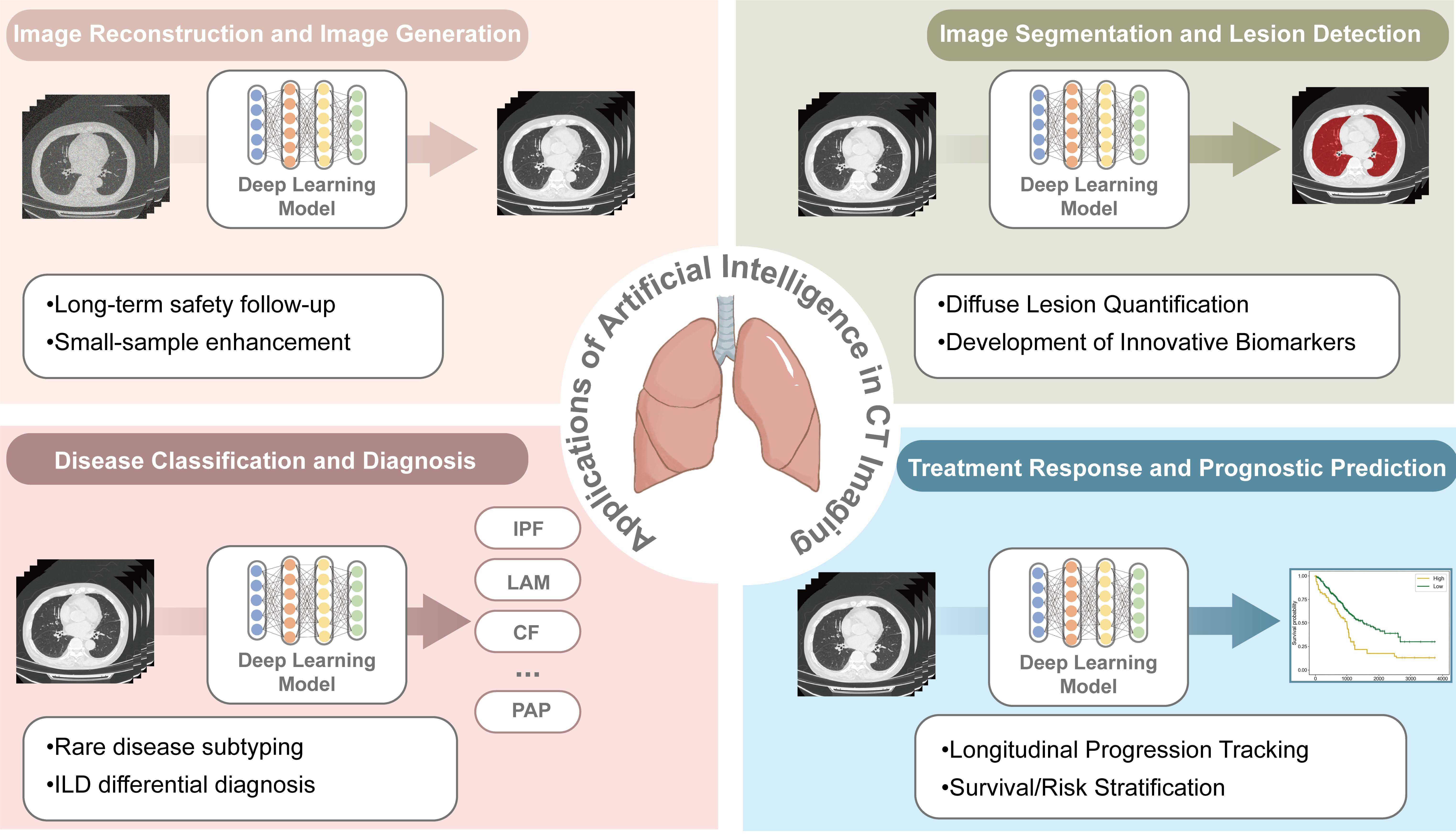
Figure 1. Applications of Deep Learning in Chest CT Imaging The framework illustrates the integration of artificial intelligence across four core clinical stages: (Top Left) Image Reconstruction and Image Generation, focusing on enhancing image quality and denoising; (Top Right) Image Segmentation and Lesion Detection, involving the automated delineation of lung structures and pathological regions; (Bottom Left) Disease Classification and Diagnosis, facilitating the differential diagnosis of various respiratory conditions such as idiopathic pulmonary fibrosis (IPF), cystic fibrosis (CF), lymphangioleiomyomatosis (LAM), and pulmonary alveolar proteinosis (PAP), and (Bottom Right) Treatment Response and Prognostic Prediction, utilizing longitudinal data for survival analysis and outcome stratification. All CT images shown are anonymized clinical data from the authors’ institution. The bar chart and survival curves are for illustration only, with no real data implication. Created in Adobe Illustrator. ILD: Interstitial lung disease; CT: computed tomography.