

fig3
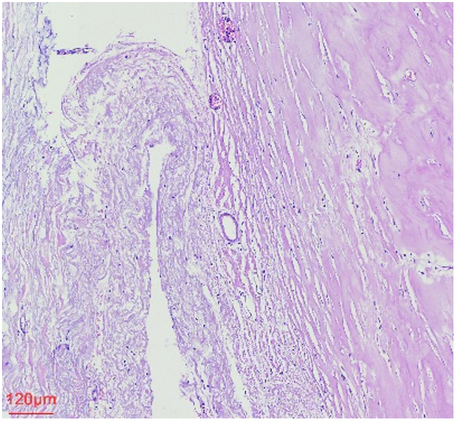
Figure 3. HE staining revealed extensive fibrous hyperplasia and hyaline degeneration (10×). In some areas, this was accompanied by lymphocytic and other inflammatory exudation.
Figure 3. HE staining revealed extensive fibrous hyperplasia and hyaline degeneration (10×). In some areas, this was accompanied by lymphocytic and other inflammatory exudation.


All published articles are preserved here permanently:
https://www.portico.org/publishers/oae/ All published articles are preserved here permanently:
https://www.portico.org/publishers/oae/