


fig2
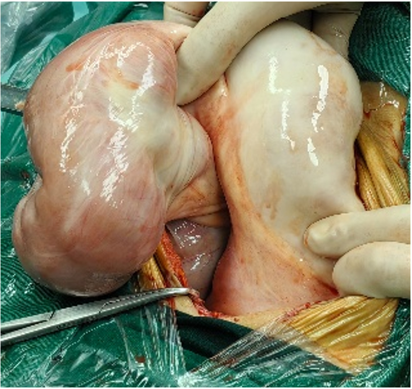
Figure 2. Intraoperative photograph showing a segment of the distal ileum. The intestinal wall is thickened and stiff, with a pale serosal surface. The affected small intestine and its mesentery appear to be encased in a whitish membranous capsule. No obvious peristalsis is observed in this segment.


