


fig1
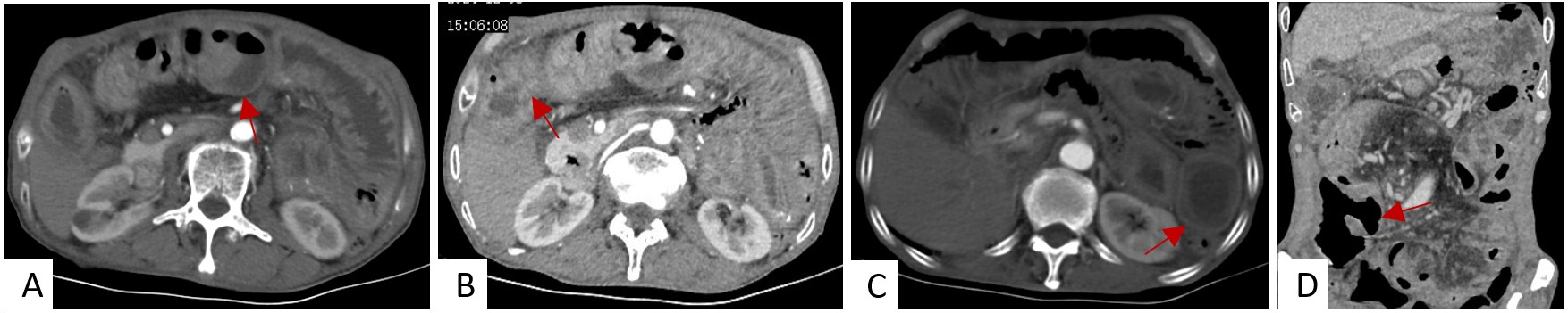
Figure 1. Contrast-enhanced CT of the abdomen shows thickening and dilatation of the small intestinal wall in the right middle and lower abdomen. The contrast-enhanced scan reveals obvious enhancement of the encapsulated intestinal tube, with a strip-like peritoneal shadow surrounding it. These findings are consistent with abdominal cocoon with low-grade incomplete intestinal obstruction. (A-C): axial view, (D): coronal view, the arrow indicated the abnormality.


