


fig6
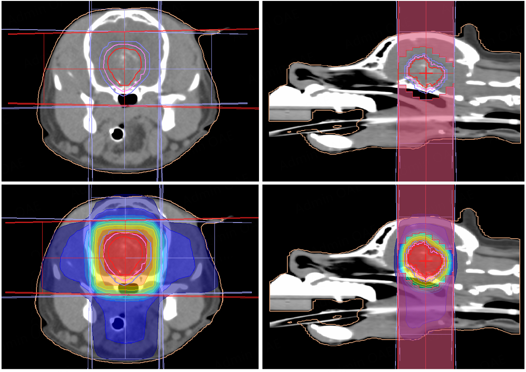
Figure 6. Four-field 3D conformal radiotherapy plan for a canine pituitary tumor. Axial and sagittal CT images show the gross tumor volume (GTV, red), clinical target volume (CTV, pink), and planning target volume (PTV, light purple). A dose color-wash is displayed from 30% (blue) to 100% (red) of the prescribed dose. The configuration of the lateral multileaf collimator is visible on the sagittal images. (Images derived from Monaco Treatment Planning System, version 5.11.03 Elektra AB, Stockholm, Sweden).


