


fig4
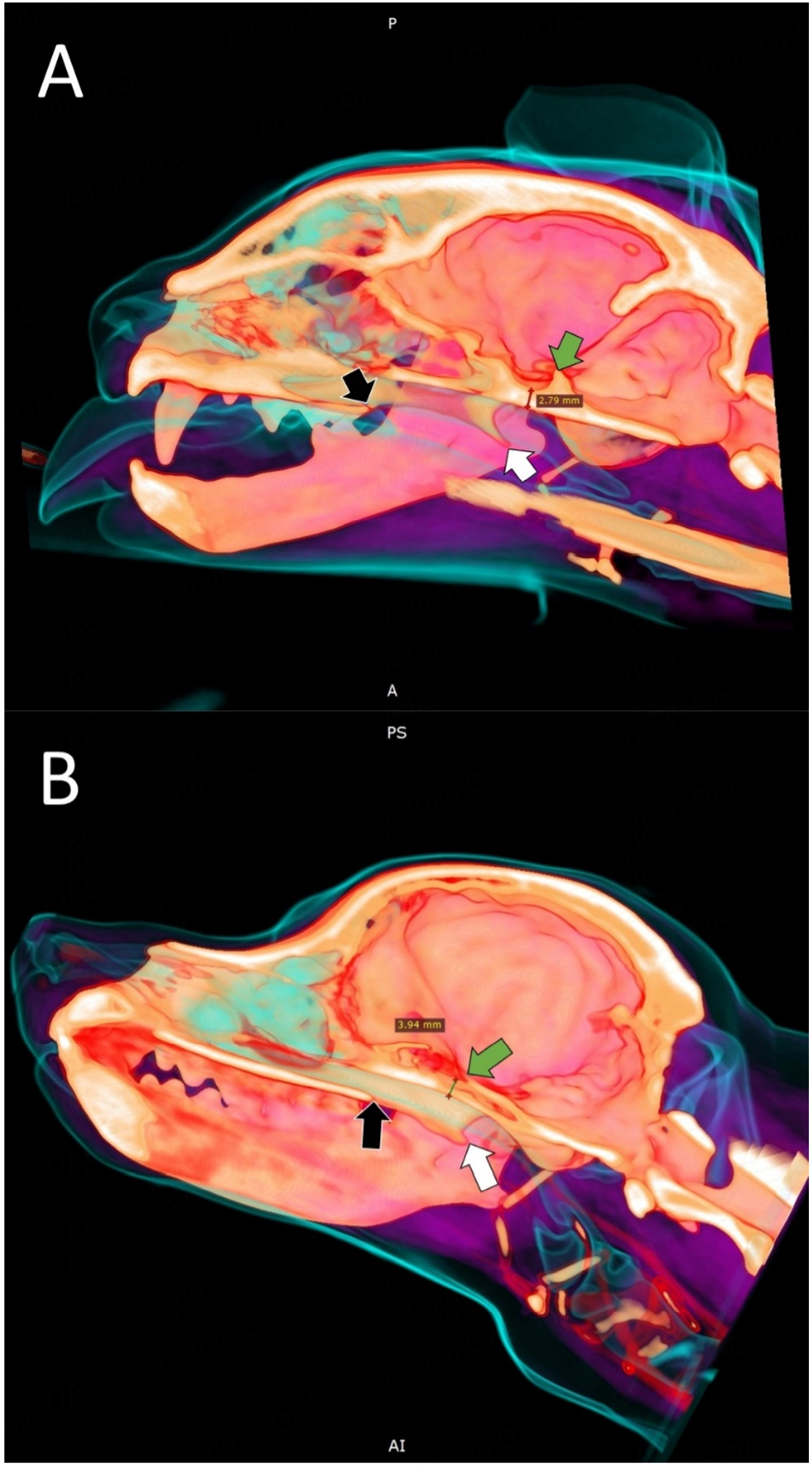
Figure 4. Sagittal 3D mock-up for in silico preparation of a transsphenoidal approach in a cat (A) and a dog (B). Anatomic landmarks for intraoperative identification of correct surgical positioning of the burr hole are marked with arrows. Green arrow: dorsum sellae. White arrow: hamular process of the pterygoid bone. Black arrow: caudal edge of the hard palate. Thickness of the sphenoid bone is measured at the deepest part of the pituitary fossa (red line in A, black line in B). (Image rendering with RadiAnt DICOM Viewer, Medixant, Courtesy van Stee L).


