


fig3
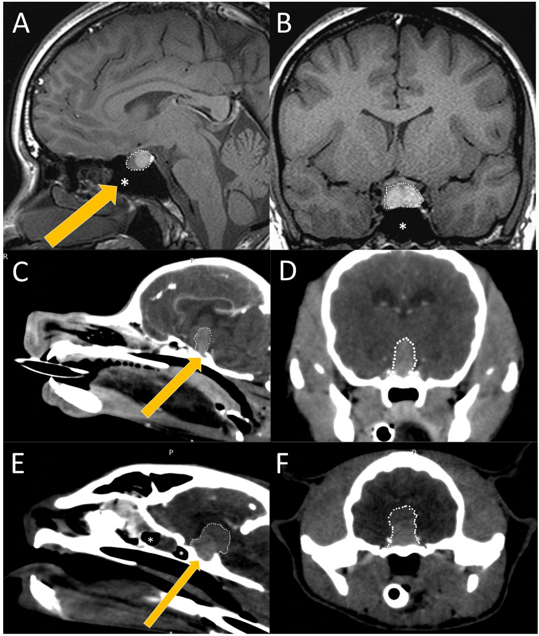
Figure 3. Radiographic anatomy of the pituitary in relation to the brain and skull of man (A and B), dog (C and D) and cat (E and F). All cases depicted, involve a large pituitary adenoma (white dotted outline). A midline sagittal (A, C, E) image at the level of the pituitary fossa and a transverse (B, D, F) image of all three species demonstrate the profound anatomical differences between the length of the maxilla and nasal bones. The presence of the large air-filled sphenoid sinus rostroventral to the pituitary fossa (white asterisk) is visible in the sagittal plane in man (A), is absent in the dog (C) and present in the cat (E), but in the cat located completely rostrally to the pituitary fossa. Mode of entrance is depicted with a yellow arrow. (Image rendering with RadiAnt DICOM Viewer, Medixant, van Stee L).


