

Dendrimer-based hydrogels for biomedical applications
 0

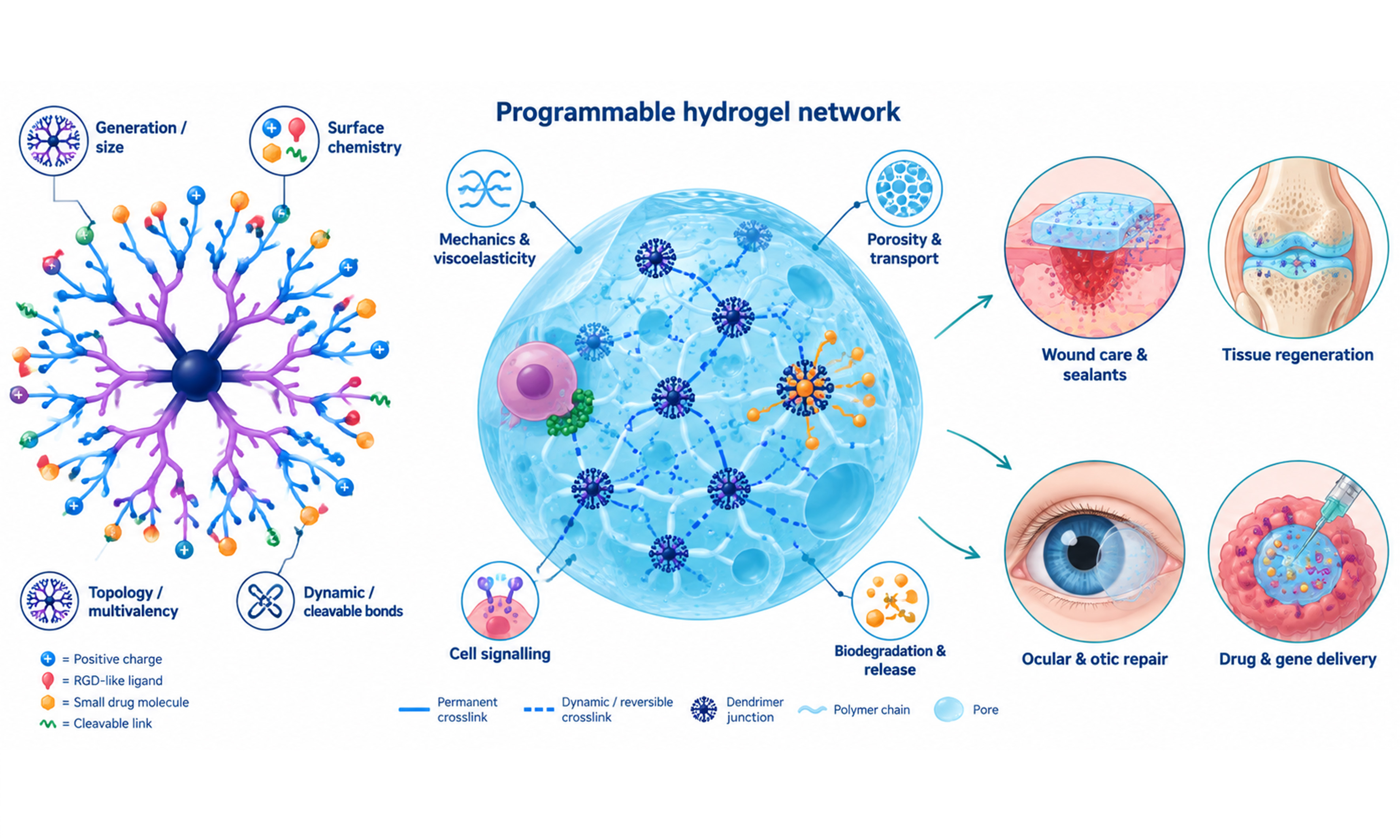
Graphical Abstract
Abstract
Dendrimers are monodisperse, highly branched macromolecules that can be integrated into hydrogels to create soft, water-rich materials tailored for biomedical use. When combined with natural or synthetic polymers, dendrimer-based hydrogels can be designed to provide hemostasis, antimicrobial activity, reactive oxygen species scavenging and controlled release of therapeutic agents, making them promising candidates for managing acute and chronic wounds as well as supporting tissue repair. By tuning dendrimer generation, surface functionality and crosslinking mechanisms, it is possible to modulate key properties of these networks, including stiffness, viscoelasticity, self-healing behavior, degradation kinetics and drug-release profiles, often within a single platform. Preclinical studies demonstrate that such systems can accelerate wound closure, enhance collagen deposition, and achieve high levels of antibacterial efficacy, in some cases without relying on conventional antibiotics, while maintaining acceptable hemocompatibility and cytocompatibility. However, issues such as potential cytotoxicity of highly cationic scaffolds, incomplete biodegradability, synthetic and sterilization complexity, and the need for robust regulatory pathways remain important barriers to clinical translation. Future progress will depend on the development of safe‑by‑design, degradable dendrimer families and their integration with technologies such as 3D bioprinting, organoid models and patient‑derived cells to create more predictive preclinical systems and personalized hydrogel therapies that can actively modulate the local microenvironment and improve healing outcomes.
Keywords


 Download PDF
INTRODUCTION
Hydrogels are three-dimensional, water-swollen polymer networks whose high water content, viscoelasticity and diffusive transport properties closely approximate those of native extracellular matrices in many soft and hydrated tissues[1-3]. These features, combined with the ability to decouple structural and biochemical cues through independent control of crosslinking chemistry, polymer composition and biofunctionalization, have positioned hydrogels as a central materials class in contemporary biomedical engineering[4-6].
From a functional point of view, hydrogels fulfill three broad tasks in biomedical applications: they act as protective or sealing barriers at tissue interfaces, as mechanically compliant scaffolds that support cell infiltration and matrix deposition, and as depots for controlled delivery of therapeutic agents ranging from small molecules and proteins to nucleic acids and whole cells[7,8]. Within this generic framework, increasingly sophisticated designs have emerged that integrate multiple functions (e.g., hemostasis, antimicrobial activity and immunomodulation) into a single hydrogel construct tailored to a particular clinical indication[9-12].
The diversity of biomedical applications has driven a corresponding diversification in hydrogel chemistries, with particular emphasis on crosslinking strategies that balance mechanical robustness, dynamic adaptability and biocompatibility. Covalent crosslinking achieved via amide coupling, thiol-ene reactions, copper-catalyzed azide-alkyne cycloaddition (CuAAC), Michael addition, enzymatic oxidative coupling or visible/UV-light-initiated polymerization remains the dominant approach where long-term stability under mechanical stress and in wet environments is required, as in load-bearing cartilage scaffolds, vascular sealants or corneal adhesives[13-15]. In parallel, dynamic covalent linkages such as Schiff bases and disulfide bonds, as well as supramolecular interactions (hydrogen bonding, π-π stacking, host-guest complexes), have been used to provide hydrogels with shear-thinning, self-healing and stimuli-responsive properties that are advantageous for minimally invasive injection and on-demand cargo release[16-19].
Modern hydrogel design increasingly seeks to decouple mechanical and transport properties from biochemical functionality, enabling independent tuning of parameters such as stiffness, viscoelasticity, degradation kinetics, ligand density and drug-release profiles within a single material platform[20-22]. This modularity is evident in nanoparticle-in-hydrogel architectures, where nanoparticles or nanogels serve as discrete drug or gene carriers embedded within a mechanically optimized matrix[23-27]. Adaptive matrices where viscoelastic dissipation can be adjusted without altering equilibrium modulus further exemplify this trend, particularly structurally dynamic hydrogels that balance macroscopic stability with microscopic bond dynamics[17,28]. Together, these advances show a shift from passive, diffusion-controlled hydrogels toward architecturally sophisticated biomaterials that integrate structural support, controlled delivery and active biological signaling in a single construct.
Dendrimers as building blocks for hydrogels
Unlike low-molecular-weight crosslinkers, dendrimers are precisely defined macromolecular objects that combine a large molecular volume with a high and controllable density of reactive terminal groups. Their incorporation into hydrogel precursor mixtures can therefore alter not only the number of network junctions, but also their spatial distribution, local functionality, chain mobility and nanoscale heterogeneity[29]. Depending on dendrimer generation, concentration, peripheral chemistry, functionality conversion and the nature of the polymer matrix, dendritic crosslinkers can regulate hydrogel stiffness, viscoelasticity, gelation kinetics, swelling, pore architecture and biological ligand presentation. Thus, dendrimers should be regarded as architectural regulators of hydrogel networks rather than simply as multifunctional crosslinkers.
Several classes of dendritic architectures have been integrated into hydrogel networks to address specific functional requirements [Figure 1]. Poly(amidoamine) (PAMAM) and 2,2-bis(hydroxymethyl)propionic acid-based dendrimers (bis-MPA) polyester dendrimers take advantage of their controllable generation, multivalent surface and ability to engage in electrostatic, hydrogen-bonding or host-guest interactions. Poly(L-lysine) dendrigrafts (DGL) and poly(L-lysine)-based dendrons provide protein-like, positively charged backbones that can serve as covalent crosslinkers for poly(ethylene glycol) (PEG) or hyaluronic acid matrices while enhancing cell adhesion and osteogenic responses, thus enabling mechanically tunable scaffolds for bone and soft-tissue regeneration. Carbosilane and phosphorus dendrimers, with robust inorganic or heteroorganic cores and functional peripheries, have been exploited to construct self-assembled physical gels and self-immolative networks in which pH-, redox- or light-triggered degradation is coupled to the release of hydrophobic drugs or nucleic acids. Finally, dendronized polymers and bis-dendron PEG architectures illustrate how dendritic motifs can be used to introduce multivalent ligand clustering, supramolecular chirality and independently tunable viscoelasticity into hydrogel matrices, opening routes toward synthetic mimics of anisotropic, hierarchically organized tissues[29-35].
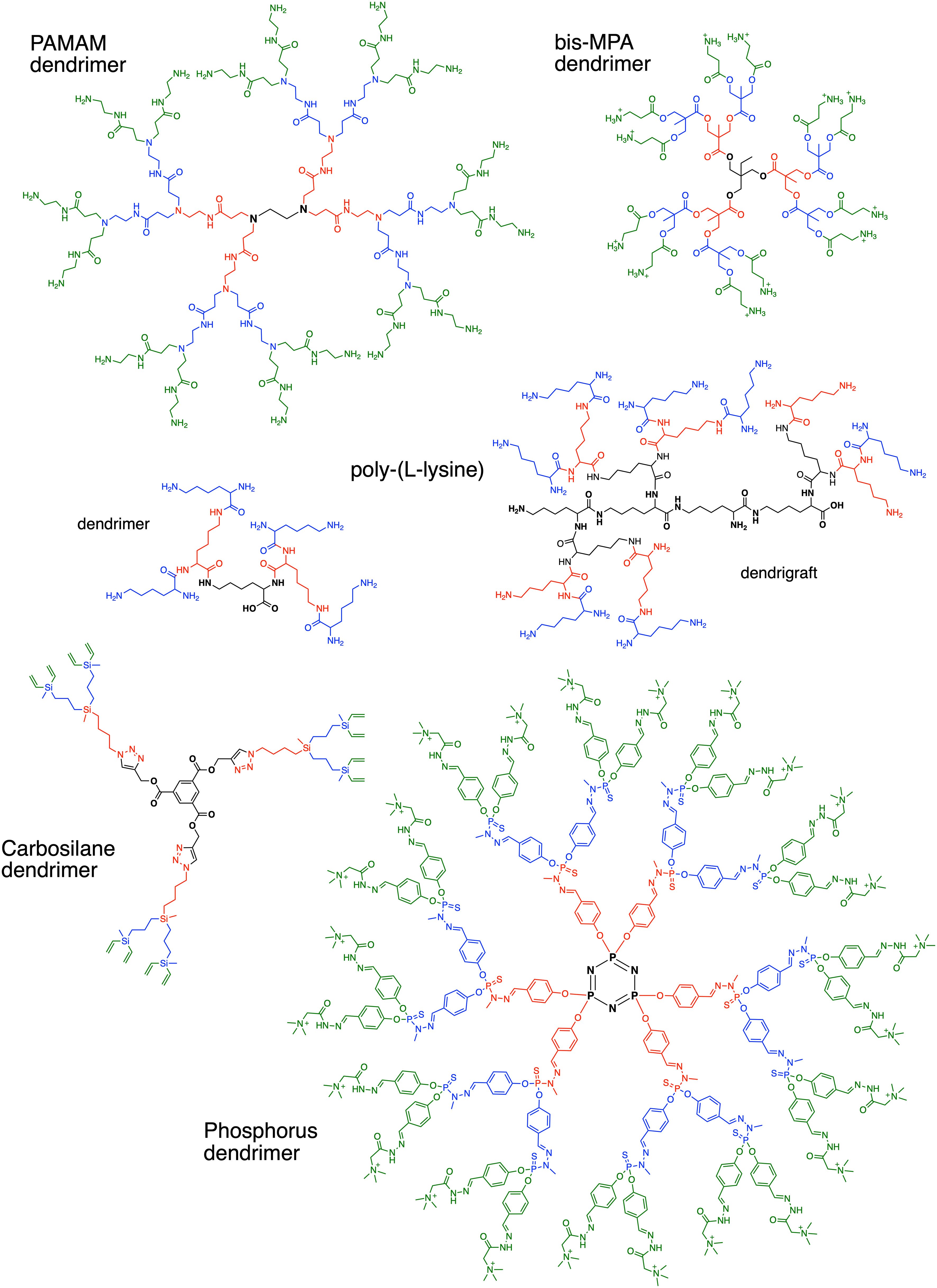
Figure 1. Dendritic architectures used for obtaining functional hydrogels. Second-generation dendritic species are presented as examples. The dendrimer core is drawn in bold; the first generation is presented in red; the second generation is presented in blue, peripheral functional groups, if different from the dendrimer branches, are presented in green. PAMAM: poly(amidoamine) dendrimer; bis-MPA: 2,2-bis(hydroxymethyl)propionic acid-based dendrimers.
Table 1 summarizes principal advantages, limitations, biodegradability, and biological properties of different dendrimer architectures and representative hydrogel applications.
Main dendrimer architectures used in biomedical hydrogels
| Dendrimer architecture | Structural features | Key advantages | Main limitations | Biodegradability | Biological profile | Representative hydrogel applications | Refs. |
| PAMAM dendrimers | Generation-defined amide/amine dendrimers; commonly amine-, carboxylate- or hydroxyl-terminated | High functional-group density; versatile crosslinking; drug loading and nucleic-acid complexation | Cationic forms may cause membrane injury, oxidative stress, hemolysis or inflammation | Limited for native scaffold; improved with cleavable linkers | Strong cell uptake and cargo binding; toxicity reduced by partial surface shielding | Sealants, wound dressings, corneal gels, musculoskeletal matrices, ocular and tumour drug delivery | [40,41,43,46,47, 55-57,64-66,69, 75,76, 80-90] |
| Poly(L-lysine) dendrimers and dendrigrafts | Lysine-based peptide dendrimers or dendrigrafts with multiple terminal amines | Cell-interactive; peptide functionalization; mechanical and viscoelastic tuning | High amine density may cause toxicity; dispersity and synthesis require control | Generally enzymatically degradable | Supports cell adhesion and osteogenic activity; assess extractables and charge effects | Bone and muscle scaffolds, injectable microspheres, bioinks and cell-instructive matrices | [44,45,51-53,67, 71-74,77] |
| Bis-MPA polyester dendrimers and copolymers | Ester-based dendrimers, often hydroxyl-functionalized; may be combined with linear PEG blocks | Degradable; click-compatible; adaptable to antimicrobial, adhesive and dynamic networks | Multi-step synthesis; cationic derivatives require cytotoxicity assessment | Generally hydrolysable; rate is architecture-dependent | Hydroxyl forms are often better tolerated; cationic groups can add antimicrobial activity | Antibacterial dressings, NIR-responsive sponges, DLD tissue adhesives and bioinks | [58-63] |
| Phosphorus dendrimers | Phosphorus-containing branches, often cyclotriphosphazene core; diverse cationic or neutral peripheries | Modular functionalization; nucleic-acid binding; physical-gel formation | Often poorly degradable; cationic variants require safety assessment | Usually limited unless cleavable motifs are introduced | Nucleic-acid binding, anticancer and immunomodulatory potential | Physical hydrogels, dendriplex-containing agarose, local gene-delivery systems | [103-106] |
| Carbosilane dendrimers | Carbon-silicon framework with modifiable hydrophobic or charged periphery | Robust chemistry; click-reactive; hydrophobic nanodomains for poorly soluble cargo | Low intrinsic degradability; cationic derivatives may be hemolytic or cytotoxic | Limited; improved with ester spacers or self-immolative motifs | Nucleic-acid binding and drug solubilization; surface shielding can reduce toxicity | Thiol-ene matrices, hydrophobic-drug depots and self-immolative networks | [92,93] |
| Dendronized polymers and bis-dendrons | Linear polymers or PEG bearing dendritic side/terminal motifs | Independent tuning of mechanics, stress relaxation, porosity and ligand clustering | Complex synthesis; functionality and grafting density require control | Backbone-dependent; PEG requires cleavable junctions for resorption | Defined multivalent ligand presentation; suitable for synthetic ECM design | Organoid matrices, adaptive hydrogels, thermogels, chiral and anisotropic networks | [78,83,97-100, 102,107,108] |
The biological behavior of dendrimers is governed by their size, generation, core and, most importantly, surface chemistry. Surface multivalency can promote cell interaction, ligand binding, cargo complexation and tissue retention; however, highly cationic surfaces may also disrupt negatively charged cell membranes, induce hemolysis, oxidative stress and inflammatory responses[36]. Neutral, anionic or partially shielded dendrimers generally show improved cytocompatibility[37], whereas acetylation, hydroxylation, PEGylation, glycosylation and covalent incorporation into hydrogel networks can reduce exposure to free cationic species. Biodistribution and elimination are similarly size- and charge-dependent: smaller dendrimers may undergo renal clearance, while larger or more hydrophobic structures can accumulate in the liver, spleen or other mononuclear-phagocyte-system organs[38,39]. Consequently, biodegradable dendritic scaffolds and systematic assessment of degradation products, local tissue response, systemic exposure and long-term clearance are essential for safe biomedical translation. A number of dendrimer species have been subjected to clinical trials and already translated into practice[34].
Dendrimers modulate mechanical properties of hydrogels
Dendritic crosslinkers provide an efficient platform for controlling hydrogel mechanics because each macromolecule presents multiple reactive groups within a confined volume. Increasing the amount of a suitably functionalized dendrimer generally increases the effective crosslink density and reduces polymer-chain mobility, thereby increasing the elastic and viscous components of the mechanical response. However, this relationship depends on the accessibility of terminal groups, stoichiometry with the complementary polymer or crosslinker, steric crowding, dendrimer flexibility and the occurrence of network defects. Dendrimer concentration and molecular architecture can be used as independent formulation variables to access a wide range of mechanical properties within a common chemical platform.
For example, hydrogels formed by crosslinking dithiol-terminated PEG with RGD peptide-functionalized PAMAM G5 dendrimers showed a marked, concentration-dependent increase in complex modulus, G*, from ca. 45 Pa at 1.2% PAMAM to 5,000 Pa at 5.1% PAMAM[40]. Similarly, RGD-functionalized PAMAM dendrimers crosslinked with PEG diacrylate (PEGDA) yielded photopolymerized matrices with storage moduli, G′, between 20 and 8,000 Pa, and loss moduli, G′′, of ca. 30-600 Pa, according to precursor composition. In that system, the stiffest formulations supported osteogenic differentiation of mesenchymal stem cells without observable cytotoxicity, illustrating how dendritic crosslinking can be used to establish mechanically instructive cell microenvironments[41].
The mechanical range accessible with dendritic building blocks is also evident in hydrogels derived from natural polymers. Low-generation multiarm PEG-polyaldehyde dendritic crosslinkers enabled the modulation of carboxymethyl chitosan hydrogels from
This capability is particularly relevant for cell-instructive matrices, because material stiffness can influence cell spreading, cytoskeletal tension, mechanotransduction and lineage commitment. Nevertheless, hydrogel mechanics should not be reduced to elastic modulus alone. Dendrimer concentration, generation and topology may simultaneously influence stress relaxation, swelling, ligand density and pore architecture. The mechanobiological interpretation therefore requires these parameters to be measured and, wherever possible, varied independently. Below, this consideration will be illustrated by DGL-based PEG and hyaluronic acid matrices, where both stiffness and viscoelastic dissipation influence osteogenic cell responses[44,45].
Dendrimers versus conventional crosslinkers
The advantage of dendrimers over conventional low-molecular-weight crosslinkers derives from their ability to create a local excess of reactive groups while maintaining a macromolecular junction size. A small bifunctional or multifunctional linker creates relatively localized junctions between adjacent polymer chains. By contrast, a dendrimer can connect multiple chains over a larger molecular volume and can establish a highly multivalent, spatially organized crosslinking node. This topology can increase network connectivity and rigidity even when the parent polymer alone would form only a weak or soft hydrogel.
A representative comparison was reported for collagen matrices crosslinked using carbodiimide, glutaraldehyde or PAMAM dendrimers. Carbodiimide, a zero-length crosslinker, and glutaraldehyde, a short molecular linker, produced collagen materials with Young’s moduli of approximately 50-100 kPa. In contrast, the macromolecular PAMAM crosslinker yielded a substantially more rigid collagen network with a Young’s modulus of approximately
An additional advantage is that crosslinking need not consume all dendrimer surface groups. In PEGDA hydrogels formed through aza-Michael addition with amino-terminated PAMAM G5 dendrimers, partial acetylation of the peripheral amines altered gelation time, network structure, rheological behavior, swelling and degradation without completely abolishing network formation[47]. Compared with unmodified PAMAM, 50% acetylation reduced network rigidity, whereas further acetylation up to 90% produced more modest changes. The corresponding gel-state storage modulus ranged from approximately 1 Pa to 10 kPa, and swelling increased to approximately 300% in dendrimer-containing systems. This result demonstrates that a proportion of terminal groups can be reserved for orthogonal functionalization with peptides, proteins, drugs, nucleic acids or imaging probes while retaining sufficient functionality for hydrogel crosslinking[41,48].
Dendrimer crosslinking controls hydrogel porosity
Because dendrimers modify the density, distribution and connectivity of hydrogel junctions, they can strongly influence pore size and pore-size distribution. This is a critical design parameter for hydrogel applications because porosity regulates diffusion, swelling, nutrient transport, cell infiltration, vascularization and the accessibility of matrix-bound signals. The direction and magnitude of the porosity change depend on the composition of the hydrogel, total solid content, dendrimer-to-polymer ratio, crosslinking kinetics and the physical state in which the material is formed or dried.
In hydrogels based on natural polymers, dendritic crosslinking frequently yields smaller but more uniform pores, consistent with formation of a denser and more regularly connected network. For instance, gelatin methacrylate hydrogels exhibited a pore diameter of 310 ± 80 µm, whereas incorporation of methacrylated PAMAM reduced the mean pore diameter to 80 ± 20 µm. This structural refinement was accompanied by an increase in compressive modulus from approximately 2.5 to 8 kPa[49]. Similar trends have been described for oxidized carboxymethylcellulose hydrogels crosslinked using PEG-poly(L-lysine) bis-dendrons[50]. Such materials may be advantageous when structural integrity and controlled molecular transport are prioritized over rapid cell migration.
In synthetic-polymer networks, the effect of dendritic components can be more complex and may be exploited to tune pore interconnectivity rather than simply reduce mean pore size. In PEG hydrogels containing lysine dendrigrafts, variation of total component concentration and dendrimer-to-polymer ratio shifted the pore-size distribution from a monomodal population centered near 100 µm to a bimodal distribution with maxima near 150 and 450 µm[51,52]. This type of hierarchical pore architecture may be favorable for cell culture and tissue formation because smaller pores can improve surface area and local cell-matrix interactions, whereas larger interconnected pores facilitate nutrient diffusion, waste removal, cell migration and vascular ingrowth.
The relationship between dendrimer incorporation and porosity should therefore be considered alongside mechanics rather than as an independent variable. Denser dendritic crosslinking may increase modulus and reduce average pore size, but formulation strategies that modify dendrimer content, functionality or crosslinking rate can generate mechanically robust networks while retaining macroporosity and transport pathways. Dendrimer-based hydrogels are consequently well suited to approaches in which pore structure and mechanical performance must be optimized simultaneously for a specific biomedical application.
Manifestation of the dendritic effect
The dendritic effect refers to the generation-dependent and often non-linear changes in macromolecular behavior that arise from increasing branching, terminal-group number, molecular size and steric organization. In hydrogel systems, this effect can manifest through altered crosslinking efficiency, terminal-group accessibility, local functionality, chain packing, polymer-dendrimer interactions and the number of effective network junctions. Importantly, increasing dendrimer generation does not necessarily produce a monotonic increase in mechanical strength. The outcome reflects the balance between multivalency, which can increase effective network connectivity, and steric shielding or reduced flexibility, which may limit the accessibility of functional groups.
A positive generation-dependent mechanical effect was observed in a peptide-dendrimer-based bioink prepared from hyaluronic acid and poly(L-lysine) (PLL) dendrimers. Increasing the PLL dendrimer generation from G1 to G3 increased G′ from
By contrast, PAMAM generation can have the opposite effect in some network chemistries. In hydrogels formed by thiol-ene coupling of methacrylated hyaluronic acid with thiol-modified PAMAM dendrimers, the complex modulus decreased from 1,240 Pa for PAMAM G2 to 583 Pa for PAMAM G3[52]. Similarly, ionic nanofibrous bioinks prepared from PAMAM dendrimers and poly(L-lactic acid) showed a decrease in Young’s modulus from approximately 95 to 70 kPa as PAMAM generation increased from G1 to G5[54]. These results suggest that in higher-generation PAMAM dendrimers, steric congestion, reduced peripheral-group accessibility, altered molecular flexibility or less efficient packing may outweigh the theoretical benefit of a higher number of terminal groups.
Generation-dependent effects are thus not merely synthetic curiosities. They provide a methodology to tailor hydrogel properties through dendrimer topology. In practice, generation should be selected according to the intended role of the dendrimer: low-generation structures may offer greater functional-group accessibility and more flexible network formation, whereas higher-generation architectures may provide greater local multivalency, larger molecular volume and enhanced capacity for ligand clustering or cargo binding. A systematic assessment of generation, surface conversion, dendrimer concentration and polymer stoichiometry is required to establish structure-property relationships and to distinguish true dendritic effects from changes caused by precursor composition or incomplete crosslinking.
HYDROGELS FOR TOPICAL USE
Dendrimer-based hydrogels are particularly well suited to topical therapy, where they can conform to irregular tissue surfaces while providing local protection, adhesion and sustained therapeutic activity [Figure 2]. By combining a hydrated matrix with programmable dendritic functionality, these materials can address key wound-healing challenges, including bleeding, infection, oxidative stress and inflammation, while limiting systemic exposure to active agents.
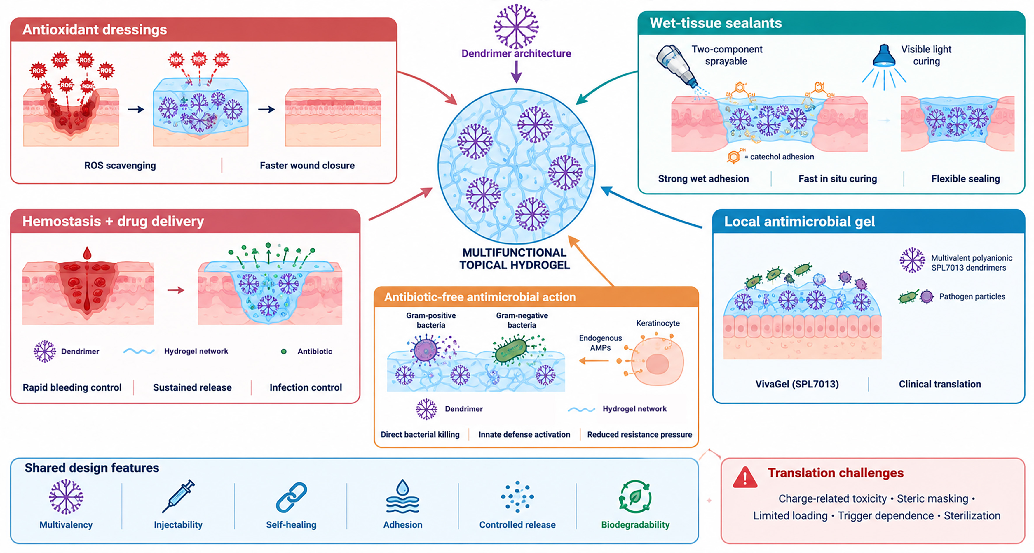
Figure 2. Dendrimer-based hydrogels for topical use. Dendrimers act as crosslinkers, carriers or bioactive components in wound dressings and tissue sealants. These hydrogels can combine hemostasis, wet-tissue adhesion, antimicrobial activity, and local therapeutic delivery. ROS: Reactive oxygen species; AMP: antimicrobial peptides.
Antioxidant and reactive oxygen species-scavenging dressings
Oxidative stress is a major driver of impaired healing in chronic wounds, particularly in diabetic patients, where excess reactive oxygen species (ROS) delay re-epithelialization and collagen deposition. Islam et al. tackled this problem by conjugating gallic acid onto a fourth-generation PAMAM dendrimer and incorporating the resulting antioxidant macromolecule into a ternary hydrogel with polyvinyl alcohol and silk fibroin, crosslinked by ionizing gamma radiation (20-35 kGy). The macroporous architecture (compressive strength 1.4 MPa at 56% strain) achieved 70% H2O2 scavenging within 24 h in vitro, while in a streptozotocin-induced diabetic mouse excision model the wound area was reduced to 30% of its original size by day 10 and reached near-complete contraction by day 16. Hemolysis remained below 5%, and cell viability was significantly boosted relative to controls. The residue-free gamma-crosslinking ensures high material purity, and the synergy between the silk scaffold and the dendrimer antioxidant is a key design advantage; the only technical constraint noted is that the radiation dose must be carefully optimized for each blend composition[55].
Multifunctional hemostatic and antibiotic hydrogels
Beyond oxidative damage, acute and infected wounds require simultaneous control of bleeding and microbial colonization. Jia et al. addressed both needs with a chemical hydrogel of sodium alginate and γ-polyglutamic acid (γ-PGA) cross-linked with a third-generation PAMAM dendrimer upon EDC/N-hydroxysuccinimide (NHS) activation. Amoxicillin (AMX) was taken as antibiotic cargo. The resulting self-healing (88% recovery) injectable network, with 200-400 µm interconnected pores, reduced hemostatic time to approximately 0.6-0.7 min (vs. over 5 min in controls), released 43% of AMX over 72 h, and achieved bactericidal rates above 96% against both E. coli and S. aureus. Applied to scald wounds in rodent models, the healing rate exceeded 98.9% after 20 days. An additional practical asset is the material's adhesion to heterogeneous substrates including pigskin, internal organs, and metal surfaces, making it suitable for emergency use in complex anatomical sites. The principal limitation is the need to optimize component ratios for wound types of varying complexity[56].
Gupta et al. took a complementary drug-delivery approach by loading hesperidin, a bioflavonoid with documented wound-healing properties but poor aqueous solubility, into G3/G4 PAMAM dendrimers physically incorporated into a chitosan-sodium alginate matrix and lyophilized into a flexible bandage. The ~12 nm dendrimer particles achieved 20% entrapment efficiency and released 86.4% of hesperidin over 24 h. In a Wistar rat full-thickness excision model, the bandage produced 98.9% wound contraction after 14 days with measurable improvements in collagen synthesis and re-epithelialization; hemolysis was below 1%. The main bottleneck is the relatively modest drug-loading capacity of the dendrimer cavities[57].
Antibiotic-free antimicrobial hydrogels
A recurring theme in recent literature is the desire to achieve effective bacterial killing without conventional antibiotics, thereby reducing resistance risk. Biscari et al. prepared hybrid hydrogels by complexing a second-generation cationic bis-MPA polyester dendrimer bearing twelve β-alanine surface groups with dopamine-functionalized gellan gum. Three crosslinking modes were exploited: physical polyelectrolyte complexation, optional ionotropic reinforcement with CaCl2 for solid gels, and dynamic covalent Schiff base/Michael addition for injectable self-healing versions. Storage moduli ranged from 0.5 (injectable) to 21.0 kPa (solid), swelling capacity exceeded 1,700%, and solid formats achieved complete (100%) bacterial killing of S. aureus and E. coli within 4 h. Injectable versions demonstrated 100% self-healing efficiency. The trade-off is a shorter functional lifetime for injectable gels (under 2 days) versus solid ones (~7 days)[58].
The same group subsequently introduced near-infrared (NIR) responsiveness by replacing the surface chemistry with cysteamine hydrochloride groups on G1 and G2 bis-MPA dendrimers, and by incorporating in situ-synthesized polydopamine (PDA) into an ethylenediamine-functionalized gellan gum matrix. The freeze-dried microporous sponge rehydrates to a bulk hydrogel, swells above 1,500%, and retains a storage modulus of ~657 Pa up to 50 °C. Against S. aureus and P. aeruginosa, G1 dendrimers eradicated bacteria even without irradiation, while G2 systems required NIR stimulation at 810 nm to loosen the hydrogel mesh. The material is non-hemolytic and shows low cytotoxicity in HaCaT keratinocytes. The dependence of G2 on external light stimulation is both its key functional feature and its primary practical constraint[59].
Fan et al. took the antibiotic-free concept further by demonstrating that hydrogels based on amino-functional hyperbranched dendritic-linear-dendritic (DLD) copolymers, a linear PEG core with G5/G6 bis-MPA-derived blocks bearing β-alanine surface groups, can not only kill bacteria directly but also modulate the host innate immune response. Covalently crosslinked DLD polymer and NHS-activated PEG cross-linkers, the porous adhesive bandages (storage moduli 638-2,969 Pa) achieved 100% killing of E. coli and P. aeruginosa and were equally active against clinical methicillin-resistant S. aureus, S. pyogenes, and P. aeruginosa strains. Uniquely, the materials upregulated expression of the endogenous antimicrobial peptides RNase 7 and psoriasin in HaCaT keratinocytes while reducing pro-inflammatory interleukin-1 beta (IL-1β) and mitochondrial ROS. Rapid degradation within 24 h prevents accumulation and resistance development, and the material adheres strongly to porcine skin[60].
Ye et al. introduced a structurally distinct concept by preparing covalently stabilized peptide nanoparticles formed via dendritic polymerization of AB2-type peptide monomers through azide-alkyne or thiol-maleimide click chemistry. These nanoparticles were used as rigid multifunctional crosslinkers within a 4-arm-PEG-azide network assembled by CuAAC click reaction. Cationic nanoparticles (zeta potential +23.1 mV) provided inherent antibacterial activity, and the resulting hydrogel (storage modulus ~4 kPa) exhibited remarkable resistance to swelling (volume expansion ~2%), achieved 100% killing of S. aureus, and drove near-complete wound closure by day 12 in a murine infected full-thickness skin defect model, accompanied by reduced IL-6 levels and promoted neovascularization (CD31 staining). The covalent assembly strategy prevents the premature disassembly that commonly limits supramolecular peptide systems, and the sequence-independent platform can be readily reformulated with different bioactive peptides[61].
Tissue sealants
Achieving reliable tissue adhesion under the wet, dynamic conditions of surgical fields or traumatic wounds requires materials combining covalent bonding chemistry with mechanical toughness.
Zhu et al. constructed a dual-bioinspired double-network hydrogel adhesive from a barbell-like dendritic lysine scaffold modified with catechol groups (53.9% substitution degree) to mimic mussel wet adhesion, combined with a noncovalent network of chitosan and carboxymethyl cellulose inspired by the energy-dissipating mucus of slugs. The covalent network was formed by HRP/H2O2-mediated π-π coupling, yielding an injectable, self-healing hydrogel (300% stretch recovery after 10 min) with a storage modulus of 2 MPa and adhesion energies of 200-900 J/m2 against porcine skin. These values substantially exceed the parameters of commercial sealants Tisseel and BioGlue. In a rabbit femoral artery damage model, hemostasis was achieved in under 30 s. Intrinsic antimicrobial activity against E. coli and S. aureus was additionally demonstrated[62].
Sanz del Olmo et al. reported a complementary on-demand-curing strategy using G2-to-G5 DLD block copolymers based on PEG and bis-MPA, with a heterofunctional surface carrying allyl groups for crosslinking and quaternary ammonium groups for antibacterial activity. Visible light-induced thiol-ene reaction cured the hydrogel in under 20 s. Storage moduli ranged from 270 to 7,024 Pa, and tensile adhesion to pig skin and bone patches reached up to 61.2 kPa, with 100% killing of Gram-negative bacteria by the optimized formulation. The technology targets wound closure adhesives, antibacterial coatings on medical devices, and surgical site infection prevention. A noted limitation is that high crosslinking density in stoichiometric formulations can sterically mask ammonium groups, reducing antibacterial efficacy[63].
Muñoz Taboada et al. demonstrated that dendrimer hydrogels can be extended to the demanding application of vascular anastomosis sealing. A two-component sprayable system was formulated by combining oxidized dextran and oxidized alginate (polyaldehyde components) with a surface-modified G5 PAMAM dendrimer (75% hydroxyl, 25% amine) that crosslinks through instantaneous covalent imine bond formation upon mixing. Delivered via an airless syringe, the hydrogel withstood supraphysiological burst pressures of ~300 mmHg, maintained cell viability above 90%, and passed genotoxicity and sensitization assessments. Crucially, unlike the rigid commercial sealant BioGlue which risks vascular stenosis, this pliable hydrogel allows the vessel to restore its natural luminal diameter after application. The material retained sealing performance after
A notable translational case of a hydrogel for topical use is VivaGel® (SPL7013), a topically administered formulation in which the dendrimer itself is the active antimicrobial agent rather than a passive crosslinker or carrier. SPL7013 is a fourth-generation poly(L-lysine) dendrimer bearing 32 terminal naphthalene disulfonate groups, which confer a highly multivalent polyanionic surface. This architecture was designed to promote local interactions with pathogens while limiting systemic exposure. VivaGel® has been evaluated clinically as a 3% vaginal microbicide for human immunodeficiency virus 1 (HIV-1) and herper simplex virus 2 (HSV-2) prevention, including phase I studies of repeated vaginal dosing and antiviral retention (NCT00331032 and NCT00740584), and as a 1% formulation for bacterial-vaginosis treatment and recurrence prevention, including phase III studies (NCT01577537 and NCT02236156). In addition, SPL7013 has been incorporated into condom lubricants marketed in several jurisdictions.
Although VivaGel® is not a dendrimer-crosslinked bulk hydrogel, it provides clinical proof that precisely defined dendritic macromolecules can be formulated into locally retained gels or device coatings and can advance through clinical development when their charge, multivalency, local retention and safety profile are matched to a specific medical indication.
HYDROGELS FOR TISSUE REGENERATION
The development of human tissues (cartilage, bone, muscle, etc.) in artificial media is the most challenging aim in regenerative medicine. Body tissues span a wide range of mechanical requirements, including load-bearing compressive stiffness of articular cartilage, anisotropic viscoelastic properties of vocal-fold lamina propria, and contractile architecture of skeletal muscle. Dendrimer-based hydrogels have been applied across these targets, serving variously as intra-articular drug depots, cell-encapsulating scaffolds, gene-delivery vehicles, mechanically tunable bone matrices, and injectable pore-forming constructs for volumetric tissue loss.
Cartilage repair
Osteoarthritis (OA) is characterized by progressive degradation of articular cartilage driven by mechanical stress and inflammatory mediators, including elevated matrix metalloproteinases (MMPs). A central challenge for intra-articular therapy is achieving adequate drug penetration into the dense, avascular cartilage matrix while restricting release to diseased tissue [Figure 3].
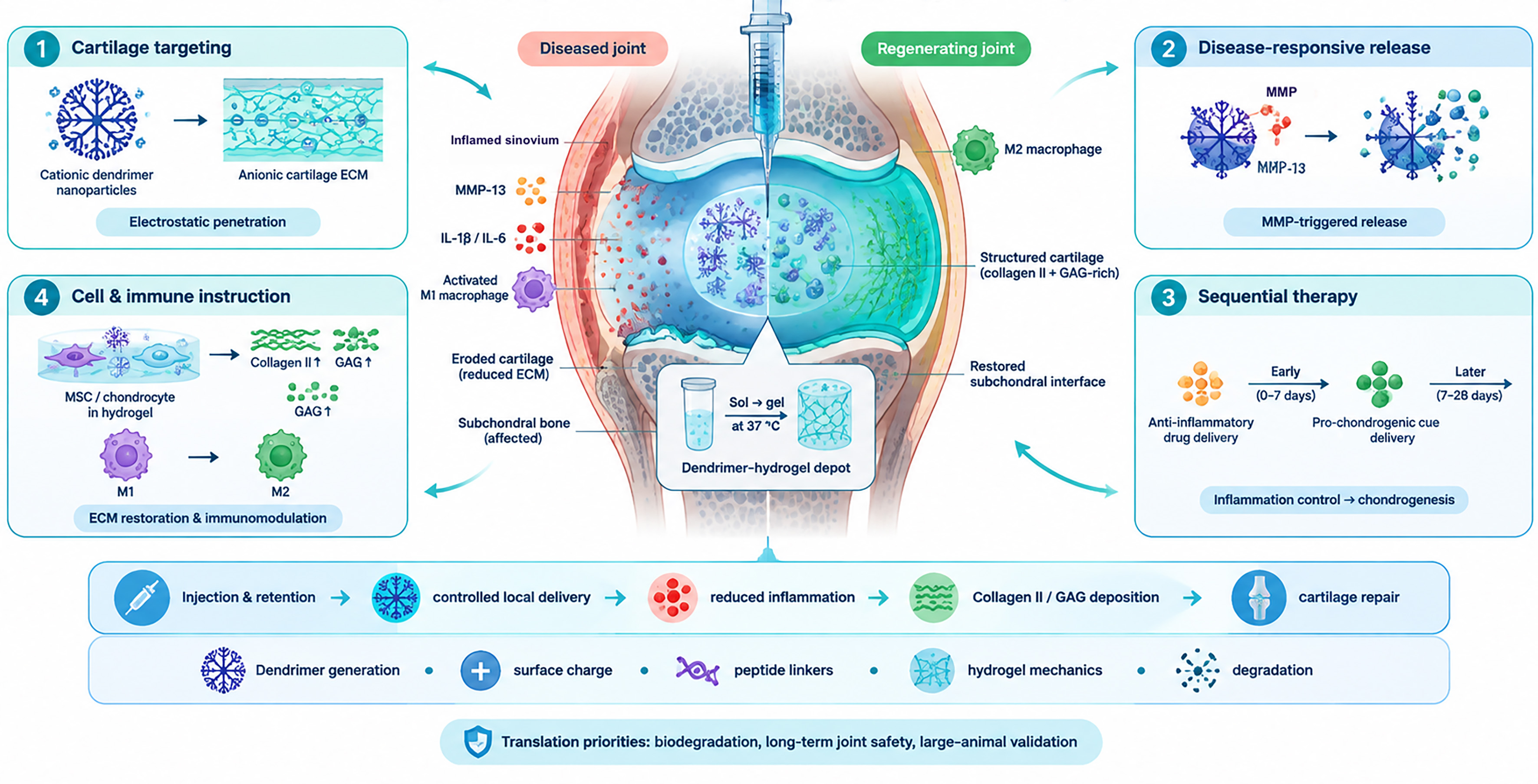
Figure 3. Dendrimer-based hydrogels for cartilage repair. Dendrimer-containing matrices provide local delivery of anti-inflammatory and regenerative agents while supporting cartilage cells and matrix restoration. ECM: Extracellular matrix; MMP: matrix metalloprotease; MSC: mesenchymal stromal cells; GAG: glycosaminoglycans; M1, M2: macrophages type 1 or 2; IL-1β: interleukin-1 beta.
Zhou et al. addressed this with a dual-responsive composite nanohydrogel in which a fifth-generation PAMAM dendrimer was conjugated to an MMP-13-responsive peptide linker and loaded with the pro-chondrogenic small molecule kartogenin. Hydrogel nanoparticles were encapsulated within a thermosensitive DLD hydrogel that gels at body temperature, enabling single-step intra-articular injection. The cationic PAMAM surface (+18.9 mV) exploited electrostatic attraction to anionic cartilage proteoglycans, achieving 67.2% cartilage penetration at
Extending the temporal dimension of cartilage therapy, Cheng et al. developed a gelatin methacrylate (GelMA)-based composite hydrogel incorporating G5 PAMAM dendrimers that carry covalently bound diclofenac on their surface and strontium ions in their interior cavities. UV photo-polymerization of the GelMA matrix provided the structural bulk, while the nanoparticles enabled sequential release: diclofenac during the initial 9 days to suppress inflammation, followed by strontium from days 3 to 15 to drive chondrogenesis. Rabbit in vivo models demonstrated significantly reduced inflammation and enhanced extracellular matrix (ECM) deposition [Collagen II and glycosaminoglycans (GAG)] at 6 weeks. The sequential release of anti-inflammatory priming and pro-regenerative signaling represents a conceptual advance over co-delivery systems, and bone marrow stem cells were identified as the preferred cellular cargo[65].
Wang et al. approached cartilage repair from a different angle, targeting concurrent treatment of bone joint tuberculosis with a multicomponent biomimetic gel formed by amine-epoxy click crosslinking between G3 PAMAM dendrimers, dendrimer-templated silver nanoparticles, amine-functionalized mesoporous silica nanoparticles, and PEG diglycidyl ether. Inclusion of silica nanoparticles and silver nanoparticles raised the tensile modulus from
Rheumatoid arthritis (RA) is a systemic autoimmune condition driving synovial inflammation and progressive joint destruction. Two complementary platforms illustrate how this immunological complexity can be addressed with dendrimer hydrogels.
Zhu et al. designed an ECM-inspired injectable hydrogel formed by reversible Schiff base crosslinking between a third-generation polylysine dendrimer and oxidized hyaluronic acid. The dynamic imine bonds conferred shear-thinning and self-healing behavior for minimally invasive intra-articular injection, and the polycationic dendrimer surface scavenged negatively charged inflammatory cytokines and cell debris. Adipose-derived stem cells encapsulated within the 3D porous network (storage modulus up to ~104.5 Pa; elastic moduli 400-1,100 kPa) showed prolonged in vivo retention of 7 days compared to bare cell injections. In a collagen-induced arthritis rat model, the system successfully reversed macrophage polarization from M1 to M2 and significantly improved bone mineral density and cartilage integrity, with no systemic organ toxicity[67].
Oliveira et al. integrated nanotechnology with organ-on-chip engineering by loading anti-TNFα monoclonal antibodies into chondroitin sulfate-modified PAMAM dendrimer nanoparticles embedded within a tyramine-functionalized gellan gum hydrogel (enzymatically crosslinked by HRP/H2O2), incorporated into a cartilage-on-a-chip microfluidic device co-culturing human monocytes and primary chondrocytes. Over 14 days, the system maintained high Collagen II expression, reduced chondrocyte death induced by inflamed macrophages, and demonstrated sustained anti-inflammatory capacity. This study was the first cartilage-on-chip model to integrate dendrimer nanotechnology and microfluidics. The use of normoxic culture conditions, while the RA joint is typically hypoxic, is identified as the principal limitation; hypoxic modeling is a stated future direction[68].
Ma et al. developed a triplex injectable composite in which siRNA targeting the stress-response gene DDIT4 was complexed with a G5 amino-terminal PAMAM dendrimer and coated with hyaluronic acid to form 100-200 nm particles, then embedded in an injectable hydrogel matrix for intradiscal delivery. In a rat needle-puncture intravertebral disc disease model, DDIT4 siRNA fluorescence persisted in discs for 35 days (vs. 21 days for naked siRNA), inhibiting nuclear pore complex-associated pyroptosis via the ROS/TXNIP/NLRP3 axis and significantly improving the disc height index. The study establishes DDIT4 as a validated therapeutic target for intravertebral disc disease and identifies biodegradable PAMAM variants as a necessary development to avoid long-term intradiscal accumulation[69].
Liu et al. extended the cartilage engineering toolkit to ear reconstruction (microtia) using a double-network hydrogel scaffold combining reversible Schiff base crosslinks (oxidized chondroitin sulfate/gelatin) and permanent methacrylate photo-polymerization, with PEG as a porogen. A G5 PAMAM dendrimer modified with guanidinobenzoic acid delivered a Runx1-expressing plasmid (pRunx1) to dedifferentiated chondrocytes. Scaffolds cast in 3D-printed ear-shaped molds and tested subcutaneously in nude mice produced mature cartilaginous tissue with a Young's modulus exceeding
The vocal fold lamina propria must sustain millions of phonation cycles while exhibiting non-linear, anisotropic, and viscoelastic behavior. Carrancá et al. constructed a fiber-reinforced composite hydrogel based on DGL cross-linked with a PEG-NHS polymer[71]. This hydrogel matrix served as an analog of the elastin-ground substance component while jet-sprayed polycaprolactone microfibers mimicked the collagen fiber network. By varying the fiber volume fraction (3%-11%) and PEG molecular weight, the tangent modulus and strength were tuned to match native vocal-fold stress-strain responses in both longitudinal tension and transverse compression. The inherently cell-interactive DGL surface eliminated the need for surface coating. The principal limitation is that jet-spraying produces straight polycaprolactone (PCL) fibers rather than the native wavy collagen architecture at the microscale[72].
Volumetric muscle loss exceeds the intrinsic regenerative capacity of skeletal muscle, leaving permanent functional deficits. Two studies from the same group developed and applied effervescent DGL/PEG hydrogels to this challenge. Griveau et al. established the foundational platform: G3 DGL was covalently crosslinked with bis-NHS-PEG by amide bonding, and during syringe injection a concurrent effervescent reaction of K2CO3 and acetic acid generated a highly interconnected macroporous bulk network
Griveau et al. then applied this platform specifically to volumetric muscle loss (VML) repair, tuning stiffness from 12 to 160 kPa by varying the DGL/PEG ratio, and showing that ~300 µm interconnected pores support contractile myotube formation by C2C12 myoblasts and primary human muscle stem cells in vitro. In a large tibialis anterior defect model in rats, injected scaffolds filled the defect, promoted neovascularization, and facilitated aligned muscle fiber regeneration at the injury periphery within 21 days. The cell-adhesive DGL surface eliminated the need for external fibronectin or laminin coatings. Limitations include restricted myotube fusion in deeper pores and the absence of functional contractility measurements; incorporation of growth factors (insulin-like growth factor and fibroblast growth factor) is identified as the key future direction[73].
Bone regeneration
Bone regeneration requires biomaterials that combine mechanical support with cues that promote stem-cell recruitment, osteogenic differentiation and vascular ingrowth. Dendrimer-based hydrogels offer a modular platform in which matrix stiffness, viscoelasticity, pore architecture and delivery of osteoinductive drugs, genes or bioactive signals can be independently tailored to support bone healing [Figure 4].
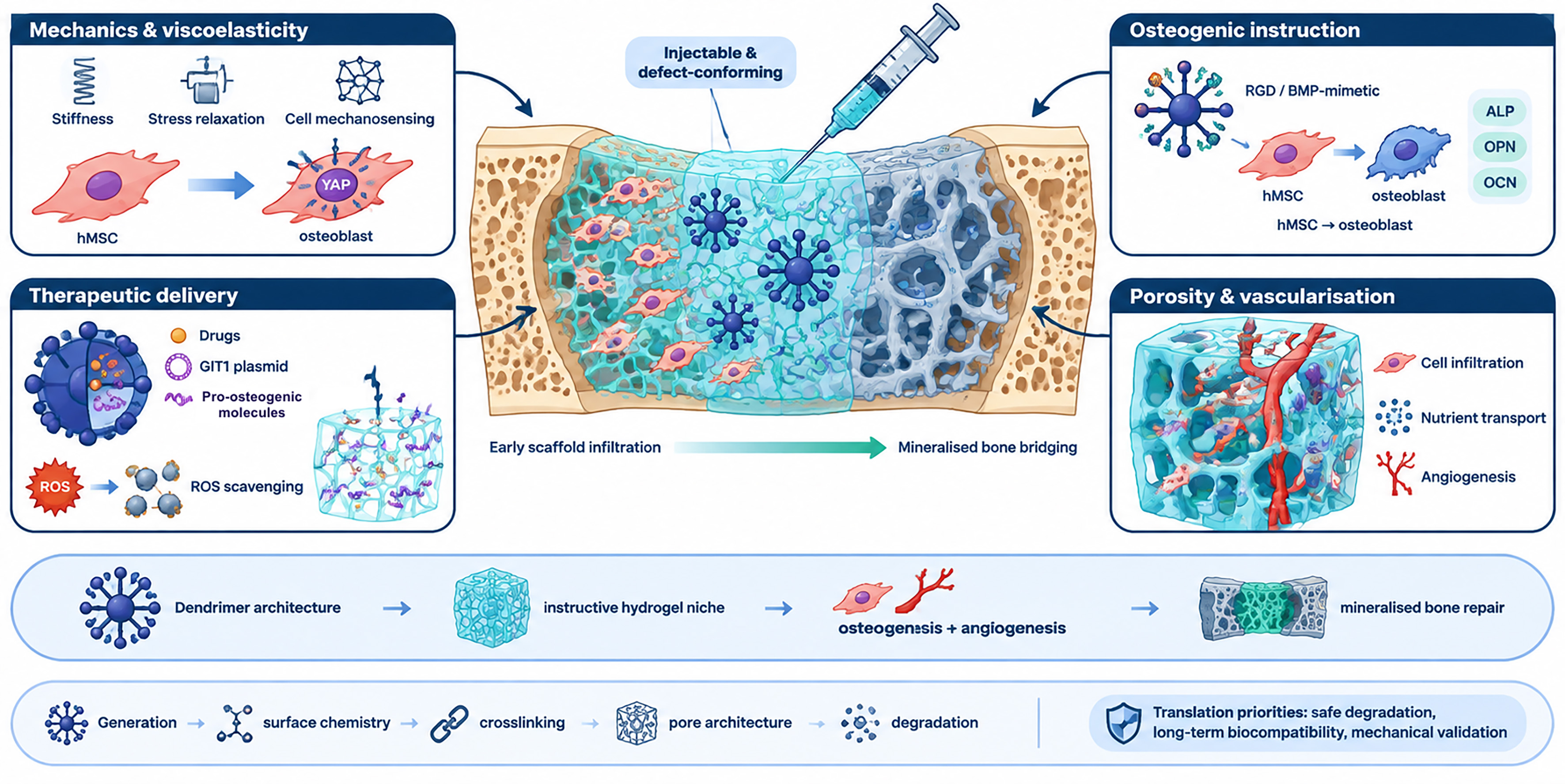
Figure 4. Dendrimer-based hydrogel for bone regeneration. Dendritic crosslinkers regulate hydrogel mechanics, porosity and the presentation or release of osteogenic cues. These properties can support stem cell recruitment and differentiation, vascularization and bone formation. YAP: Yes-associated protein; hMSC: human mesenchymal stromal cells; ROS: reactive oxygen species; BMP: bone morphogenetic protein; ALP: alkaline phosphatase; OPN: osteopontin; OCN: osteocalcin; GIT1: G protein-coupled receptor kinase-interacting protein 1.
Valeo et al. reported the first use of G5 DGL as covalent crosslinkers in PEG hydrogels for bone tissue engineering. EDC-mediated amide bonding between DGL terminal amines and dicarboxylic acid-PEG yielded bulk hydrogels with Young’s modulus tunable from 8 to over 30 kPa [15-100 kPa by atomic force microscopy (AFM)] by varying the NH2/COOH stoichiometric ratio. Dual surface functionalization with RGD and bone morphogenetic protein 2 (BMP-2) biomimetic peptides promoted human mesenchymal stromal cells (hMSC) adhesion and osteogenic differentiation: on stiffer gels (22-30 kPa), a two-fold upregulation of osteopontin was observed after two weeks. No leachable DGL cytotoxicity was detected[45].
Apartsin et al. extended this work using hyaluronic acid as the matrix component, demonstrating that in DGL G5/hyalurobic acid hydrogels, stiffness and stress relaxation timescales can be independently controlled. Lower-stiffness formulations with higher viscoelastic dissipation drove a 3-fold upregulation of the early osteocyte marker E11/podoplanin, and Yes-associated protein (YAP) nuclear localization was governed more by viscoelastic dissipation than by nominal stiffness thus providing evidence that viscoelastic properties, rather than elastic modulus alone, should be a primary design parameter. The platform enables orthogonal tuning of stiffness and viscous dissipation within a single chemical system[44].
Roumani et al. showed that DGL/PEG-NHS microspheres (G3 DGL; 22,000 g/mol; 10-1,600 µm diameter; water-in-oil emulsion fabrication) possess intrinsic osteogenic activity without cargo loading. In co-culture with human umbilical vein endothelial cells (HUVEC), the microspheres triggered a 5-fold increase in transforming growth factor β1 (TGF-β1) and a 3-fold increase in Complement C5a, driving a 2.5-fold increase in hMSC recruitment and a 3-fold increase in alkaline phosphatase (ALP) activity and BMP-2 secretion. The injectable microsphere format conforms to irregular defect geometries; elucidation of the precise molecular mechanism underlying the intrinsic osteoinductive effect is identified as a future direction[74].
Kurian et al. integrated G3 PAMAM dendrimer-functionalized nanoceria into photo-crosslinked GelMA to address oxidative stress in critical-sized bone defects. The nanoparticles provided enzymatic-like ROS scavenging [superoxide dismutase (SOD) activity], protecting MSCs from oxidative stress, while supramolecular interactions between dendrimer amines and GelMA carboxyl groups raised the compressive modulus to ~16.1 kPa. In a rat calvaria defect model at 12 weeks, mature bone formed with high Collagen I, osteopontin (OPN), osteocalcin, and CD31 expression through canonical Wnt pathway activation, with excellent tissue integration and minimal inflammatory response[75].
Yesbeck et al. confirmed the feasibility of G5 PAMAM/PEGDA hydrogels for sustained cefazolin release in periodontal bone regeneration, retaining ~75% of the antibiotic at 72 h and demonstrating compatibility with co-applied particulate bone allografts. Longer monitoring periods and osteoblast cytotoxicity assessment are identified as priorities[76]. Attik et al. further applied the effervescent DGL/PEG platform combined with aluminum-free borosilicate glass particles to dental tissue regeneration, achieving ~79% porosity (313 µm average pore size), enhanced calcium deposition, and cell colonization to a depth of 200 µm in human gingival fibroblasts[77].
Cheng et al. used an injectable thermoresponsive dendronized chitosan/PEGDA hydrogel to deliver G protein-coupled receptor kinase-interacting protein 1 (GIT1) plasmids for gene therapy of bone defects. The dendronized chitosan backbone provided both mechanical stability via hydrophobic interactions and DNA condensation via cationic surface charge, achieving in vivo transfection efficiency more than 3000-fold above lipoplex controls at day 3 and significant improvements in trabecular bone volume at four weeks through Notch-pathway-driven neovascularization[78].
Shrestha et al. used G2 PAMAM dendrimers to coat porous silicon nanoparticles, enabling loading of three pro-osteogenic miRNAs (miR-29b-3p, miR-101-3p, miR-125b-5p) within a norbornene-modified gelatin/PEG-dithiol visible-light photo-crosslinked hydrogel. Transfection efficiency in the 3D hydrogel reached 97.9%, which is substantially higher and more spatially uniform than 2D conditions, thus driving a 2.7-fold increase in hBMSC mineralization over seven days. The degradation of nanoparticles to orthosilicic acid provided an additional direct stimulus for mineralization. In vivo validation remains to be completed[79].
HYDROGELS FOR OCULAR AND OTIC APPLICATIONS
The eye and inner ear impose exceptionally demanding constraints on biomaterials. Ocular formulations must be optically transparent, mechanically compliant, non-inflammatory, and capable of withstanding fluctuating intraocular pressure, while otic systems must navigate the tortuous anatomy of the middle and inner ear and achieve sustained gene or drug delivery to delicate sensory cells without triggering local inflammation [Figure 5].
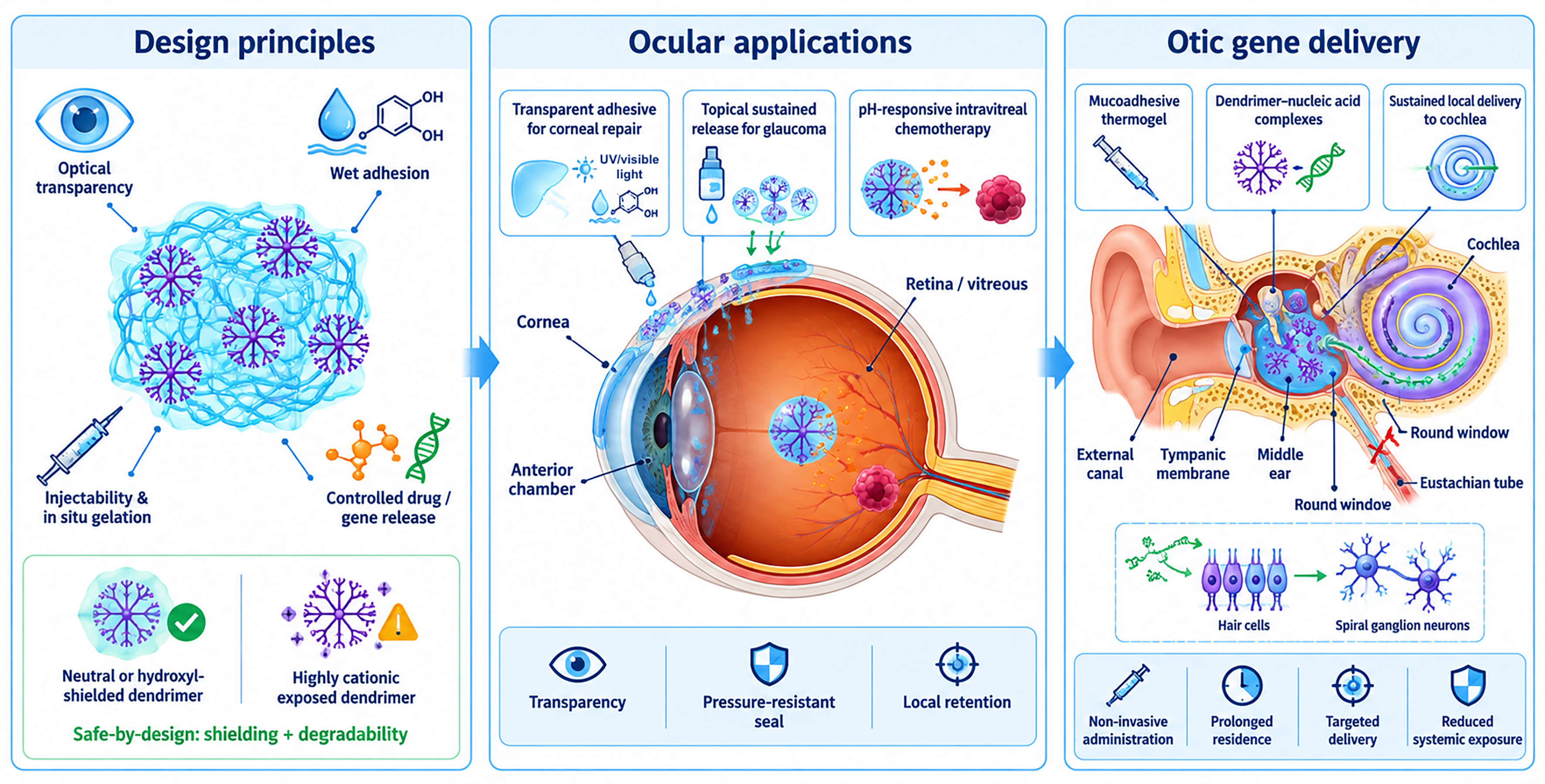
Figure 5. Dendrimer-based hydrogel for ocular and otic applications. Transparent, adhesive or thermogelling dendrimer hydrogels support corneal sealing and repair while enabling sustained ocular drug delivery. Dendrimer carriers can also provide local chemotherapy in the eye or protect and retain gene-delivery complexes after intratympanic administration.
Conventional corneal repair relies on sutures or rigid cyanoacrylate glues, both of which can distort corneal architecture, induce inflammation, and compromise visual outcomes. Three dendrimer hydrogel platforms have been developed to address these shortcomings through instant adhesion and optically compatible formulations.
Zhang et al. reported a triple-crosslinked double-network hydrogel for sutureless keratoplasty, combining three distinct mechanisms: UV-initiated photo-crosslinking of methacrylated alginate, Schiff base formation between the terminal amino groups of a G3 PAMAM dendrimer and aldehyde-dextran, and secondary physical reinforcement via Ca2+ chelation. This layered crosslinking strategy yielded a tensile Young’s modulus of 468.9 kPa and a storage modulus of 116.9 kPa, with adhesive strength at least two-fold higher than non-dendrimer controls and hemolysis below 0.5%. In a rabbit model of sutureless lamellar keratoplasty, an 80% surgical success rate was achieved with normal corneal ultrastructure maintained at 90 days. The high mechanical compliance and strong wet tissue adhesion are the principal advantages; a 20% failure rate attributed to graft dislocation or infection in the animal model identifies the cohesion-adhesion balance as a target for optimization[80].
Kambhampati et al. developed OcuPair, a two-component system comprising an injectable sodium hyaluronate filler to restore globe volume and an in situ photo-curable adhesive hydrogel formed by UV crosslinking between methacrylated G4-OH PAMAM and methacrylated hyaluronic acid at a 30:70 ratio. The transparent, flexible bandage withstood intraocular pressures above 60 mmHg, which is above the physiological range and reached adhesion strengths of approximately 252 kPa. In an in vivo rabbit corneal injury model, the material proved non-toxic over 30 days and supported stromal integration and epithelial regrowth in a 4-month partial keratoplasty model, demonstrating compatibility with long-term tissue remodeling. The system is designed to be battlefield-ready and optically clear for intraocular inspection, offering superior comfort relative to cyanoacrylate glues. A practical manufacturing consideration is that the D-MA precursor requires low-pH storage (pH 5.5) and an inhibitor [mequinol (MEHQ)] during synthesis to prevent premature polymerization[81].
Chen et al. extended the OcuPair concept to full corneal regeneration, using the same photo-crosslinked hydrogel as a sutureless alternative to conventional lamellar keratoplasty. In rabbit anterior lamellar keratoplasty models addressing both shallow and deep stromal defects and large 7 mm corneal lesions, the hydrogel achieved near-complete corneal regeneration within 6-10 months. Corneal transparency reached approximately 70% and thickness returned to presurgical levels within 2 weeks. A particularly notable finding is that the hydrogel spontaneously delaminates from intact, fully healed epithelium within 48 h while remaining on the wound bed providing natural self-limiting residence. Variability in clearance time (5-7.5 months) and the absence of specific incorporated growth factors to accelerate healing are identified as areas for further development[82].
Feng et al. took a cell-free scaffold approach to corneal tissue engineering using thermo-gelling dendronized chitosans in which oligoethylene glycol-based G1 dendrons were grafted onto a chitosan backbone. The material undergoes a sol-gel transition at physiological temperature (above
Glaucoma management relies on sustained intraocular pressure reduction, typically via topical eye drops, but conventional formulations suffer from poor corneal penetration, rapid clearance, and the inconvenience of frequent dosing. Wang et al. developed nano-in-nano dendrimer hydrogel particles by crosslinking G5 PAMAM dendrimers with PEGDA via aza-Michael addition in an inverse emulsion, producing either nanoscale (~200 nm) or microscale (3-9 µm) particles. These particles were loaded with two anti-glaucoma drugs, brimonidine tartrate and timolol maleate, and administered as topical eye drops. Nanoparticles increased corneal permeability by 17-fold compared to plain drug solutions and achieved zero-order sustained release kinetics. In normotensive rats and in a genetic glaucoma mouse model (A1 cKO), daily dosing produced a 4-fold stronger intraocular pressure (IOP) reduction than standard eye drops at day 7. Hemolysis was below 1% and the particles were non-irritating to ocular tissues. The system also enables high-ratio combination therapy by confining drugs in distinct particulate compartments. A reported limitation is drug loss during the particle washing step, which affects loading efficiency[84].
Retinoblastoma is the most common intraocular malignancy in children, and its first-line chemotherapeutic agent, melphalan, is practically insoluble in aqueous media which limits intravitreal bioavailability. Cooper et al. developed a pH-responsive injectable hydrogel by crosslinking G5 PAMAM dendrimers surface-modified with acid-sensitive acetal-dibenzocyclooctyne (DBCO) linkers with PEGDA via aza-Michael addition. The mild crosslinking retained a flowable liquid state suitable for intravitreal injection, while the acetal linkages provided pH-triggered release of conjugated melphalan at the acidic pH of the tumor microenvironment (pH 5.3), supplemented by simultaneous physical release of encapsulated free drug. In a Y79 tumor xenograft model, the conjugated formulation achieved 80% animal survival compared to only 20% with free drug. The coordinated dual-mode delivery significantly improves melphalan bioavailability by overcoming its solubility limitations. Long-term ocular safety evaluation is identified as a prerequisite for further translational development[85].
Gene therapy for sensorineural hearing loss requires delivery of nucleic acids to the hair cells and spiral ganglion neurons of the cochlea, which are challenging targets owing to the tight anatomical compartmentalization of the inner ear and the rapid clearance of intratympanic injections through the Eustachian tube. Le et al. addressed this by complexing plasmid DNA with a second-generation PAMAM dendrimer doubly modified with histidine and arginine and dispersing the polyplexes in a
HYDROGELS FOR DRUG DELIVERY
Dendrimer-based hydrogels have been extensively engineered as drug and gene delivery depots, addressing two persistent limitations of conventional chemotherapy and biologics: the poor solubility of hydrophobic payloads and the lack of spatiotemporal control over release [Figure 6].
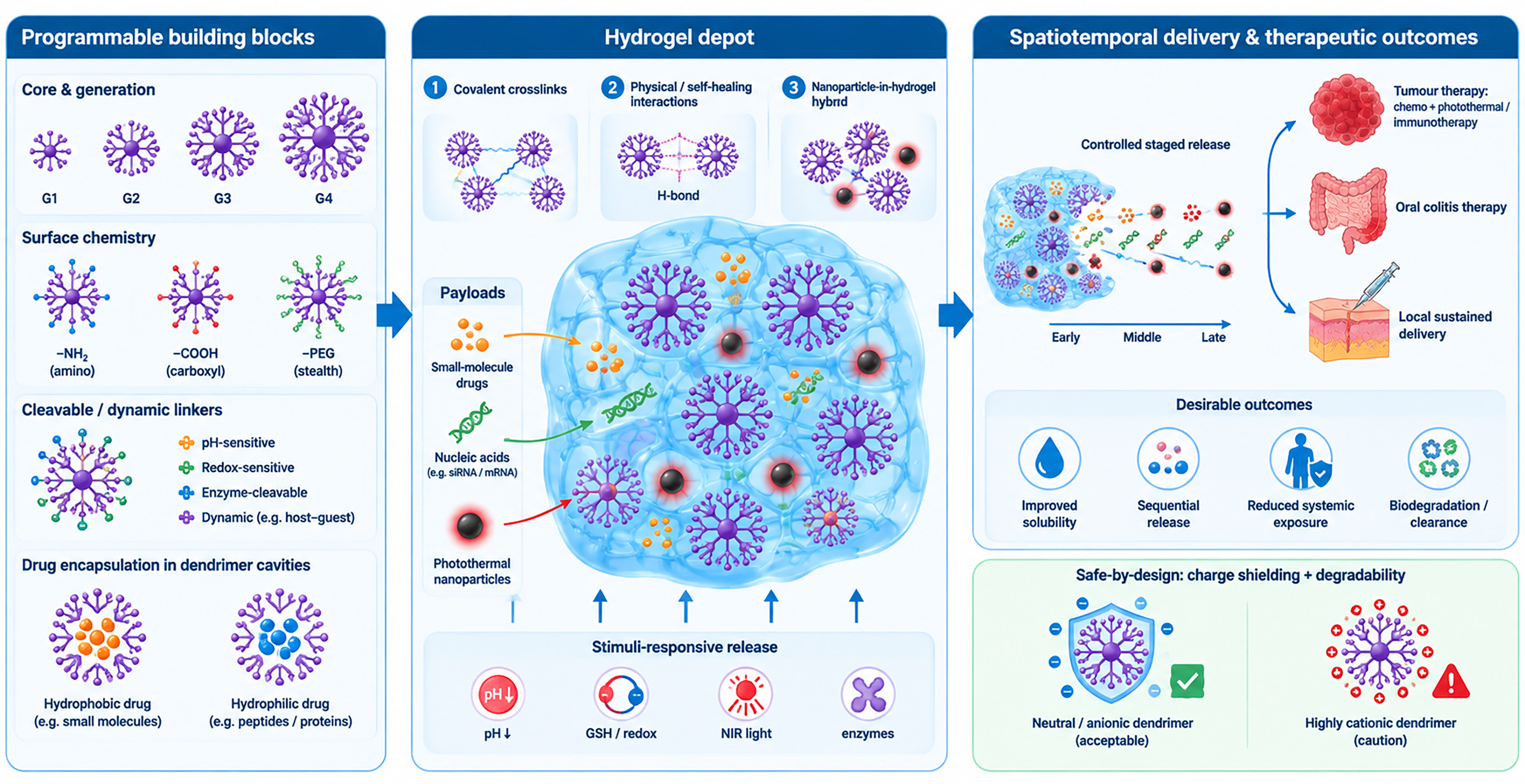
Figure 6. Dendrimer-based hydrogels for drug delivery. Dendrimer cavities and functionalized surfaces bind therapeutic cargoes, while the surrounding hydrogel acts as a local depot. Release can be sustained or triggered by chemical or physical factors, enabling spatial and temporal control of treatment. PEG: Poly(ethylene glycol); GSH: glutathione; NIR: near-infrared.
Jia et al. constructed a pH-responsive intratumoral hydrogel by crosslinking amino-terminated G3 PAMAM dendrimer with γ-PGA described above[56], but here applying it specifically to colon cancer therapy. The swelling rate reached up to 40-fold in simulated tumor environments (pH 5.4), and the gel was fully biodegradable in vivo within 7 days. The dual cargo strategy loaded hydrophilic doxorubicin (DOX) into the matrix network and hydrophobic camptothecin into the dendrimer hydrophobic cavities, thus overcoming its poor solubility. In a BALB/c-Nude mouse CT26 colon tumor model, the system achieved a 100% survival rate, significant tumor volume reduction, and elevated pro-apoptotic Bax expression. Hemolysis remained below 5%. The biodegradable design eliminates the need for post-treatment surgical removal, and the spatiotemporal co-delivery of two drugs with opposing solubility profiles minimizes systemic toxicity[87].
Jia and Wang developed a multifunctional tumor hydrogel by covalently crosslinking γ-PGA with G5 PAMAM dendrimers through EDC/NHS-mediated amide formation and incorporating PDA nanoparticles as photothermal agents. DOX was physically encapsulated in the porous γ-PGA/G5/PDA network, which gelled rapidly and accommodated drug loading up to 50 mg/mL. Under 808 nm NIR irradiation, the PDA component provided a photothermal conversion efficiency of 45.6%, while mild hyperthermia at 50 °C increased cumulative DOX release to 79% within 72 h under acidic tumor-mimicking conditions. The hydrogel degraded completely within 14 days in vitro and within 10 days after subcutaneous implantation in mice, while maintaining low hemolysis (< 5%) and cell viability above 85%. In a CT26 colon tumor model, the combined DOX-loaded hydrogel and NIR treatment achieved an 82.3% tumor-inhibition rate with no evident systemic toxicity, demonstrating the potential of γ-PGA/G5/PDA hydrogels for biodegradable chemo-photothermal tumor therapy[88].
An injectable hybrid hydrogel was assembled from a G4.5 PAMAM dendrimer, sodium deoxycholate, and mannitol to form a self-healing, thixotropic network through non-covalent interactions, with DOX loaded into the dendrimer interior and resveratrol distributed within the matrix. Sequential release was achieved: resveratrol liberated more rapidly (~70% in 7 days) to prime the tumor microenvironment and mitigate DOX cardiotoxicity via synergistic sensitization, followed by sustained DOX release (~54% in 7 days) at pH 6.5. In HeLa xenograft models, dual loading achieved the highest tumor inhibition rate at 68.4%. The thixotropic nature of the gel, which the authors identify as limiting release duration beyond 7 days, is flagged as the primary engineering target for future development[89].
Wang et al. developed a zwitterionic injectable hydrogel combining covalent thiol-ene crosslinking of methacrylated dextran with a sulfobetaine methacrylate-based thiol precursor as the matrix, into which G5 PAMAM monomolecular micelles co-loaded with DOX and the indoleamine 2,3-dioxygenase (IDO) immune checkpoint inhibitor 1-methyl-tryptophan, further coated with macrophage membranes for biomimetic tumor targeting, were dispersed. The zwitterionic dextran matrix provided strong anti-protein adsorption and biodegradability. 1-Methyl-tryptophan was released rapidly to reverse immunosuppression, and DOX was released slowly to induce immunogenic cell death. This led to an 89.2% postoperative recurrence inhibition rate and a 91.7% reduction in lung metastasis in melanoma models. The macrophage membrane coating provides tumor-homing activity, and the combination minimizes systemic DOX toxicity while activating cytotoxic T lymphocytes[90].
Ooya and Lee reported that polyglycerol dendrimers generations G3 and G4 crosslinked with ethylene glycol diglycidyl ether produce highly swollen hydrotropic hydrogels (swelling ratio up to 36.2) whose dense hydroxyl periphery creates favorable solvation microenvironments for paclitaxel, a clinically critical but practically insoluble antitumor agent. Paclitaxel loading capacity scaled proportionally with the swelling ratio, and 60% of loaded drug was released within 90-150 min under sink conditions without crystallization or precipitation known as a common failure mode of paclitaxel (PTX) formulations. The all-hydroxyl surface chemistry avoids the cytotoxicity concerns of cationic PAMAM dendrimers, and the hydrotropic mechanism represents an inherent solubilization strategy independent of external stimuli. Future directions include fabricating nanogels via click chemistry or miniemulsion to enable oral chemotherapy delivery[91].
Recio-Ruiz et al. introduced a platform based on G1-G3 carbosilane dendrimers crosslinked with dithiothreitol via thiol-ene reaction, producing nanostructured bulk hydrogels with aromatic nanodomains capable of solubilizing poorly water-soluble compounds through π-π stacking and lipophilic interactions. Low swelling [swelling degree (SD) 6%-16%] provided dimensional stability. Ibuprofen and curcumin were delivered as model cargos; curcumin covalently attached via fluoride-promoted esterification to a newly designed biodegradable trimesic acid core variant was 4-10 times more efficient than physical encapsulation, achieving 77%-79% cell death in PC3 prostate cancer cells after 6-24 h in dynamic assays. The platform’s pH-responsive release profiles and synthetic precision offer an orthogonal design route to PAMAM-based systems[92].
Muñoz-Sánchez et al. extended the carbosilane dendrimer toolkit to true stimuli-triggered network disassembly by crosslinking G2/G3 bifunctional carbosilane dendrimers bearing pendant hydroxyl groups and alkyne termini with self-immolative poly(ethyl glyoxylate) via CuAAC. Protonation of the poly(ethyl glyoxylate) acetal linkages at low pH initiates a “head-to-tail” depolymerization cascade that progressively dismantles the entire network, releasing curcumin in a controlled manner targeted to the acidic tumor microenvironment. NMR monitoring confirmed clean degradation products. The simultaneous structural collapse and payload liberation provides a conceptually distinct release mechanism compared to diffusion-dominated hydrogels, and the biodegradable ester bonds in the trimesic acid-core dendrimers provide additional degradability. The principal limitation is that cleavage of three ester bonds simultaneously may slow full network dissolution[93].
Guo et al. designed a polysaccharide-dendrimer hydrogel forming a shear-thinning self-assembled porous network through intermolecular hydrogen bonding and electrostatic interactions between rhubarb polysaccharides and a fourth-generation PAMAM dendrimer that encapsulates berberine in the hydrophobic cavity via host-guest inclusion to overcome its poor solubility. The oral formulation showed significant colonic accumulation for up to 18 h following gavage in dextran sulfate sodium-induced colitis models, reducing inflammation scores by 66.7%, promoting M2 macrophage polarization, and restoring gut microbiota diversity, notably increasing the abundance of the beneficial commensal Akkermansia. No pathological organ alterations were detected. Future directions include personalizing the hydrogel viscoelasticity and degradation profiles for patient-specific colitis phenotypes[94].
Zheng et al. assembled a fully natural-origin pH-responsive hydrogel fabricated from enzymatically assembled dendrimers of a glucan (a highly branched polysaccharide from sweet corn) and probed its use for lysozyme delivery. The 3D network formed through glycosidic bond entanglement is responsive to luminal pH. The protonation of carboxymethyl groups at gastric pH (1.2) suppresses swelling and protects cargo, while deprotonation at intestinal pH (7.4) drives swelling and release. Lysozyme loading efficiency approached 98%, and release reached 88% under intestinal conditions vs. only 25% in gastric fluid. The high resistant glucan content (72%) confers a low glycaemic index, making this system particularly suited to infant nutrition and diabetic populations. A sparser network structure relative to unmodified polyglucan, resulting from the carboxymethyl modification, is identified as a structural limitation[95].
Arenhoevel et al. reported mucin-inspired sulfated dendronized polymer fibers based on a G1 oligoglycerol dendron grafted onto a polyacrylamide backbone. The surface degree of sulfation was 94%, yielding highly extended ~53 nm single-chain fibers with a strongly negative zeta potential (~-54 mV) that mimic the heparan sulfate proteoglycan decoration of mucus glycoproteins. These fibers inhibited Herpes Simplex Virus Type 1 (HSV-1) with IC50 values in the lower nanomolar range (< 3 nM) through reversible competitive blocking of viral heparan sulfate binding sites, while anticoagulant activity was at least 10-fold lower than heparin. Although the current study characterizes the fibers rather than a crosslinked hydrogel, the pyridine-disulfide end-groups present on each chain enable thiol-disulfide exchange-mediated gelation, pointing to a direct route toward topical antiviral hydrogel formulations. The inhibitory activity depends critically on precise sulfate density, which sets a tight synthetic quality requirement[96].
NEW CONCEPTS IN DESIGNING BIOACTIVE HYDROGELS
The works discussed in this section do not target a single clinical indication but instead advance the conceptual and chemical design space of dendrimer-based hydrogels. They introduce new crosslinking logics, novel dendrimer chemistries, topological design principles, and organizational strategies.
Dendrimer-based hydrogels as defined alternatives to Matrigel for organoid culture
3D organoid culture relies heavily on Matrigel and related basement-membrane extracts that contain a complex, variable mixture of basement-membrane proteins, growth factors and other bioactive constituents. Their composition, mechanical properties and growth-factor content may vary both within and between batches, limiting reproducibility and preventing independent control of biochemical and biophysical variables[97-99].
Dendrimer-based hydrogels offer a potentially valuable route towards chemically defined synthetic ECMs. Their generation-defined molecular architecture, controlled number of terminal groups and modular surface chemistry enable the preparation of reproducible matrices in which network connectivity, ligand density and local multivalency can be rationally designed.
Apartsin et al. designed adaptive PEG bis-dendron hydrogels in which PEG chains of 2, 4, or 10 kDa carry first-generation branching units (dimethyl 5-hydroxyisophthalate) decorated with norbornene groups, covalently crosslinked with 4-arm-PEG-thiol via UV-induced thiol-ene reaction. The bis-dendron topology clusters bioactive cues at branch points, promoting multivalent integrin engagement, a feature unavailable in linear or branched PEG systems. Stiffness was tunable from G' = 30 Pa to
Peptide-dendrimer-reinforced bioinks for 3D bioprinting
Bioprinting of functional tissue models requires hydrogel inks that are simultaneously processable, mechanically stable after deposition, cytocompatible, and capable of sustaining multiple cell types in distinct spatial zones. Zhou et al. developed a dual-network bioink based on lysine-derived peptide dendrimers G1-G3 grafted onto a PEG backbone and end-modified with norbornene groups, crosslinked via thiol-ene photoclick chemistry with cysteamine-modified hyaluronic acid and DTT under 405 nm visible light. Storage modulus scaled with dendrimer generation, reaching ~6.4 kPa for G3, and HC-PDN showed a significant reduction in ROS accumulation compared to standard hyaluronic acid methacrylate bioinks. Used as a support bath for 3D embedded bioprinting of heterogeneous hepatic models co-encapsulating HepG2-C3A hepatocytes, LX-2 stellate cells, and EA.hy.926 endothelial cells, the system reproduced functional endothelium barrier properties, glycogen storage, and albumin and urea secretion. A key design advantage is that the fully degradable network offers a route to future in vivo translation; the main limitation is the mechanical weakness of standard natural bioinks that the dendronized system overcomes, widening the biofabrication window[53].
Self-immolative dendron hydrogels
A fundamental limitation of diffusion-controlled hydrogel release is that payload liberation continues even when the therapeutic need is absent. Self-immolative architectures, in which a single stimulus triggers complete network disassembly via a cascade fragmentation mechanism, offer a conceptually distinct solution.
Gill et al. reported the first self-immolative dendron hydrogels, in which G1 and G2 polycarbamate dendrons bearing a photocleavable o-nitrobenzyl carbamate trigger at their focal point and alkyne groups on their periphery were covalently crosslinked with 4-arm-PEG-azide via CuAAC. UV irradiation at 365 nm cleaved the focal-point trigger, initiating the “one trigger, many bonds” cascade that dismantled the entire network: G1 gels degraded more than 40% within one day of irradiation, while G2 gels were slower (18%) owing to higher crosslinking density. Gel content exceeded 88%, G2 moduli reached 144 kPa, and no cytotoxic leachable species were detected in C2C12 myoblast cultures. The principal limitations are the slow complete fragmentation timescale (10-18 days) and restricted UV penetration in thick bulk samples, which together constrain use to near-surface or thin-geometry applications[101].
Dynamic and supramolecular dendritic hydrogels
Beyond covalently crosslinked networks, dendritic building blocks enable hydrogel formation through reversible covalent and non-covalent interactions, including hydrogen bonding, electrostatic association, dynamic imine or disulfide linkages, etc. Because dendrimers present multiple interacting groups within a confined, generation-defined architecture, they can amplify weak molecular interactions into stable three-dimensional networks while retaining reversibility at the individual junction level. This combination can impart shear-thinning, self-healing, injectable and stimulus-responsive behavior that is difficult to achieve using conventional linear polymers alone. The following examples illustrate how dendritic topology has been exploited to create self-healing, physically assembled, chiral and stimuli-responsive hydrogel networks for biomedical and bioinspired applications.
Li et al. built a self-healing hydrogel from a thiol-rich glutamic acid-cysteine dendrimer and oxidized sodium alginate, crosslinked through dual dynamic linkages: Schiff base formation between dendrimer amines and alginate aldehydes (pH-responsive), and disulfide bond formation between dendrimer thiols (redox-responsive via GSH). The combination of these two reversible chemistries provided autonomous self-healing and pH/GSH dual-stimuli-triggered doxorubicin release: 76% of DOX was liberated at pH 5.5 with 10 mM GSH, matching the tumor microenvironment, while release was substantially suppressed at physiological conditions. Water retention capacity reached 25 times the hydrogel's original mass. Cell viability in normal L929 fibroblasts remained high while HepG-2 hepatocellular carcinoma cells showed comparable cytotoxicity to free DOX. The multi-dynamic crosslinking approach, which confers structural robustness alongside stimuli-responsiveness, is the platform’s defining conceptual contribution[102].
Apartsin et al. synthesized cyclotriphosphazene-core phosphorus dendrimers G1-G3 bearing peripheral trimethylammonium or pyridinium acetohydrazone groups. These polycationic dendrimers self-assembled into physical hydrogels through hydrogen bonding between surface hydrazone groups upon incubation at 65 °C in the presence of biocompatible additives (glucose, glycine, PEG), with gelation kinetics and gel rigidity depending on dendrimer generation, cation type, and biocompatible additive. Owing to their strong cationic surface charge, the hydrogels efficiently bound a model fluorescein-labeled oligonucleotide, achieving loading capacities of 1.5-3 µmol/g depending on dendrimer type and additive, but cargo release studied across pH 4.5-7.0 was slow and pH-dependent (higher below pH 6). The authors conclude that these biocompatible-additive-based phosphorus dendrimer hydrogels represent a novel, well-characterized nanomaterial platform for nucleic acid loading, and suggest that reducing dendrimer surface charge density could improve control over cargo release kinetics for future drug- or gene-delivery applications[103].
These authors have also developed another hydrogel platform using phosphorus dendrimers. They obtained neutral, biocompatible agarose hydrogels impregnated with polyelectrolyte complexes (dendriplexes) formed between anticancer Mcl-1 siRNA and polycationic G3 phosphorus dendrimers bearing piperidinium, trimethylammonium acetohydrazone, or pyridinium acetohydrazone peripheral groups. These dendrimers and dendriplexes have been shown to possess anti-tumor activity[104,105]. Dendriplexes were uniformly round, roughly 100 nm particles regardless of dendrimer composition, and they were embedded into 2% agarose hydrogels. Release kinetics from the hydrogels depended strongly on dendrimer surface chemistry: complexes of siRNA with acetohydrazone-terminated dendriplexes were retained in a hydrogel due to hydrogen bonding between the hydrazone moieties and the agarose scaffold. By mixing dendrimers in different ratios, the authors demonstrated that the dendriplexes release rate can be finely tuned to achieve programmed, sustained local delivery of therapeutic nucleic acids for applications such as tumor treatment, wound healing, or tissue engineering as the hydrogel bioresorbs over time[106].
Phan et al. used asymmetric polyglycerol dendrimers G1-G3 as multi-functional initiators for N-carboxyanhydride ring-opening polymerization of lysine to create star-shaped poly(L-lysine) architectures with 6, 12, and 24 arms, whose side chains were subsequently grafted with indole or phenyl groups. Gelation occurred through non-covalent hydrogen bonding, π-π stacking, and hydrophobic interactions among the grafted moieties, producing shear-thinning, rapidly recovering physical networks. The 24-armed indole-grafted variant exhibited the strongest mechanics and the lowest critical gelation concentration (2.9 wt%), with polypeptide chains adopting β-sheet/β-turn conformations (50%-60%) that drove network formation. The key design insight is that asymmetric topology (more arms, greater steric diversity) amplifies the gelation tendency[107].
The introduction of chirality and helical order into hydrogel matrices opens pathways toward chiral recognition, asymmetric catalysis, and biomimetic scaffolds that replicate the structural anisotropy of native cartilage and muscle. Two studies establish the principles using dendronized polymer systems. Mei et al. reported that G1 dendronized amphiphiles - three-fold dendritic oligoethylene glycol units bridged by a phenylalanine-glycine dipeptide and terminating in diacetylene groups - self-assembled in water into thermoresponsive helical fibers whose supramolecular chirality was controllable by stirring direction (vortex-dependent chirality). UV irradiation at 254 nm triggered topochemical polymerization of the diacetylene segments, covalently locking the helical conformation and converting the physical gel into a robust material: storage modulus rose from ~20 Pa to >10 kPa post-polymerization. The combination of programmable supramolecular assembly and covalent fixation enables hierarchical structural organization. A limitation is the low mechanical strength of the prefixation network[108].
Shen et al. extended the principle to helical poly(phenylacetylene) copolymers carrying dendritic oligoethylene glycol pendants, crosslinked by dynamic covalent arylhydrazone linkages. Gelation temperature determined network architecture: room-temperature gelation produced homogeneous gels, above-cloud-point gelation yielded heterogeneous networks, and cryo-gelation at -20°C via ice-templating generated macroporous lamellar structures with G' reaching 30 kPa. The lamellar gels exhibited sponge-like compressive behavior: extruding 80% of their water under load and recovering to 90% strain over 50 fatigue cycles. This feature positions them as synthetic mimetics of cartilage or muscle. Crucially, chirality was fixed and stabilized after gelation, preventing the dynamic helical inversion seen in precursor solutions. Precise temperature control during fabrication is the process constraint[109].
DISCUSSION
Across biomedical applications, dendrimer hydrogels have demonstrated that a single architectural motif can simultaneously provide structural integrity, bioactivity and controlled delivery, yet no single formulation satisfies all requirements at once[35]. A recurring strength of dendrimer hydrogels is their ability to integrate multiple therapeutic functions into a single construct, such as combining hemostasis, antibiotic-free antimicrobial action and wet adhesion in vascular sealants, or co-delivering anti-inflammatory and pro-regenerative cues in musculoskeletal depots.
Dendrimers should not be considered interchangeable multifunctional additives, but rather programmable nanoscale junctions whose architecture determines hydrogel performance. The dendrimer core, generation, branching density, peripheral chemistry and internal microenvironment jointly regulate network formation, mechanics, porosity, ligand presentation, cargo association and degradation behavior. In turn, these hydrogel-level properties influence clinically relevant functions, including tissue adhesion, cell-matrix signaling, antimicrobial activity, controlled drug or gene delivery, bioprinting and organoid culture. Figure 7 summarizes this architecture-to-function relationship, whereas Table 2 links individual dendrimer structural features to representative hydrogel properties, biological functions and examples discussed throughout this review. This framework provides a basis for selecting dendritic platforms according to the dominant requirement of a given application: for example, high crosslinking functionality for tissue sealants, clustered biochemical cues for synthetic ECMs, hydrophobic nanodomains for drug delivery, or dynamic/cleavable motifs for injectable and biodegradable matrices.
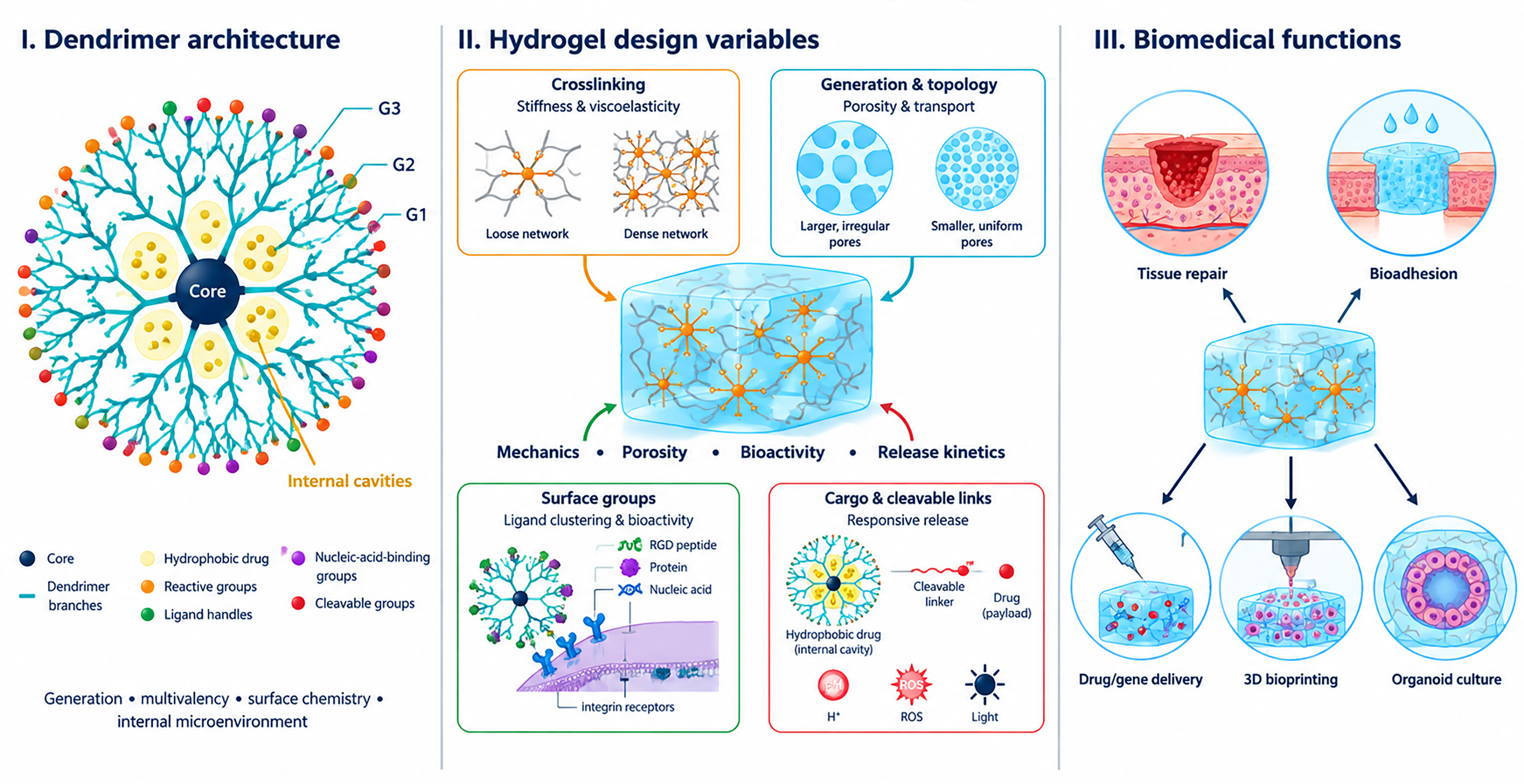
Figure 7. From dendrimer structure to hydrogel function. Core chemistry, generation, branching and surface groups determine how dendrimers crosslink networks, present ligands and bind cargo. These molecular parameters affect hydrogel mechanics, porosity, degradation and release, which in turn influence the biological performance of hydrogels. G1, G2, G3: Dendrimer generation 1, 2, 3; ROS: reactive oxygen species.
Relationship between dendrimer structural features, hydrogel properties and biomedical functions.
| Dendrimer structural feature | Role at molecular level | Role at hydrogel-level | Biological or biomedical function | Representative examples | Refs. |
| Core chemistry | Defines molecular rigidity, polarity, degradability and, in selected architectures, internal microenvironment | Influences assembly, cargo affinity and network-degradation pathway | Hydrophobic-drug solubilization; programmed release; biodegradable or responsive matrices | Polyglycerol dendrimer hydrotropic gels for paclitaxel; carbosilane dendrimer hydrogels with hydrophobic nanodomains; self-immolative carbosilane networks | [91-93] |
| Generation and branching density | Alters molecular size, terminal-group number, local functional density and steric crowding | Modulates crosslinking efficiency, stiffness, viscoelasticity and mesh architecture; response may be non-monotonic | Mechanical matching; tuning of stem-cell microenvironments; bioink optimization | PLL peptide dendrimer bioinks: increasing generation increased G and G; PAMAM-containing HA and PLLA systems: higher generation decreased modulus in selected formulations | [52-54] |
| Multivalent reactive periphery | Creates a high local density of crosslinkable groups | Forms multi-chain junctions; increases network connectivity; modulates swelling and pore structure | Mechanical reinforcement; structural stabilization; tissue repair | PAMAM/PEG and PAMAM/alginate-dextran networks; PAMAM-crosslinked collagen stiffer than EDC- or glutaraldehyde-crosslinked collagen | [40-43,46] |
| Peripheral functional groups | Determine charge, hydrophilicity, crosslinking reactivity and interaction with cells, proteins or nucleic acids | Control gelation mechanism, cell interaction, antimicrobial behavior and molecular binding | Cell adhesion; cytokine scavenging; antimicrobial action; nucleic-acid complexation | Cationic PLL dendrimer/oxidized HA hydrogel for rheumatoid arthritis; β-alanine-functional bis-MPA and DLD hydrogels with antibacterial activity; PAMAM polyplexes for gene delivery | [58-60,67,86] |
| Residual orthogonal groups | Preserve non-crosslinking sites after network formation | Enable independent incorporation of biochemical or diagnostic signals without changing bulk formulation | Presentation of adhesion peptides and osteogenic cues; drug, protein or nucleic-acid conjugation | RGD-functionalized PAMAM/PEG hydrogels; partially acetylated PAMAM/PEGDA networks; DGL/PEG matrices functionalized with RGD and BMP-2 peptides | [44] |
| Internal cavities or hydrophobic nanodomains | Sequester hydrophobic cargo or enable host-guest association | Provides local drug reservoirs within aqueous networks; enables separate loading of hydrophilic and hydrophobic payloads | Dual-drug delivery; improved solubility; sequential release | PAMAM systems carrying diclofenac and strontium; doxorubicin/camptothecin hydrogel; doxorubicin/resveratrol composite; carbosilane and polyglycerol hydrotropic systems | [65,87,89,91-93] |
| Ligand clustering at dendritic branch points | Places multiple ligands within nanoscale proximity and enhances local avidity | Enables controlled multivalent cell-matrix and biomolecular interactions | Integrin engagement; synthetic ECM design; organoid culture and stem-cell regulation | Adaptive PEG bis-dendron hydrogel with clustered RGD peptides and independently tunable mechanics, relaxation and pore size | [102] |
| Dynamic or cleavable branches/linkers | Introduce hydrolysis-, enzyme-, pH-, redox- or light-sensitive bond cleavage | Enables remodeling, self-healing, triggered disassembly and programmed release | Injectable depots; pH/redox-responsive delivery; biodegradable implants | Dual dynamic thiol-rich dendrimer/oxidized alginate hydrogel; self-immolative dendron and carbosilane hydrogels; dendritic thermogels | [88,93,103,106] |
However, this multifunctionality also amplifies design complexity and can obscure mechanistic attribution: in many studies, it remains difficult to disentangle the relative contributions of mechanical environment, dendrimer surface chemistry, drug payload and degradation products to the observed biological outcomes. This is particularly evident in musculoskeletal systems where stiffness, stress-relaxation and ligand presentation all influence osteogenic or chondrogenic differentiation, yet are rarely varied independently within a given study, and in immune-modulatory platforms where dendrimer-induced cytokine scavenging, innate immune activation and drug action are tightly coupled. Future work will benefit from more systematic, factorized experimental designs and from integrating biophysical readouts with molecular markers of cell state[110-112].
From a translational perspective, several practical limitations emerge. Many of the most innovative systems, as self-immolative networks, highly dynamic dual-responsive matrices, or NIR-triggered antibacterial sponges, have been validated only in small animal models and often under conditions that do not fully recapitulate human loading, fluid flow or immune landscapes. Long-term biodegradation, clearance and potential off-target accumulation of dendrimer fragments remain insufficiently characterized in large animals, particularly for intra-articular, intradiscal, intravitreal and intratumoral deposits where local confinement and repetitive mechanical stress could alter degradation pathways[113]. Furthermore, manufacturing and sterilization constraints are only intermittently addressed: several platforms rely on UV or visible-light curing in situ, precise stoichiometry of multi-component systems, or radiation doses that may be challenging to scale, and relatively few studies evaluate stability under clinically relevant sterilization regimes and storage conditions[114,115].
At the conceptual level, the most forward-looking studies suggest that dendrimer hydrogels are moving from simple drug reservoirs toward architecturally encoded, bio-instructive materials that can orchestrate cell fate and immune responses over time. Adaptive PEG bis-dendron matrices, DGL-based viscoelastic bone scaffolds and chirally ordered dendritic hydrogels collectively demonstrate that stiffness, viscoelastic dissipation, ligand clustering and even supramolecular chirality can be treated as independent design axes rather than fixed material properties. Yet this sophistication also demands new standards for characterization: rheology alone is no longer sufficient, and there is a need for multi-scale, time-resolved analyses that link molecular architecture to emergent tissue-level function, especially under physiologically relevant loading and in complex co-culture or organ-on-chip systems[22,116,117]. The field also lacks head-to-head comparisons between dendrimer-based and non-dendritic hydrogels in the same indication, which would clarify when dendrimers are genuinely necessary and when simpler architectures might be enough[11].
CONCLUSION AND OUTLOOK
Dendrimer-based hydrogels have evolved from niche materials into a coherent platform for wound care, musculoskeletal regeneration, ocular and otic repair, and local drug and gene delivery. Their distinctive value lies in the architectural control provided by generation, valency, topology and surface chemistry, which enables the coordinated tuning of network mechanics, viscoelasticity, degradation, ligand presentation, molecular transport and release behavior in ways that are difficult to reproduce with linear polymers or low-molecular-weight crosslinkers[30,31]. This versatility has enabled antibiotic-free antimicrobial dressings and sealants, mechanically tunable cartilage and bone scaffolds, transparent corneal adhesives, long-acting ocular depots, and self-immolative or nanoparticle-in-hydrogel systems for spatiotemporally programmed delivery.
Importantly, the advantage of dendritic crosslinking extends beyond increasing hydrogel stiffness. By changing the size, functionality and topology of network junctions, dendrimers can modify optical transparency, pore architecture, molecular permeability, cell-matrix interactions and material biocompatibility. However, dendrimers are not universally superior to conventional crosslinkers: their performance depends on the compatibility between dendrimer generation, flexibility, surface charge, reactive-group density, matrix polymer and gelation chemistry. Highly cationic or high-generation dendrimers, particularly PAMAM-based systems, may induce membrane-associated cytotoxicity, oxidative stress, inflammatory responses or long-term accumulation if they are insufficiently shielded, covalently incorporated or degraded. Accordingly, meaningful comparisons with non-dendritic materials should extend beyond modulus to include gel fraction, swelling, degradation, transport, extractables, cytocompatibility, immune response and long-term stability.
Future progress will therefore require biodegradable, safe-by-design dendritic families whose generation, surface chemistry and degradation pathways can be tailored to balance functional performance with biological safety. Integration with 3D bioprinting, organoid culture and organ-on-chip technologies could provide more predictive preclinical models and enable systematic structure-function studies, while mechanobiology-informed modeling, high-dimensional immunoprofiling and patient-derived cells or microbiota may support the development of more personalized hydrogel therapies. Translation beyond proof-of-concept will additionally require standardized characterization protocols, benchmarking against best-in-class non-dendritic hydrogels, scalable and sterilization-compatible manufacturing processes, and long-term large-animal studies that assess efficacy, degradation, clearance and local and systemic safety under clinically relevant conditions. If these challenges are addressed, dendrimer-based hydrogels could become an important class of next-generation biomaterials, providing not only finely tuned mechanics and release kinetics but also programmable, context-responsive interfaces capable of actively directing therapeutic outcomes.
DECLARATIONS
Authors’ contributions
Conception and preparation of the review: Apartsin E, Durrieu MC
Availability of data and materials
Not applicable.
AI and AI-assisted tools statement
During the preparation of this manuscript, the AI tool Perplexity Pro (version GPT-5.6 Sol, released 2026-07-09) was used for language editing and preparation of the graphical abstract and Figures 2-7. The tool did not influence the study design, data collection, analysis, interpretation, or the scientific content of the work. Both authors take full responsibility for the accuracy, integrity, and final content of the manuscript.
Financial support and sponsorship
This article is issued from the BOTS project co-financed by the Interreg Atlantic Area Programme through the European Regional Development Fund.
Conflicts of interest
Both authors declared that there are no conflicts of interest.
Ethical approval and consent to participate
Not applicable.
Consent for publication
Not applicable.
Copyright
© The Author(s) 2026.
REFERENCES
1. Choi H, Choi WS, Jeong JO. A review of advanced hydrogel applications for tissue engineering and drug delivery systems as biomaterials. Gels. 2024;10:693.
2. Sui J, Pragnere S, Kurniawan NA. Revisiting the biophysical aspects of extracellular-matrix-mimicking hydrogels: what cells see vs. what cells feel. Biomater Sci. 2025;13:5297-324.
3. Rijns L, Rutten MGTA, Vrehen AF, Aldana AA, Baker MB, Dankers PYW. Mimicking the extracellular world: from natural to fully synthetic matrices utilizing supramolecular biomaterials. Nanoscale. 2024;16:16290-312.
4. Ansari M, Darvishi A, Sabzevari A. A review of advanced hydrogels for cartilage tissue engineering. Front Bioeng Biotechnol. 2024;12:1340893.
5. Kohar R, Ghosh M, Sawale JA, Singh A, Rangra NK, Bhatia R. Insights into translational and biomedical applications of hydrogels as versatile drug delivery systems. AAPS PharmSciTech. 2024;25:17.
6. Li ZY, Li TY, Yang HC, et al. Design and fabrication of viscoelastic hydrogels as extracellular matrix mimicry for cell engineering. Chem Bio Eng. 2024;1:916-33.
7. Kaur H, Gogoi B, Sharma I, et al. Hydrogels as a potential biomaterial for multimodal therapeutic applications. Mol Pharm. 2024;21:4827-48.
8. Tanveer H, Sarfraz A, Fatima A, Sarwar S. Multifunctional hydrogels for biomedical applications. Nano Biomed Eng. 2024;16:558-73.
9. Zöller K, To D, Bernkop-Schnürch A. Biomedical applications of functional hydrogels: Innovative developments, relevant clinical trials and advanced products. Biomaterials. 2025;312:122718.
10. Tang S, Feng K, Yang R, et al. Multifunctional adhesive hydrogels: from design to biomedical applications. Adv Healthc Mater. 2025;14:2403734.
11. Dong S, An S, Saiding Q, et al. Therapeutic hydrogels: properties and biomedical applications. Chem Rev. 2025;125:8835-920.
12. Nagay BE, Mamizadeh Janghour L, El-Khordagui LK, et al. Multifunctional implantable hydrogels: smart platforms at the forefront of biomedical innovation. Mater Today Bio. 2026;37:102940.
13. Luo Y, Tan J, Zhou Y, et al. From crosslinking strategies to biomedical applications of hyaluronic acid-based hydrogels: a review. Int J Biol Macromol. 2023;231:123308.
14. Munyiri CN, Madivoli ES, Kisato J, Gichuki J, Kareru PG. Biopolymer based hydrogels: crosslinking strategies and their applications. Int J Polym Mater Polym Biomater. 2025;74:625-40.
15. Yan X, Huang H, Bakry AM, Wu W, Liu X, Liu F. Advances in enhancing the mechanical properties of biopolymer hydrogels via multi-strategic approaches. Int J Biol Macromol. 2024;272:132583.
16. Singh D, Kumar A. A comprehensive review on supramolecular polymer hydrogels for localized and long-term therapeutic delivery. J Macromol Sci B. 2026;65:876-86.
17. Maity S, Mahata K, Meshram B, Banerjee S. Smart polymer-derived injectable hydrogels: current status and future perspectives. ACS Polym Au. 2025;5:680-711.
18. Gao H, Wang Z, Lin T, Zhang G, Han X, Cheng C. Dynamic covalent bonding based polysaccharide hydrogels and their applications in wound dressings: a review. Colloids Surf B Biointerfaces. 2025;254:114878.
19. Yin B, Gosecka M, Bodaghi M, et al. Engineering multifunctional dynamic hydrogel for biomedical and tissue regenerative applications. Chem Eng J. 2024;487:150403.
20. Madl CM, Katz LM, Heilshorn SC. Tuning bulk hydrogel degradation by simultaneous control of proteolytic cleavage kinetics and hydrogel network architecture. ACS Macro Lett. 2018;7:1302-7.
21. Richbourg N, Wechsler ME, Rodriguez-Cruz JJ, Peppas NA. Model-based modular hydrogel design. Nat Rev Bioeng. 2024;2:575-87.
22. Yang J, Zhao Y, Jeang WJ, et al. A modular hydrogel system with independent control of bioadhesion, fibrosis, and stiffness. Sci Adv. 2026;12:eaee3894.
23. Al-Hussain SA, Sultan A, Rubab L, et al. Nanoparticle-reinforced polysaccharide hydrogels in drug delivery systems: a review of recent progress. Int J Biol Macromol. 2026;335:149238.
24. Yoon MS, Lee JM, Jo MJ, et al. Dual-drug delivery systems using hydrogel-nanoparticle composites: recent advances and key applications. Gels. 2025;11:520.
25. Fan Y, Han Q, Li H, et al. Recent developments in nanoparticle‐hydrogel hybrid materials for controlled release. Adv Sci. 2025;12:e07209.
26. Ahmad N, Bukhari SNA, Hussain MA, Ejaz H, Munir MU, Amjad MW. Nanoparticles incorporated hydrogels for delivery of antimicrobial agents: developments and trends. RSC Adv. 2024;14:13535-64.
27. Choi W, Kohane DS. Hybrid nanoparticle-hydrogel systems for drug delivery depots and other biomedical applications. ACS Nano. 2024;18:22780-92.
28. Zhang K, Feng Q, Fang Z, Gu L, Bian L. Structurally dynamic hydrogels for biomedical applications: pursuing a fine balance between macroscopic stability and microscopic dynamics. Chem Rev. 2021;121:11149-93.
29. Apartsin E, Caminade A. Single-component physical hydrogels of dendritic molecules. J. Compos Sci. 2023;7:26.
30. Kaga S, Arslan M, Sanyal R, Sanyal A. Dendrimers and dendrons as versatile building blocks for the fabrication of functional hydrogels. Molecules. 2016;21:497.
31. Ghobril C, Rodriguez EK, Nazarian A, Grinstaff MW. Recent advances in dendritic macromonomers for hydrogel formation and their medical applications. Biomacromolecules. 2016;17:1235-52.
32. Apartsin E, Caminade AM. Supramolecular self‐associations of amphiphilic dendrons and their properties. Chemistry. 2021;27:17976-98.
33. Dzmitruk V, Apartsin E, Ihnatsyeu-Kachan A, Abashkin V, Shcharbin D, Bryszewska M. Dendrimers show promise for siRNA and microRNA therapeutics. Pharmaceutics. 2018;10:126.
34. Kisakova LA, Apartsin EK, Nizolenko LF, Karpenko LI. Dendrimer-mediated delivery of DNA and RNA vaccines. Pharmaceutics. 2023;15:1106.
35. Bhowmik DD, Gupta JS, Kamandar PA, Gholap AD, Rojekar S, Hatvate NT. Dendrimers-based hydrogels and nanogels for drug delivery. Curr Pharm Biotechnol. 2026;27:542-63.
36. Sadekar S, Ghandehari H. Transepithelial transport and toxicity of PAMAM dendrimers: Implications for oral drug delivery. Adv Drug Deliv Rev. 2012;64:571-88.
37. Vu MT, Bach LG, Nguyen DC, et al. Modified carboxyl-terminated PAMAM dendrimers as great cytocompatible nano-based drug delivery system. Int J Mol Sci. 2019;20:2016.
38. Kharwade R, Badole P, Mahajan N, More S. Toxicity and surface modification of dendrimers: a critical review. Curr Drug Deli. 2022;19:451-65.
39. Wang J, Li B, Qiu L, Qiao X, Yang H. Dendrimer-based drug delivery systems: history, challenges, and latest developments. J Biol Eng. 2022;16:18.
40. Bi X, Maturavongsadit P, Tan Y, et al. Polyamidoamine dendrimer-PEG hydrogel and its mechanical property on differentiation of mesenchymal stem cells. Biomed Mater Eng. 2019;30:111-23.
41. Wang Y, Zhao Q, Zhang H, Yang S, Jia X. A novel poly(amido amine)‐dendrimer‐based hydrogel as a mimic for the extracellular matrix. Adv Mater. 2014;26:4163-7.
42. Wang H, Yang J, Tian W, et al. A sodium alginate/carboxymethyl chitosan dual-crosslinked injectable hydrogel scaffold with tunable softness/hardness for bone regeneration. Int J Biol Macromol. 2024;257:128700.
43. Muñoz Taboada G, Dosta P, Edelman ER, Artzi N. Sprayable hydrogel for instant sealing of vascular anastomosis. Adv Mater. 2022;34:2203087.
44. Apartsin E, Valeo M, Marie S, Granier F, Durrieu M. Bioactive hyaluronic acid hydrogels based on lysine dendrigrafts as crosslinkers: impact of stiffness and viscoelasticity on the osteogenic differentiation of mesenchymal stem cells. Mater Adv. 2026;7:7581-94.
45. Valeo M, Marie S, Rémy M, et al. Bioactive hydrogels based on lysine dendrigrafts as crosslinkers: tailoring elastic properties to influence hMSC osteogenic differentiation. J Mater Chem B. 2024;12:12508-22.
46. Duan X, Sheardown H. Dendrimer crosslinked collagen as a corneal tissue engineering scaffold: Mechanical properties and corneal epithelial cell interactions. Biomaterials. 2006;27:4608-17.
47. Wang J, He H, Cooper RC, Yang H. In situ-forming polyamidoamine dendrimer hydrogels with tunable properties prepared via aza-michael addition reaction. ACS Appl Mater Interfaces. 2017;9:10494-503.
48. Bi X, Amie Luckanagul J, Allen A, et al. Synthesis of PAMAM dendrimer-based fast cross-linking hydrogel for biofabrication. J Biomater Sci Polym Ed. 2015;26:669-82.
49. Liu F, Wang X, Li Y, et al. Dendrimer-modified gelatin methacrylate hydrogels carrying adipose-derived stromal/stem cells promote cartilage regeneration. Stem Cell Res Ther. 2022;13:26.
50. Zhu H, Mei X, He Y, et al. Fast and high strength soft tissue bioadhesives based on a peptide dendrimer with antimicrobial properties and hemostatic ability. ACS Appl Mater Interfaces. 2020;12:4241-53.
51. Griveau L, Lafont M, Le Goff H, et al. Design and characterization of an in vivo injectable hydrogel with effervescently generated porosity for regenerative medicine applications. Acta Biomater. 2022;140:324-37.
52. Maturavongsadit P, Bi X, Metavarayuth K, Luckanagul JA, Wang Q. Influence of cross-linkers on the in vitro chondrogenesis of mesenchymal stem cells in hyaluronic acid hydrogels. ACS Appl Mater Interfaces. 2017;9:3318-29.
53. Zhou K, Ding R, Tao X, et al. Peptide-dendrimer-reinforced bioinks for 3D bioprinting of heterogeneous and biomimetic in vitro models. Acta Biomater. 2023;169:243-55.
54. Memarian P, Solouk A, Bagher Z, Akbari S, Nazarpak MH. Ionic conductive nanocomposite based on poly(l-lactic acid)/poly(amidoamine) dendrimerelectrospun nanofibrous for biomedical application. Biomed Mater. 2022;17:015007.
55. Islam MR, Manir MS, Razzak M, et al. Silk-enriched hydrogels with ROS-scavenging dendrimers for advanced wound care. Int J Biol Macromol. 2024;280:135567.
56. Jia X, Fan P, Guo J, et al. Multifunctional polyamidoamine dendrimer/sodium alginate/γ-polyglutamic acid/amoxicillin hydrogel with enhanced hemostatic, antibacterial and wound healing properties for biomedical applications. Colloids Surf B Biointerfaces. 2026;265:115742.
57. Gupta P, Sheikh A, Abourehab MAS, Kesharwani P. Amelioration of full-thickness wound using hesperidin loaded dendrimer-based hydrogel bandages. Biosensors. 2022;12:462.
58. Biscari G, Fan Y, Namata F, et al. Antibacterial broad‐spectrum dendritic/gellan gum hybrid hydrogels with rapid shape‐forming and self‐healing for wound healing application. Macromol Biosci. 2023;23:2300224.
59. Biscari G, Sanz Del Olmo N, Palumbo FS, et al. Antimicrobial NIR-responsive hydrogels based on gellan gum and bis-MPA polyester dendrimers. ACS Appl Mater Interfaces. 2025;17:22448-63.
60. Fan Y, Mohanty S, Zhang Y, et al. Dendritic hydrogels induce immune modulation in human keratinocytes and effectively eradicate bacterial pathogens. J Am Chem Soc. 2021;143:17180-90.
61. Ye H, Gao Z, Liu Y, et al. Peptide dendritic polymerization-enabled stable and tailorable nanoparticles as crosslinkers for fabricating multifunctional hydrogels with enhanced wound healing efficacy. J Mater Chem B. 2026;14:5734-44.
62. Zhu H, Xu G, He Y, et al. A dual‐bioinspired tissue adhesive based on peptide dendrimer with fast and strong wet adhesion. Adv Healthc Mater. 2022;11:2200874.
63. Sanz Del Olmo N, Molina N, Fan Y, Namata F, Hutchinson DJ, Malkoch M. Antibacterial hydrogel adhesives based on bifunctional telechelic dendritic-linear-dendritic block copolymers. J Am Chem Soc. 2024;146:17240-9.
64. Zhou K, He X, Shi K, et al. A dual responsive nanohydrogel system for sustained drug delivery and cartilage penetration in osteoarthritis therapy. J Control Release. 2026;389:114494.
65. Cheng P, Yang J, Wu S, et al. Temporal modulation of inflammation and chondrogenesis through dendritic nanoparticle-mediated therapy with diclofenac surface modification and strontium ion encapsulation. J Biomater Sci Polym Ed. 2024;35:2049-67.
66. Wang J, Li B, Pu X, et al. Injectable multicomponent biomimetic gel composed of inter-crosslinked dendrimeric and mesoporous silica nanoparticles exhibits highly tunable elasticity and dual drug release capacity. ACS Appl Mater Interfaces. 2020;12:10202-10.
67. Zhu H, Wu X, Liu R, Zhao Y, Sun L. ECM‐inspired hydrogels with ADSCs encapsulation for rheumatoid arthritis treatment. Adv Sci. 2023;10:2206253.
68. Oliveira IM, Carvalho MR, Fernandes DC, et al. Modulation of inflammation by anti-TNF α mAb-dendrimer nanoparticles loaded in tyramine-modified gellan gum hydrogels in a cartilage-on-a-chip model. J Mater Chem B. 2021;9:4211-8.
69. Ma M, Zhang C, Zhong Z, et al. siRNA incorporated in slow-release injectable hydrogel continuously silences DDIT4 and regulates nucleus pulposus cell pyroptosis through the ROS/TXNIP/NLRP3 axis to alleviate intervertebral disc degeneration. Bone Joint Res. 2024;13:247-60.
70. Liu Y, Chen X, Tan X, et al. Double network hydrogels encapsulating genetically modified dedifferentiated chondrocytes for auricular cartilage regeneration. J Mater Chem B. 2025;13:1823-44.
71. Carrancá M, Griveau L, Remoué N, et al. Versatile lysine dendrigrafts and polyethylene glycol hydrogels with inherent biological properties: in vitro cell behavior modulation and in vivo biocompatibility. J Biomed Mater Res. 202;109:926-37.
72. Ferri-Angulo D, Yousefi-Mashouf H, Michel M, et al. Versatile fiber-reinforced hydrogels to mimic the microstructure and mechanics of human vocal-fold upper layers. Acta Biomater. 2023;172:92-105.
73. Griveau L, Bouvet M, Christin E, et al. Synthetic injectable and porous hydrogels for the formation of skeletal muscle fibers: Novel perspectives for the acellular repair of substantial volumetric muscle loss. J Tissue Eng. 2024;15:20417314241283148.
74. Roumani S, Jeanneau C, Giraud T, et al. Osteogenic potential of a polyethylene glycol hydrogel functionalized with poly-lysine dendrigrafts (DGL) for bone regeneration. Materials. 2023;16:862.
75. Kurian AG, Mandakhbayar N, Singh RK, Lee J, Jin G, Kim H. Multifunctional dendrimer@nanoceria engineered GelMA hydrogel accelerates bone regeneration through orchestrated cellular responses. Mater Today Bio. 2023;20:100664.
76. Yesbeck N, Huang D, Carrico C, Madurantakam P, Yang H. Antibiotic-loaded dendrimer hydrogels in periodontal bone regeneration: an in vitro release feasibility study. Gels. 2024;10:593.
77. Attik N, Basri I, Sohier J, Gauthier R, Villat C, Goutaudier C. Aluminum-free borosilicate glass functionalized hydrogels for enhanced dental tissue regeneration. Materials. 2024;17:5862.
78. Cheng L, Zhou Z, Li Q, et al. Dendronized chitosan hydrogel with GIT1 to accelerate bone defect repair through increasing local neovascular amount. Bone Rep. 2023;19:101712.
79. Shrestha S, Tieu T, Wojnilowicz M, Voelcker NH, Forsythe JS, Frith JE. Delivery of miRNAs using porous silicon nanoparticles incorporated into 3D hydrogels enhances MSC osteogenesis by modulation of fatty acid signaling and silicon degradation. Adv Healthc Mater. 2024;13:2400171.
80. Zhang W, Liu S, Wang L, et al. Triple-crosslinked double-network alginate/dextran/dendrimer hydrogel with tunable mechanical and adhesive properties: a potential candidate for sutureless keratoplasty. Carbohydr Polym. 2024;344:122538.
81. Kambhampati SP, Sharma R, Lin H, et al. OcuPair, a novel photo‐crosslinkable PAMAM dendrimer‐hyaluronic acid hydrogel bandage/bioadhesive for corneal injuries and temporary corneal repair. Adv Sci. 2025;12:2417731.
82. Chen M, Sharma A, Simons X, et al. Near complete corneal regeneration after lamellar keratoplasty in rabbits enabled by an in situ photo-crosslinkable dendrimer-hyaluronic acid hydrogel. Ocul Surf. 2026;40:239-53.
83. Feng L, Liu R, Zhang X, et al. Thermo-gelling dendronized chitosans as biomimetic scaffolds for corneal tissue engineering. ACS Appl Mater Interfaces. 2021;13:49369-79.
84. Wang J, Li B, Huang D, et al. Nano-in-nano dendrimer gel particles for efficient topical delivery of antiglaucoma drugs into the eye. Chem Eng J. 2021;425:130498.
85. Cooper RC, Wang J, Yang H. Injectable dendrimer hydrogel delivers melphalan in both conjugated and free forms for retinoblastoma. Biomacromolecules. 2024;25:5928-37.
86. Le TP, Le TT, Jin S, et al. Injectable polyplex-loaded glycol chitosan thermogel for efficient and safe inner ear gene delivery. J Control Release. 2025;380:1095-108.
87. Jia X, Wei Y, Zhang X, et al. Dual‐drug loaded γ‐polyglutamic acid hydrogel with hydrophilic doxorubicin and hydrophobic camptothecin for enhanced tumor therapy. Chem Asian J. 2025;20:e00058.
88. Jia X, Wang S. A multifunctional γ-polyglutamic acid hydrogel for combined tumor photothermal and chemotherapy. Gels. 2025;11:217.
89. Mekonnen TW, Andrgie AT, Darge HF, et al. Bioinspired composite, pH-responsive sodium deoxycholate hydrogel and generation 4.5 poly(amidoamine) dendrimer improves cancer treatment efficacy via doxorubicin and resveratrol co-delivery. Pharmaceutics. 2020;12:1069.
90. Wang R, Hu Q, Huang S, et al. Zwitterionic injectable hydrogel-combined chemo- and immunotherapy medicated by monomolecular micelles to effectively prevent the recurrence of tumor post operation. ACS Appl Mater Interfaces. 2024;16:4071-88.
91. Ooya T, Lee J. Hydrotropic hydrogels prepared from polyglycerol dendrimers: enhanced solubilization and release of paclitaxel. Gels. 2022;8:614.
92. Recio-Ruiz J, Carloni R, Ranganathan S, et al. Amphiphilic dendritic hydrogels with carbosilane nanodomains: preparation and characterization as drug delivery systems. Chem Mater. 2023;35:2797-807.
93. Muñoz-Sánchez S, Gong J, de la Mata FJ, Gillies ER, García-Gallego S. Functional self-immolative hydrogels with dendritic cross-linkers for controlled drug delivery. Chem Mater. 2025;37:5814-24.
94. Guo M, Li B, Li H, et al. Colon‐targeted natural polysaccharide‐berberine armored hydrogel for the treatment of colitis. Adv Healthc Mater. 2025;14:2404908.
95. Zheng J, Zhou R, Jin Z, Zhang T, Miao M. Structural characterizations and potential delivery platform of glucan dendrimer-derived hydrogel. Carbohydr Polym. 2025;370:124432.
96. Arenhoevel J, Schmitt AC, Kerkhoff Y, et al. Mucin‐inspired polymeric fibers for herpes simplex virus type 1 inhibition. Macromol Biosci. 2024;24:2400120.
98. Cruz-Acuña R, Quirós M, Farkas AE, et al. Synthetic hydrogels for human intestinal organoid generation and colonic wound repair. Nat Cell Biol. 2017;19:1326-35.
99. Gan Z, Qin X, Liu H, Liu J, Qin J. Recent advances in defined hydrogels in organoid research. Bioact Mater. 2023;28:386-401.
100. Apartsin E, Richard N, Habenstein B, Loquet A, Lecomte S, Durrieu M. Adaptive PEG bis-dendron hydrogels with tunable mechanics and bioactivity. Chem Mater. 2026;38:4319-33.
102. Li L, Lei D, Zhang J, et al. Dual-responsive alginate hydrogel constructed by sulfhdryl dendrimer as an intelligent system for drug delivery. Molecules. 2022;27:281.
103. Apartsin EK, Grigoryeva AE, Malrin-Fournol A, et al. Hydrogels of polycationic acetohydrazone-modified phosphorus dendrimers for biomedical applications: gelation studies and nucleic acid loading. Pharmaceutics. 2018;10:120.
104. Ionov M, Lazniewska J, Dzmitruk V, et al. Anticancer siRNA cocktails as a novel tool to treat cancer cells. Part (A). Mechanisms of interaction. Int J Pharm. 2015;485:261-9.
105. Ihnatsyeu-Kachan A, Dzmitruk V, Apartsin E, et al. Multi-target inhibition of cancer cell growth by siRNA cocktails and 5-fluorouracil using effective piperidine-terminated phosphorus dendrimers. Colloids Interfaces. 2017;1:6.
106. Apartsin E, Venyaminova A, Majoral J, Caminade A. Dendriplex-impregnated hydrogels with programmed release rate. Front Chem. 2022;9:780608.
107. Phan THM, Yang Y, Tsai Y, et al. Synthesis and hydrogelation of star-shaped graft copolypetides with asymmetric topology. Gels. 2022;8:366.
108. Mei W, Li W, Zhang A. Supramolecular assembly of dendronized diacetylenes into thermoresponsive chiral fibers and their covalent fixation through topochemical polymerization. J Colloid Interface Sci. 2024;669:314-26.
109. Shen L, Cao Y, Wang L, Zhang X, Zhang A, Li W. Compressible hydrogels with stabilized chirality from thermoresponsive helical dendronized poly(phenylacetylene)s. Angew Chem Int Ed Engl. 2024;63:e202407552.
110. López-Serrano C, Côté-Paradis Y, Habenstein B, et al. Integrating mechanics and bioactivity: a detailed assessment of elasticity and viscoelasticity at different scales in 2D biofunctionalized PEGDA hydrogels for targeted bone regeneration. ACS Appl Mater Interfaces. 2024;16:39165-80.
111. Yang C, Cai W, Xiang P, et al. Viscoelastic hydrogel combined with dynamic compression promotes osteogenic differentiation of bone marrow mesenchymal stem cells and bone repair in rats. Regen Biomater. 2025;12:rbae136.
112. Li P, Liu Y, Liu S, Zhou M, Wang X, Yu F. Hydrogels with reversible stiffness modulation: new materials for studying dynamic mechanical cues that regulate cell behavior. Front Bioeng Biotechnol. 2025;13:1710978.
114. Alonso JM, Andrade Del Olmo J, Perez Gonzalez R, Saez-Martinez V. Injectable hydrogels: from laboratory to industrialization. Polymers. 2021;13:650.
115. Bento CSA, Gaspar MC, Coimbra P, de Sousa HC, Braga MEM. A review of conventional and emerging technologies for hydrogels sterilization. Int J Pharm. 2023;634:122671.
116. Shaikh A, Kesharwani P, Gajbhiye V. Dendrimer as a momentous tool in tissue engineering and regenerative medicine. J Control Release. 2022;346:328-54.
Cite This Article
 Open Access
How to Cite
Apartsin E, Durrieu MC. Dendrimer-based hydrogels for biomedical applications. Nanomed Ther. 2026;1:8. https://dx.doi.org/10.20517/nmt.2026.07
Download Citation
If you have the appropriate software installed, you can download article citation data to the citation manager of your choice. Simply select your manager software from the list below and click on download.
Export Citation File
Type of Import
Tips on Downloading Citation
Citation Manager File Format
Type of Import
Direct Import: When the Direct Import option is selected (the default state), a dialogue box will give you the option to Save or Open the downloaded citation data. Choosing Open will either launch your citation manager or give you a choice of applications with which to use the metadata. The Save option saves the file locally for later use.
Indirect Import: When the Indirect Import option is selected, the metadata is displayed and may be copied and pasted as needed.
About This Article
Special Topic
Copyright

Data & Comments
Data
0







Comments
Comments must be written in English. Spam, offensive content, impersonation, and private information will not be permitted. If any comment is reported and identified as inappropriate content by OAE staff, the comment will be removed without notice. If you have any queries or need any help, please contact us at [email protected].