

fig1
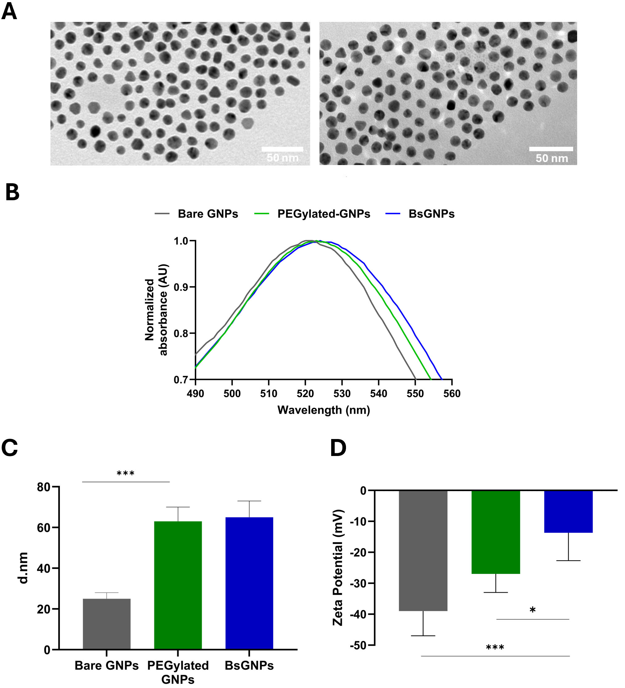
Figure 1. Physicochemical characterization of bispecific gold nanoparticles (BsGNPs). (A) Representative TEM images of bare GNPs (left) and BsGNPs (right) showing uniform, spherical morphology (scale bar = 50 nm); (B) Normalized UV-visible absorption spectra of bare GNPs, PEGylated GNPs, and BsGNPs, demonstrating characteristic red-shifts in the surface plasmon resonance following sequential surface functionalization; (C) Hydrodynamic diameters measured by dynamic light scattering (DLS) after each modification step, indicating increased particle size upon PEGylation and antibody conjugation. (D) Zeta potential measurements showing progressive modulation of surface charge during nanoparticle functionalization. In (C and D), data are presented as mean ± SD of three independent nanoparticle preparations analyzed by one-way ANOVA followed by Tukey’s multiple-comparisons test; *P < 0.05, ***P < 0.001. GNP: gold nanoparticle; PEG: polyethylene-glycol; TEM: transmission electron microscopy; SD: Standard deviation; ANOVA: analysis of variance.

