

fig9
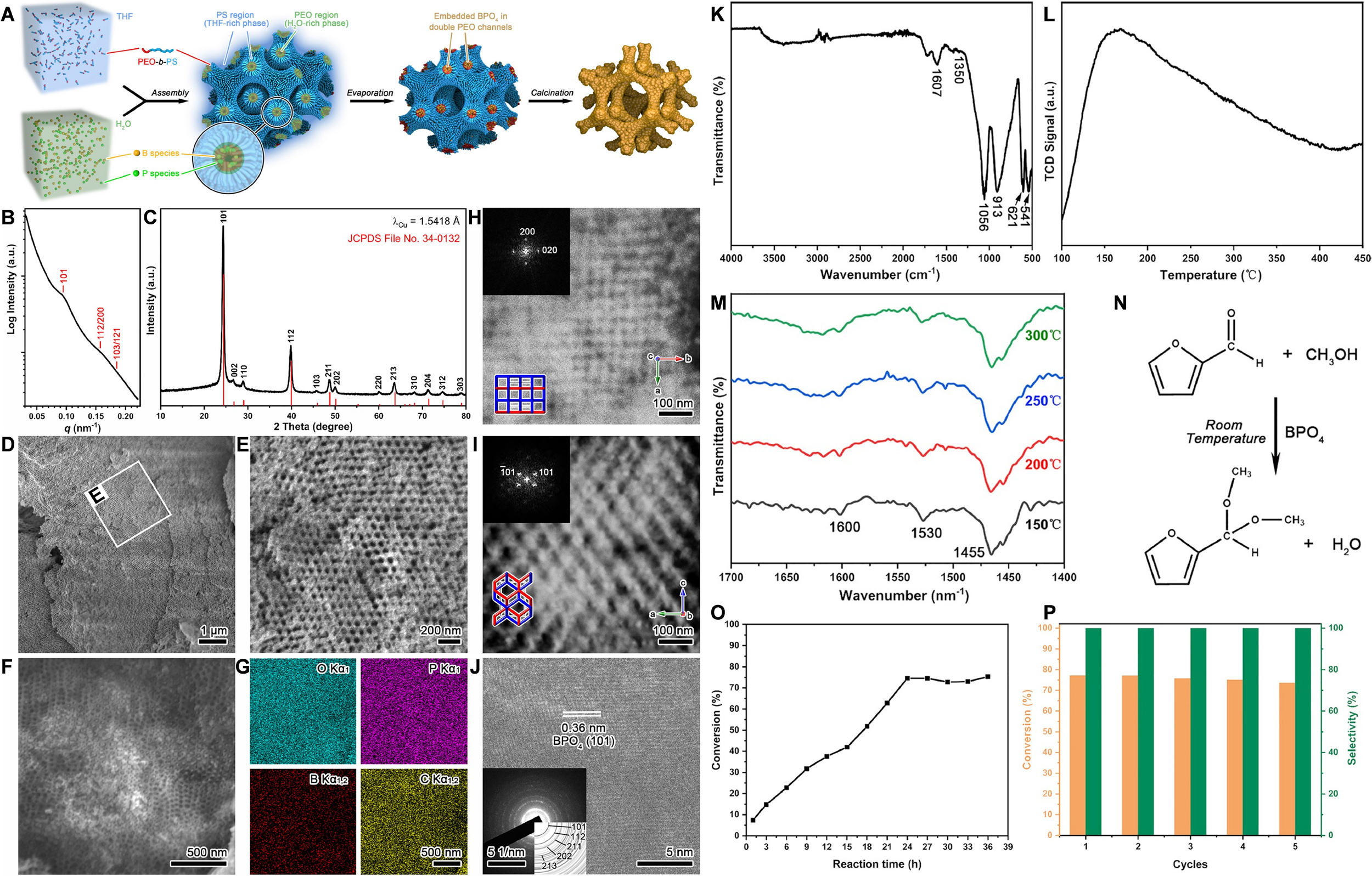
Figure 9. (A) Schematic drawing of the synthetic strategy of crystalline SDD BPO4 scaffold; (B-M) Characterizations of crystalline SDD BPO4 scaffold. (B) SAXS pattern. (C) Wide-angle XRD pattern. (D) Low-magnification SEM image. (E) High-magnification SEM image as shown in the white box in (D). (F and G) EDS mapping. (H and I) STEM images and the corresponding Fourier diffractograms of SDD BPO4 scaffold taken from the [001] and [010] directions, respectively. (J) High-resolution TEM (HRTEM) image of SDD BPO4 scaffold corresponding to the (101) plane of BPO4. The inset shows the selected area electron diffraction with the polycrystalline ring pattern. (K) FTIR spectrum. (L) Temperature-programmed desorption of ammonia profile. (M) Pyridine-adsorption FTIR spectrum; (N) Catalytic acetalization of furfural and methanol to 2-(dimethoxymethyl)furan; (O) Effect of reaction time on the acetalization; (P) Reusability tests of SDD BPO4 for the acetalization. This figure is reproduced with permission[218]. Copyright 2024, Science China Press. THF: Tetrahydrofuran; PEO-b-PS: poly(ethylene oxide)-b-polystyrene; PEO: poly(ethylene oxide); SDD: shifted double diamond; SAXS: small-angle X-ray scattering; XRD: X-ray diffraction; SEM: scanning electron microscopy; STEM: scanning transmission electron microscopy; FTIR: Fourier transform infrared; EDS: energy dispersive X-ray spectroscopy.

