

fig1
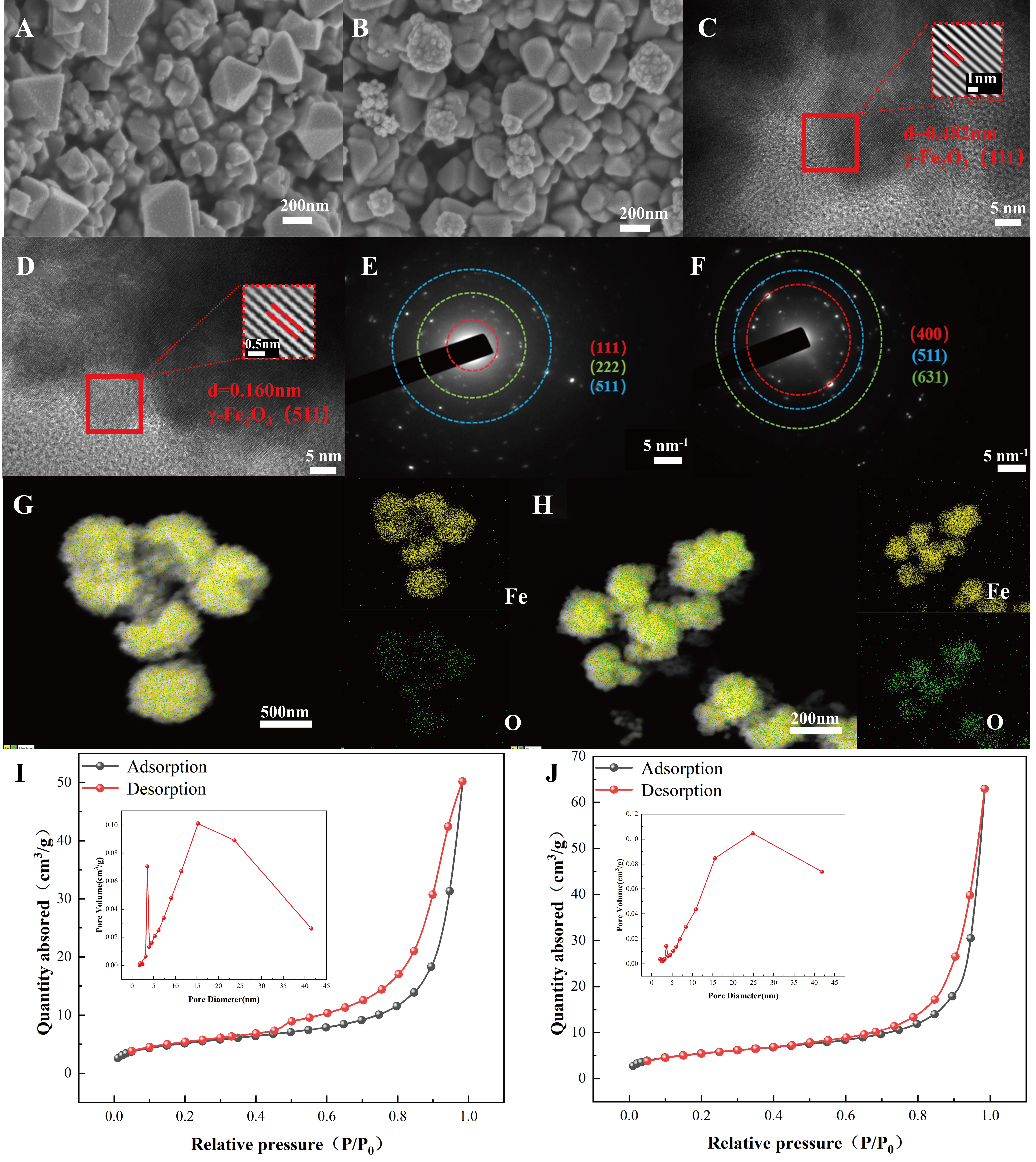
Figure 1. (A and B) SEM images of Fe400 and 15%-Fe400; (C and D) HRTEM images of Fe400 and 15%-Fe400; (E and F) SAED patterns of Fe400 and 15%-Fe400; (G and H) EDS elemental mapping of Fe and O in Fe400 and 15%-Fe400 samples; The BET specific surface area and pore-size distribution of Fe400 and 15%-Fe400 are shown in panels (I and J), respectively. All subfigures are original. Panels (A and D) were directly exported from the SEM instrument; panels (B, C, E and F) were directly exported from the TEM instrument; panels (G and H) were directly exported from the EDS mapping system; panels (I and J) were plotted using Origin 2024. SEM: Scanning electron microscopy; HRTEM: high-resolution transmission electron microscopy; EDS: energy-dispersive X-ray spectroscopy; SAED: selected-area electron diffraction; BET: Brunauer-Emmett-Teller; TEM: transmission electron microscopy.

