

fig4
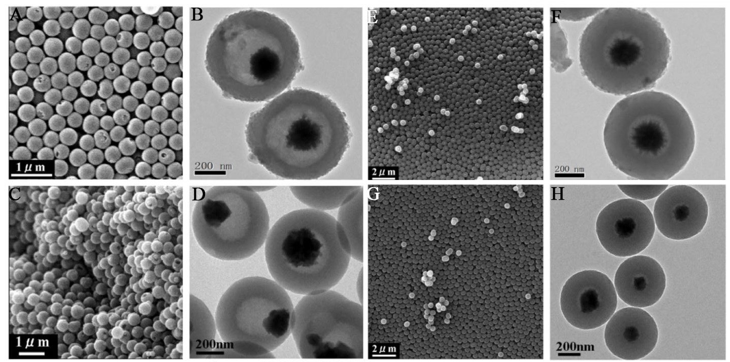
Figure 4. Morphology evolution of Fe3O4@C microspheres obtained with external pre-curing at 100 °C before calcination under nitrogen. (A and B) SEM and TEM images after 5 min of pre-curing; (C and D) SEM and TEM images after 1 h of pre-curing; (E and F) SEM and TEM images after 2.5 h of pre-curing; (G and H) SEM and TEM images after 8 h of pre-curing; (B, D, F and H) TEM images illustrating the structural evolution from yolk-shell to solid core-shell configurations with increasing pre-curing time. SEM: Scanning electron microscopy; TEM: transmission electron microscopy.

