




Eu3+-doped GAGG scintillating transparent ceramics: optimization of optical, thermal, and X-ray imaging performance
 0
0 
Abstract
Scintillators, which are critical for X-ray detection, are widely used in medical imaging, radiation detection, and high-energy physics. In this study, Eu3+-doped Gd3Al3Ga2O12 (GAGG: xEu3+) transparent ceramic scintillators were fabricated using a solid-state reaction method combined with pressureless atmosphere sintering. The effect of Eu3+ concentration on the optical quality, scintillation performance, and thermal stability of the ceramics was systematically investigated, and their X-ray imaging performance was preliminarily evaluated. The as-prepared ceramics exhibited high optical transmittance and a uniform grain-size distribution. Photoluminescence (PL) spectra revealed characteristic Eu3+ emission peaks at approximately 590 nm, corresponding to the dominant 5D0 → 7F1 magnetic dipole transition. The optimal Eu3+ concentration was determined to be 2 at%, and the concentration quenching behavior was predominantly governed by electric dipole-dipole interactions. Furthermore, the GAGG: xEu3+ ceramics demonstrated satisfactory thermal stability and radioluminescence performance, with the radioluminescence intensity reaching its maximum at a doping concentration of 3 at%. The discrepancy between the optimal concentrations obtained from PL and radioluminescence measurements is attributed to the different excitation mechanisms involved. These results indicate that GAGG: xEu3+ transparent ceramics are promising candidates for X-ray detection and imaging applications.
Keywords
INTRODUCTION
Scintillators are functional materials capable of converting high-energy particles or radiation into detectable ultraviolet or visible light[1]. Owing to this unique property, they are widely used in medical imaging, radiation detection, and national security applications[2,3]. During X-ray detection, scintillators absorb incident high-energy radiation and become excited, subsequently emitting fluorescence. The emitted fluorescence is then converted into electrical signals by photodetectors, such as photomultiplier tubes (PMTs), photodiode arrays, and charge-coupled device/complementary metal-oxide-semiconductor (CCD/CMOS) detectors, thereby transforming otherwise invisible high-energy radiation into displayable signals or images for further analysis[4]. Therefore, scintillators play a crucial role in X-ray detection systems.
Garnet-type scintillation materials have attracted considerable attention owing to their excellent optical transparency, compositional flexibility, and tunable luminescence properties[5]. Gd3Al3Ga2O12 (GAGG)-based ceramics, which belong to the cubic garnet crystal system, are particularly promising for X-ray detection due to their high density (~6.7 g·cm-3), large effective atomic number (Zeff = 55), strong absorption capability for high-energy photons, and high light yield[6].
Research on GAGG-based materials began with single-crystal forms. Kuwano et al.[7] first grew GAGG: Nd single crystals and demonstrated their potential for laser materials. Kamada et al.[8,9] successfully fabricated
To overcome the limitations associated with single crystals, GAGG-based ceramics have attracted increasing attention owing to their shorter fabrication cycles, lower production costs, ability to achieve homogeneous high-concentration doping, and suitability for large-scale manufacturing[13]. Kanai et al.[6] were the first to synthesize GAGG: Ce ceramics via a solid-state reaction method and investigated the effect of compositional deviation on afterglow, although the resulting samples exhibited poor optical quality. Seeley et al.[14] prepared a series of GYGAG: Ce ceramics and systematically examined the effects of calcination and sintering atmospheres on their scintillation performance. Yanagida et al.[15] fabricated GAGG: Ce ceramics with a thickness of 1 mm and a doping concentration of 1 at%, achieving scintillation performance that partially exceeded that of single crystal counterparts, although the optical transmittance remained low. Wu et al.[16] compared the scintillation properties of Ce: (Lu,Gd)3(Ga,Al)5O12 ceramics and GAGG: Ce single crystals. The results showed that ceramics exhibited a higher light output (48,200 Ph/MeV) than single crystals
Studies have demonstrated that incorporation of rare-earth ions (RE3+) into host lattices can effectively tailor their optical and electronic properties, thereby broadening their functional applications[17,18]. Liu et al.[19,20] prepared GAGG: Ce ceramics with a luminous efficiency of 388 lm·W-1, making them suitable for
In this study, GAGG: xEu3+ (x = 1~4 at%) transparent ceramics were fabricated via an optimized solid-state reaction route followed by pressureless sintering. The effects of Eu3+ content on the optical transmittance, photoluminescence, and scintillation properties of the ceramics were systematically investigated. In addition, the luminescence thermal stability and defect-related characteristics were comprehensively analyzed through temperature-dependent emission spectroscopy and thermoluminescence measurements. Based on their excellent optical quality, the radioluminescence behavior and potential applicability of GAGG: Eu3+ ceramics in X-ray imaging were also evaluated.
MATERIALS AND METHODS
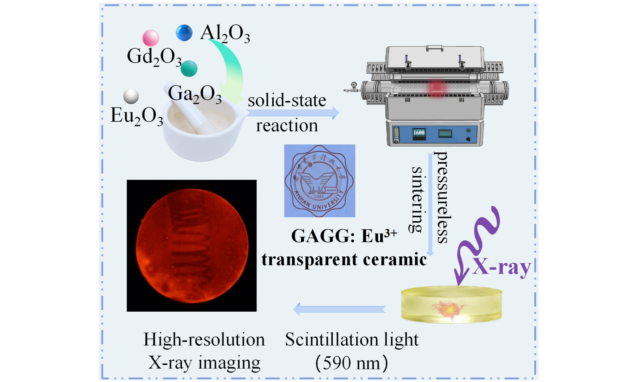
(EuxGd1-x)3Al3Ga2O12 scintillation ceramics (abbreviated as GAGG: xEu3+, x = 1, 2, 3, 4 at%) were fabricated using a solid-state reaction method combined with pressureless sintering. High-purity raw oxides, including gadolinium oxide (Gd2O3, 99.9%, Macklin), alumina (Al2O3, 99.9%, Aladdin), gallium oxide (Ga2O3, 99.99%, Aladdin), and europium oxide (Eu2O3, 99.9%, Aladdin), were accurately weighed as starting materials. The raw materials were milled in a planetary ball mill for 12 h. After drying the slurry at 80 °C, the resulting powder was sieved through a 200-mesh screen. The sieved powder was subsequently compacted into pellets by uniaxial pressing and further densified via cold isostatic pressing at 250 MPa. The obtained green body was then calcined at 1,000~1,300 °C for 4 h in an air atmosphere. After calcination, the samples were sintered at 1,600~1,620 °C for 10 h in an oxygen atmosphere to obtain dense ceramics.
The phase composition of the samples was analyzed by X-ray powder diffraction (XRD, D8 Advance, Bruker, Germany). The microstructural and elemental distribution were examined using transmission electron microscopy (TEM, F200X, Thermo Fisher Scientific, USA) and scanning electron microscopy (SEM, JSM-7500F, JEOL, Japan). The optical transmittance spectra were measured using an ultraviolet-visible-near-infrared (UV-Vis-NIR) spectrophotometer (Lambda950, PerkinElmer, USA). Photoluminescence excitation (PLE) and photoluminescence (PL) spectra were recorded using a fluorescence spectrometer (FLS-1000, Edinburgh Instruments, UK). Radioluminescence spectra were obtained using an X-ray excitation and emission spectrometer (XEL, OmniFLUO990-Xray, Zhuoli, China). The defect states of the ceramics were evaluated using thermally stimulated luminescence (TSL) measurements using a three-dimensional thermoluminescence spectrometer (TOSL-3DS, Radiation Technology, China).
RESULTS AND DISCUSSION
Phase and morphology analysis
The XRD patterns of the GAGG: xEu3+ transparent ceramics, shown in Figure 1A, confirm that all diffraction peaks can be well indexed to the standard Gd3Al3Ga2O12 (PDF#46-0448), with no detectable secondary phases. This result indicates that the incorporation of Eu3+ ions does not alter the garnet crystal structure of the host lattice. To further verify the phase purity and structural characteristics, Rietveld refinements were performed using GSAS-Ⅱ software[26]. The corresponding refinement profiles are presented in Figure 1B-E. The obtained reliability factors (Rwp = 5.72%~8.47%) demonstrate satisfactory fitting quality. The refined structural parameters are summarized in Figure 1F and Table 1. As the Eu3+ concentration increases, both the lattice constant and unit-cell volume exhibit a slight increase, suggesting that Eu3+ ions are successfully incorporated into the lattice through substitution at the Gd3+ sites. The bulk densities (D) of GAGG: xEu3+ ceramics, measured by the Archimedes method, are approximately 6.4 g·cm-3, corresponding to more than 98% of the theoretical density. These results indicate that highly densified ceramics were successfully obtained.
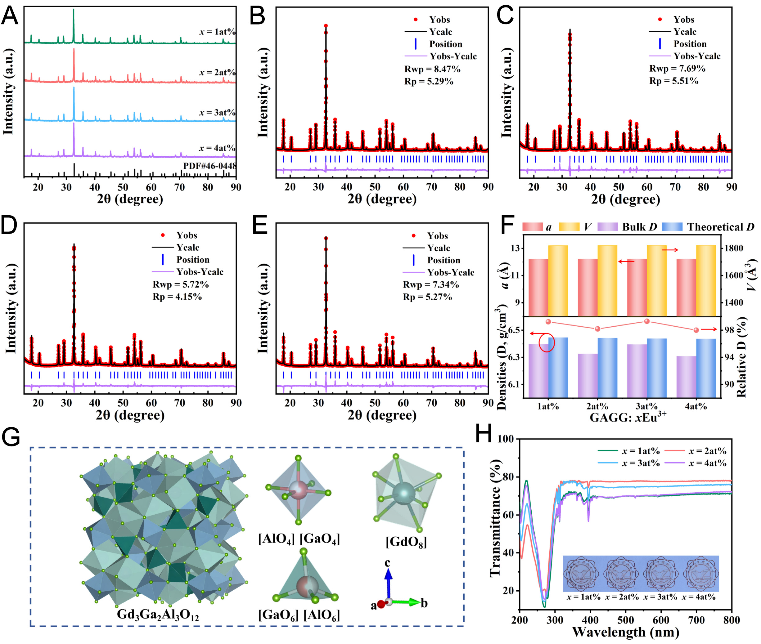
Figure 1. (A) XRD patterns, (B-E) Rietveld refinement results, (F) lattice parameters and densities, (G) schematic crystal structure, (H) optical transmittance spectra and photographs (inset) of GAGG: xEu3+ (x = 1~4 at%) ceramics. GAGG: Gd3Al3Ga2O12.
Lattice and refinement parameters of GAGG: xEu3+ ceramics
| Parameters | x = 1 at% | x = 2 at% | x = 3 at% | x = 4 at% |
| R p (%) | 5.29 | 5.51 | 4.15 | 5.27 |
| R wp (%) | 8.47 | 7.69 | 5.72 | 7.34 |
| a = b = c (Å) | 12.213 | 12.214 | 12.215 | 12.216 |
| V (Å3) | 1,821.659 | 1,822.106 | 1,822.554 | 1,823.002 |
| Density (g·cm-3) | 6.446 | 6.443 | 6.439 | 6.437 |
The schematic illustration of the GAGG crystal structure is presented in Figure 1G. GAGG crystallizes in a cubic garnet structure with space group Oh10-Ia3d. In this structure, Gd3+ ions occupy the dodecahedral
As shown in Figure 1H, the polished GAGG: xEu3+ transparent ceramics (thickness = 1 mm) exhibit excellent transparency without visible defects, confirming good optical quality. The prepared ceramics demonstrate optical transmittance values of approximately 70% within the 400-800 nm range. Among the investigated samples, the ceramic doped with the 2 at% Eu3+ exhibits the highest value of 77%, which is comparable to that reported for GAGG single crystals. Several sharp absorption peaks are observed in the visible region around 394, 466, and 527 nm, which correspond to Eu3+-related 4f-4f transitions[29].
To further investigate the influence of Eu3+ doping on the microstructural evolution of GAGG ceramics, polished samples were thermally etched at 1,300 °C in air. The resulting SEM micrographs are shown in Figure 2. All samples exhibit dense microstructures with well-defined grain boundaries, and no obvious pores or secondary phases are observed. These results indicate a high degree of densification, which is consistent with the XRD and density measurements discussed above. To quantitatively evaluate the microstructural characteristics, grain-size distributions were analyzed, as in the inset of Figure 2. The average grain size shows a non-monotonic dependence on the Eu3+ concentration, increasing initially and then decreasing. The maximum average grain size of 8.32 μm occurs at 2 at% Eu3+, which also exhibits the highest optical transmittance. This enhancement is attributed to reduced light scattering at grain boundaries due to grain growth, effectively lowering optical losses and improving transparency in the ceramics.
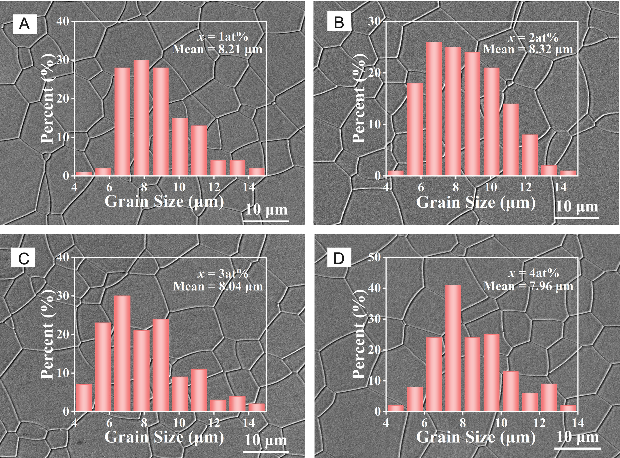
Figure 2. SEM images and grain size distributions of GAGG: xEu3+ transparent ceramics. GAGG: Gd3Al3Ga2O12; SEM: scanning electron microscopy.
The microstructure of the optimal GAGG: 2 at% Eu3+ ceramic was further characterized by TEM. The selected area electron diffraction (SAED) pattern shown in Figure 3A exhibits well-defined diffraction spots along the [211] zone axis, indicating the high crystallinity of the GAGG phase.
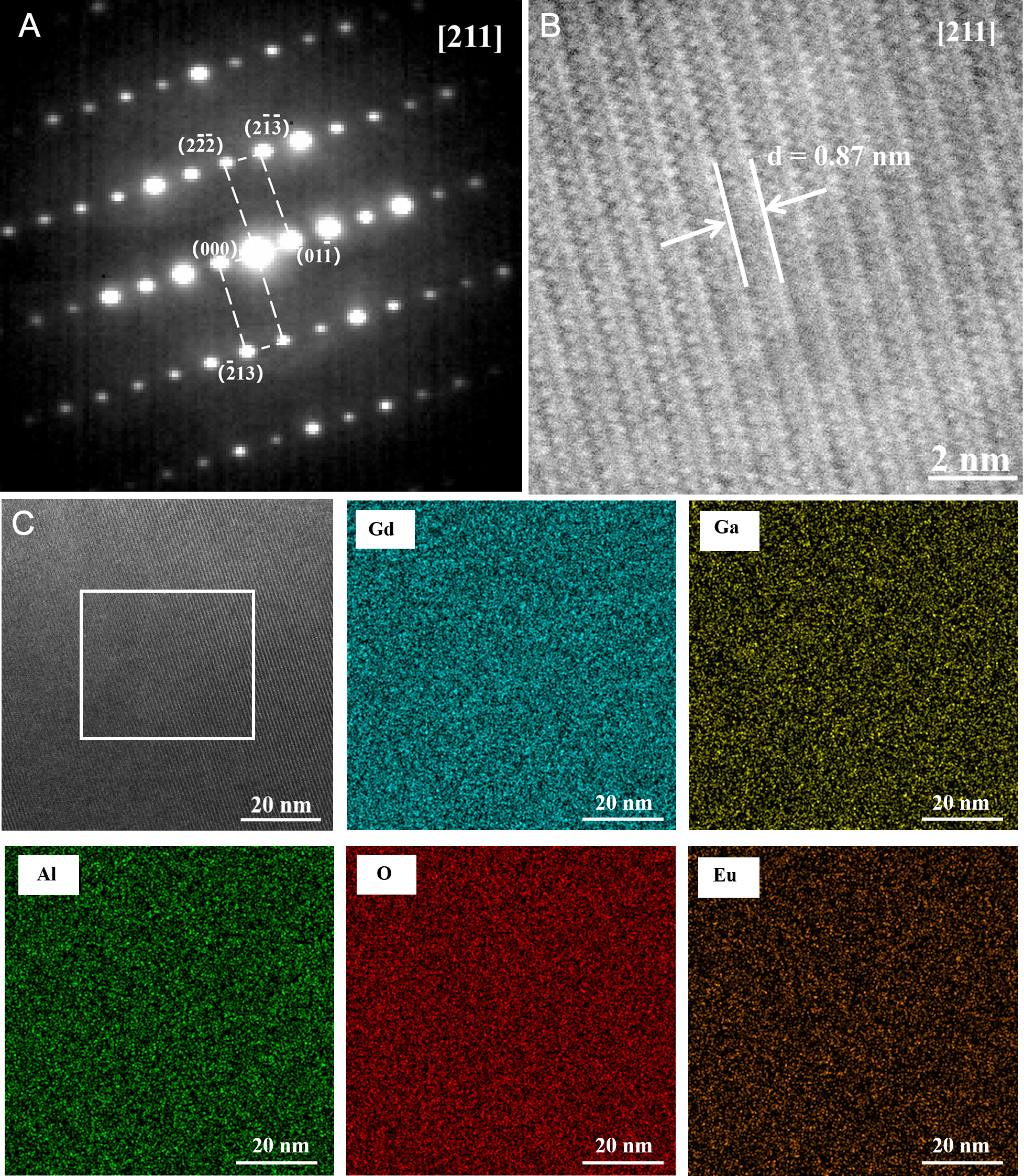
Figure 3. TEM characterization of GAGG: 2 at% Eu3+ ceramic: (A) SAED pattern, (B) HAADF-STEM image, and (C) EDS elemental mapping. GAGG: Gd3Al3Ga2O12; TEM: transmission electron microscopy; HAADF-STEM: high-angle annular dark-field scanning transmission electron microscopy; SAED: selected area electron diffraction; EDS: energy dispersive spectroscopy.
The high-angle annular dark-field scanning transmission electron microscopy (HAADF-STEM) image acquired along the [211] direction is presented in Figure 3B. Clear lattice fringes can be observed, demonstrating the excellent crystallinity of the ceramic. The measured interplanar spacing is 0.87 nm, corresponding to the (
Optical properties
The PLE spectrum of the GAGG: 2 at% Eu3+ ceramic is shown in Figure 4A. Five distinct excitation bands are observed in 350-500 nm, which can be assigned to the following intra-4f transitions of Eu3+ ions: 7F0 → 5D4, 7F0 → 5L7, 7F0 → 5L6, 7F0 → 5D3, and 7F0 → 5D2 [30]. The PL spectra of GAGG: xEu3+ ceramics with different Eu3+ concentrations are presented in Figure 4B. A dominant orange-red peak is observed at 590 nm, corresponding to the 5D0 → 7F1 magnetic dipole transition of Eu3+ ions. Additional emission peaks located at 610 and 630 nm are attributed to the 5D0 → 7F2 electric dipole transition, while the weaker emission bands at 653, 695, and 710 nm originate from the 5D0 → 7F3 and 5D0 → 7F4 transitions, respectively[31]. Based on the Eu3+ emission spectrum, a schematic energy-level diagram illustrating the photoluminescence process is proposed, as shown in Figure 4C.
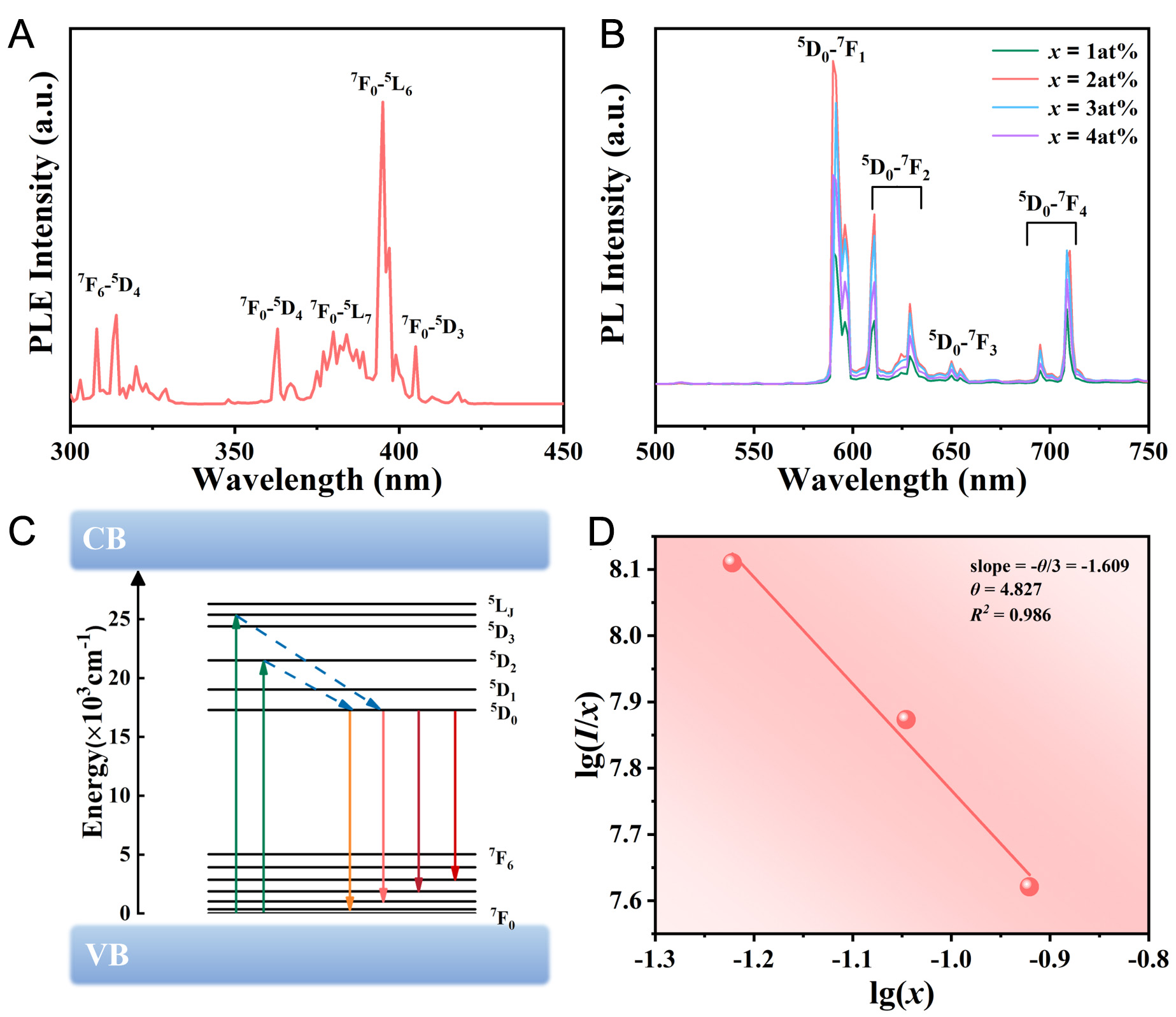
Figure 4. (A) PLE spectrum of GAGG: 2at% Eu3+ ceramic; (B) Emission spectra of GAGG: xEu3+; (C) Schematic diagram of the Eu3+ photo-luminescence energy-level transitions; (D) Relationship between lg(I/x) and lg(x) for GAGG: xEu3+ ceramics. GAGG: Gd3Al3Ga2O12; PLE: photoluminescence excitation.
Studies have shown that the luminescence behavior of Eu3+ ions is highly sensitive to their local crystal environment. In the PL spectrum, the 5D0 → 7F1 magnetic dipole transition remains dominant, indicating that Eu3+ ions occupy sites with relatively high symmetry in the host lattice[32] The emission intensity initially increases with increasing Eu3+ concentration and reaches a maximum at 2 at%, beyond which concentration quenching becomes apparent. This quenching behavior is likely associated with reduced interionic distance between Eu3+ ions at higher doping concentrations, which facilitates energy transfer among activator ions and increases the probability of excitation energy being trapped by defect states or impurity levels. The trapped energy may subsequently be dissipated through non-radiative relaxation or transferred to impurity ions, thereby reducing the luminescence efficiency of Eu3+ ions[33,34]. The non-radiative energy transfer between rare earth ions can occur through two primary mechanisms: exchange interaction and electric multipole interaction[35]. According to the Blasse model[36], the critical distance (Rc) for doped ions can be calculated using Equation (1):
Where (Xc) is the critical doping concentration; (V) is the unit-cell volume and (N) represents the number of available substitutional sites per unit cell. For the GAGG ceramics, a = b = c = 12.214 Å, N = 8, and V = abc = 1,822.106 Å3, the calculated (Rc) is 22.15 Å. When Rc < 5 Å, energy transfer is predominantly governed by exchange interaction. The calculated (Rc) is significantly larger than 5 Å, indicating that the concentration quenching behavior is primarily attributed to electric multipolar interaction rather than exchange interaction[37]. To further clarify the electric multipolar interaction mechanism, Dexter’s theory[38] was employed to analyze the relationship between the luminescence intensity (I) and the Eu3+ concentration (x), which can be expressed by Equation (2):
where (I) represents the luminous intensity of the 5D0 → 7F1 transition, while (C) and (θ) are constants. The type of multipolar interaction can be identified according to the value of (θ), where θ = 6, 8, and
To evaluate the luminescence stability of GAGG: xEu3+ ceramics, temperature-dependent PL spectra of the GAGG: 2 at% Eu3+ sample were recorded over the temperature range of 300~550 K, as shown in Figure 5A. The luminescence intensity of Eu3+ gradually decreases with increasing temperature and retains 50.28% of its initial intensity when the temperature reaches 550 K. The decrease in luminescence intensity with increasing temperature is attributed to thermal quenching. As the temperature rises, electrons in the excited states of the luminescence centers can be thermally activated to higher vibrational levels and subsequently return to the ground state through non-radiative relaxation processes, leading to a reduction in luminescence intensity[41]. The thermal quenching is closely related to the strength of phonon-electron coupling[42]. The activation energy (ΔEa) for thermal quenching was calculated by fitting the temperature-dependent luminescence intensity using the Arrhenius[43] Equation (3):
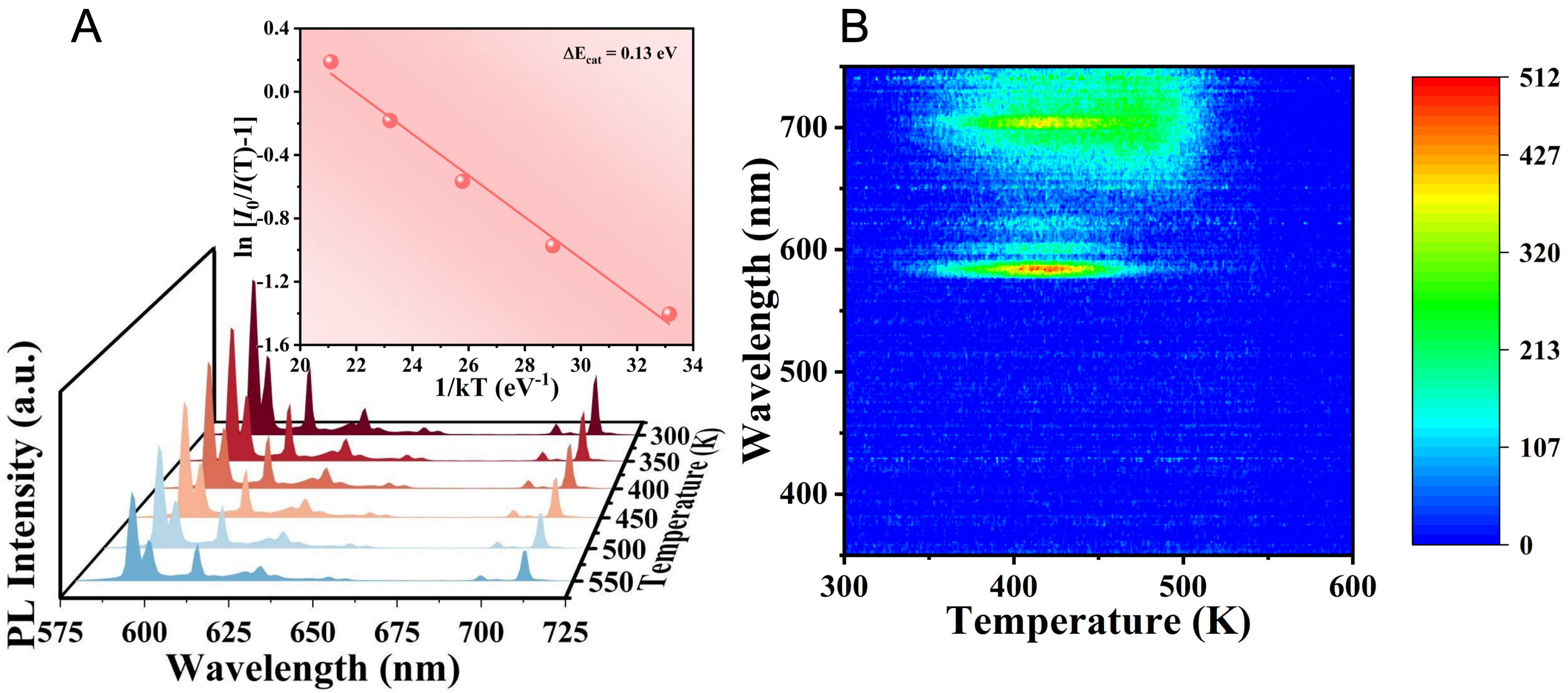
Figure 5. GAGG: 2at% Eu3+ ceramic: (A) Temperature-dependent PL spectra, and the Arrhenius relationship between ln[I0/I(T)-1] and 1/kT; (B) Thermally stimulated luminescence spectra. GAGG: Gd3Al3Ga2O12; PL: photoluminescence.
where (I0) is the initial luminescence intensity, I(T) is the luminescence intensity at a given temperature; (B) is a constant and (k) is the Boltzmann constant. The fitted curve for ΔEa across the temperature range of 300-550 K is shown in the inset of Figure 5A. The fitting process yields ΔEa = 0.13 eV. A higher ΔEa indicates enhanced stability, as it corresponds to a higher non-radiative energy barrier that must be overcome during the transition from the excited state to the ground state. This elevated barrier effectively suppresses nonradiative relaxation, thereby confirming the excellent thermal stability of the GAGG: 2 at% Eu3+ ceramic[44].
To further examine the influence of defect states on the thermal stability of radioluminescence in ceramics, TSL measurements were performed on the 2 at% Eu3+ ceramic, and the corresponding spectra are presented in Figure 5B. Unlike the sharp Eu3+ emission lines observed in the PL spectra, the TSL spectrum exhibits a broad red emission band in the 650-750 nm range, indicating that TSL process does not simply follow the PL emission mechanism. During TSL measurements, thermally released charge carriers undergo recombination through defect-related or activator-related radiative pathways. Therefore, the broad emission band centered at approximately 700 nm is likely associated with defect-mediated recombination processes within the GAGG lattice, involving oxygen vacancies, antisite defects, and local lattice distortions, together with a possible contribution from the weak Eu3+ 5D0 → 7F4 transition. Continuous trap distributions are observed over the temperature range of 340~550 K. This is likely due to the stronger lattice vibrations at higher temperatures, which cause the release of electrons (or holes) from traps, leading to light emission. Their release difficulty depends on both the depth of the trap and the temperature of the material. Shallow trap levels can release charge carriers at relatively low temperatures, whereas deep trap levels require higher temperatures for carrier release. The relationship between the temperature and the trap depth (E) can be calculated using Equation (4)[45]:
where (Tm) is the temperature corresponding to the TSL peak, (s) is the frequency factor, (β) is the heating rate, and (k) is the Boltzmann constant. In this measurement, (β) was fixed at 1 °C/s. Considering that the frequency factor (s) typically falls within the range of 1010~1014, a characteristic value of 1012 was adopted to simplify the calculation while maintaining reasonable accuracy. The calculated trap depths of the GAGG:
Scintillation properties
To evaluate the high-energy X-ray detection capability of GAGG: xEu3+ transparent ceramics, as shown in Figure 6A. The GAGG: Eu ceramics exhibit X-ray absorption capability comparable to that of commercial scintillators. The scintillation properties were further characterized by X-ray-excited emission spectroscopy, and the corresponding spectra for different Eu3+ concentrations are presented in Figure 6B. The radioluminescence spectra are generally consistent with the PL spectra. Among the prepared GAGG: xEu3+ ceramics, the highest luminescence intensity is observed at 3 at%. When the Eu3+ concentration exceeds this value, concentration quenching becomes apparent. The quenching phenomenon observed under X-ray excitation differs from that observed in the PL spectra. The discrepancy in the optimal Eu3+ concentration between PL and X-ray-excited luminescence can be attributed to the different excitation and energy-transfer pathways. Under UV excitation, Eu3+ ions are mainly excited through localized 4f-4f transitions, and the emission intensity is strongly influenced by concentration-dependent energy migration among Eu3+ ions. In contrast, under X-ray excitation, the scintillation process involves X-ray absorption by the host lattice, generation of secondary electrons and holes, carrier transport, defect trapping and release, and subsequent energy transfer to Eu3+ emission centers. Therefore, the scintillation performance is jointly influenced by Eu3+ concentration, defect states, and optical transparency[39]. The CIE 1931 chromaticity coordinates derived from X-ray-excited emission spectra are shown in Figure 6C. All GAGG: xEu3+ ceramics exhibit red emission in the visible region, indicating their potential for optoelectronic applications.
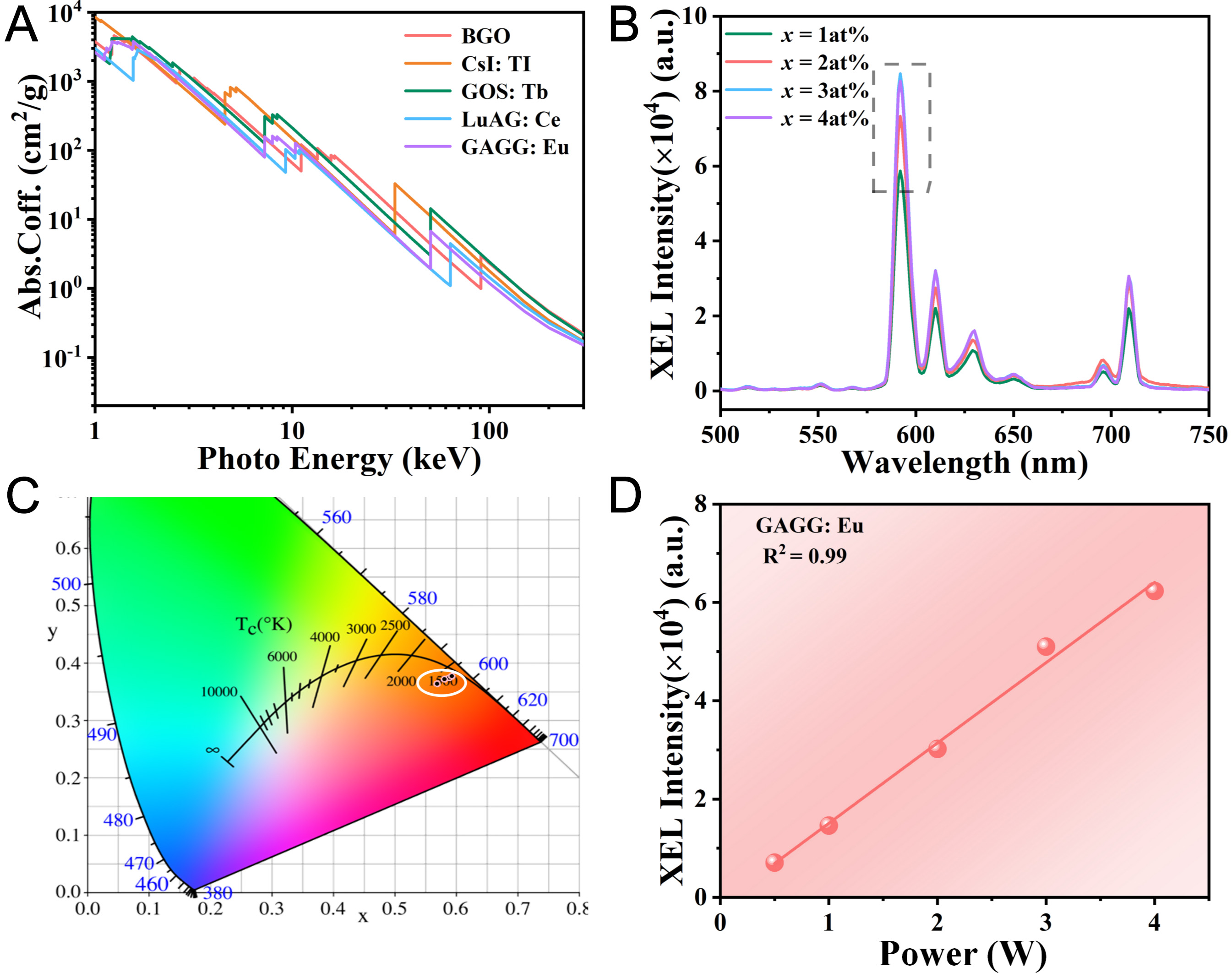
Figure 6. (A) Comparison of X-ray absorption coefficients among BGO, CsI: Tl, GOS: Tb, LuAG: Ce and GAGG: Eu3+; (B) X-ray-excited emission spectra, and (C) CIE chromaticity coordinates of GAGG: xEu3+ transparent ceramics; (D) Relationship between X-ray power and luminescence intensity of GAGG: 2at% Eu3+ ceramic. GAGG: Gd3Al3Ga2O12; CIE: International Commission on Illumination; XEL: X-ray excited luminescence; BGO: Bi4Ge3O12, Bismuth Germanate; CsI:Tl: thallium-doped cesium iodide; GOS:Tb: Gd2O2S:Tb, terbium-doped gadolinium oxysulfide; LuAG:Ce: Lu3Al5O12:Ce, cerium-doped lutetium aluminum garnet.
In addition, the dependence of the radioluminescence intensity of the GAGG: 2 at% Eu3+ ceramic on X-ray tube power was investigated, as shown in Figure 6D. The results show that the luminescence intensity increases steadily with increasing X-ray power. This enhancement is attributed to the increased flux of incident X-ray photons at higher power levels. These photons interact with the scintillator through processes such as the photoelectric effect and Compton scattering, generating a larger number of secondary electrons. The generated secondary electrons subsequently transfer energy to the luminescent centers, resulting in enhanced visible light emission. These findings suggest that the prepared ceramic scintillator holds promise for applications in X-ray detection.
To explore the potential of the prepared GAGG: xEu3+ transparent ceramics as scintillation layers for X-ray imaging, an X-ray imaging system was employed, as illustrated in Figure 7A. In this system, X-rays generated by an X-ray tube penetrate the object and subsequently irradiate the ceramic scintillator, where the incident X-rays are converted into visible photons. The emitted visible light is then captured by a digital camera and processed to generate the corresponding image. In the experiment, the GAGG: 2 at% Eu3+ transparent ceramic scintillator was selected for imaging, as shown in Figure 7B. A capsule containing a miniature spring was used as the imaging target [Figure 7C]. Under a tube voltage of 50 kV, the X-ray intensity was varied by adjusting the tube current from 20 μA to 80 μA. As shown in Figure 7D, both the image brightness and clarity improved with increasing current. At lower currents of 20 μA, the X-ray intensity was insufficient, resulting in a blurred image of the spring. As the current increased, the image quality gradually improved. At 80 μA, the scintillator exhibited the highest imaging brightness, and the internal structure of spring could be clearly distinguished. These preliminary imaging results demonstrate the feasibility of using GAGG: Eu3+ transparent ceramics as scintillation converters for X-ray imaging.
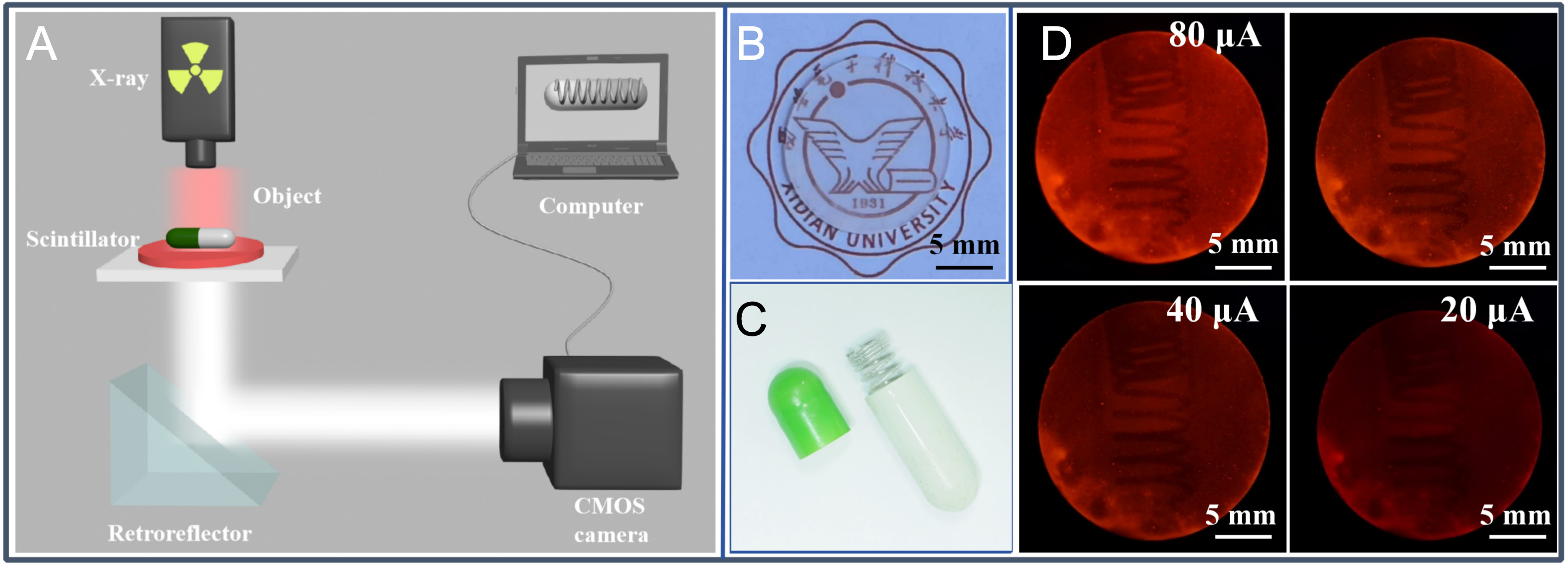
Figure 7. (A) Schematic diagram of the X-ray imaging system; (B) Photograph of GAGG: 2 at% Eu3+ ceramic, and (C) photograph of the capsule containing the sub-piece spring under ambient light; (D) X-ray images captured under different X-ray tube currents. GAGG: Gd3Al3Ga2O12.
CONCLUSIONS
In summary, a series of GAGG: xEu3+ (x = 1~4 at%) transparent ceramic scintillators with narrow-band red emission were successfully fabricated by a solid-state reaction method combined with pressureless sintering. The prepared ceramics exhibited excellent optical properties, with a maximum transmittance of 77% in the 400~800 nm. Photoluminescence investigations demonstrated efficient Eu3+ emission and good thermal stability over a wide temperature range. Under X-ray excitation, the GAGG: xEu3+ ceramics exhibited intense radioluminescence, and their emission bands exhibited favorable spectral matching with silicon-based photodetectors, indicating their potential for radiation detection applications. The X-ray imaging experiments performed using a self-developed imaging system successfully revealed the internal structure of target objects, highlighting the capability of these ceramic scintillators for X-ray imaging. Overall, the GAGG: Eu3+ transparent scintillation ceramics, featuring narrow-band red emission, high transmittance, excellent thermal stability, and effective X-ray imaging performance, represent promising candidates for future applications in X-ray detection and imaging technologies.
DECLARATIONS
Authors’ contributions
Conceived and designed the experiment, writing-original draft: Dong, G.; Yang, X.
Performed the experiments and recorded the results: Yang, X.; Bai, R.; Su, H.
Experimental testing and data processing: Yang, X.; Lv, C.; Zhang, B.
Supervision & conceptualization & data curation: Dong, G.; Liu, X.; Wang, T.; Jie, W.
Availability of data and materials
The original contributions presented in this study are included in the article. Further inquiries can be directed to the corresponding author.
AI and AI-assisted tools statement
Not applicable.
Financial support and sponsorship
Supported by the Key Research and Development Program of Shaanxi (2025GH-YBXM-047), the Technology Innovation Leading Program of Shaanxi (2025QCY-KXJ-002), the Fundamental Research Funds for the Central Universities (QTZX26014, ZYTS24120), and the State Key Laboratory of Solidification Processing at NWPU (SKLSP202213).
Conflicts of interest
Dong, G. is a Junior Editorial Board Member of the journal Microstructures, but was not involved in any steps of editorial processing, notably including reviewer selection, manuscript handling, and decision-making, while the other authors have declared that they have no conflicts of interest.
Ethical approval and consent to participate
Not applicable.
Consent for publication
Not applicable.
Copyright
© The Author(s) 2026.
REFERENCES
1. Nikl, M.; Yoshikawa, A. Recent R&D trends in inorganic single-crystal scintillator materials for radiation detection. Adv. Opt. Mater. 2015, 3, 463-81.
2. Ma, J.; Zhu, W.; Lei, L.; et al. Highly efficient NaGdF4:Ce/Tb nanoscintillator with reduced afterglow and light scattering for high-resolution X-ray Imaging. ACS. Appl. Mater. Interfaces. 2021, 13, 44596-603.
3. Wang, T.; Hu, S.; Ji, T.; et al. High-temperature X-Ray imaging with transparent ceramics scintillators. Laser. Photonics. Rev. 2024, 18, 2300892.
4. Ou, X.; Qin, X.; Huang, B.; et al. High-resolution X-ray luminescence extension imaging. Nature 2021, 590, 410-5.
5. Zhu, D.; Nikl, M.; Chewpraditkul, W.; Li, J. Development and prospects of garnet ceramic scintillators: a review. J. Adv. Ceram. 2022, 11, 1825-48.
6. Kanai, T.; Satoh, M.; Miura, I. Characteristics of a nonstoichiometric Gd3+δ(Al,Ga)5-δO12:Ce garnet scintillator. J. Am. Ceram. Soc. 2008, 91, 456-62.
7. Kuwano, Y.; Saito, S.; Hase, U. Crystal growth and optical properties of Nd: GGAG. J. Cryst. Growth. 1988, 92, 17-22.
8. Kamada, K.; Endo, T.; Tsutumi, K.; et al. Composition engineering in cerium-doped (Lu,Gd)3(Ga,Al)5O12 single-crystal scintillators. Cryst. Growth. Des. 2011, 11, 4484-90.
9. Kamada, K.; Yanagida, T.; Endo, T.; et al. 2Inch diameter single crystal growth and scintillation properties of Ce:Gd3Al2Ga3O12. J. Cryst. Growth. 2012, 352, 88-90.
10. Kunikata, T.; Watanabe, K.; Kantuptim, P.; et al. Dopant concentration dependence on optical and scintillation properties of Eu-doped Gd3Al2Ga3O12 single crystals. Jpn. J. Appl. Phys. 2024, 63, 01SP18.
11. Endo, Y.; Kunikata, T.; Hayashi, N.; et al. Optimization of Al/Ga ratio for light yield of Eu-doped GAGG single-crystal scintillators. Opt. Mater. 2026, 177, 118181.
12. Lee, G.; Struebing, C.; Wagner, B.; et al. Synthesis and characterization of a BaGdF5:Tb glass ceramic as a nanocomposite scintillator for X-ray imaging. Nanotechnology 2016, 27, 205203.
13. Zhao, W.; Xu, T.; Wang, Y.; et al. Sintering mechanism and optical properties of (Lu1-xScxEu0.05)2O3 scintillation ceramics. J. Eur. Ceram. Soc. 2024, 44, 4631-8.
14. Seeley, Z. M.; Cherepy, N. J.; Payne, S. A. Expanded phase stability of Gd-based garnet transparent ceramic scintillators. J. Mater. Res. 2014, 29, 2332-7.
15. Yanagida, T.; Kamada, K.; Fujimoto, Y.; Yagi, H.; Yanagitani, T. Comparative study of ceramic and single crystal Ce:GAGG scintillator. Opt. Mater. 2013, 35, 2480-5.
16. Wu, Y.; Luo, Z.; Jiang, H.; Meng, F.; Koschan, M.; Melcher, C. L. Single crystal and optical ceramic multicomponent garnet scintillators: a comparative study. Nucl. Instrum. Methods. Phys. Res. Sect. A. 2015, 780, 45-50.
17. Drdlikova, K.; Klement, R.; Hadraba, H.; Drdlik, D.; Galusek, D.; Maca, K. Luminescent Eu3+-doped transparent alumina ceramics with high hardness. J. Eur. Ceram. Soc. 2017, 37, 4271-7.
18. Zhang, C.; Shi, Y.; Wang, X.; Qi, J.; Lu, T. Mechanical, photoluminescent properties and energy transfer mechanism of highly transparent (Y0.99-xGdxSm0.01)2O3 ceramics for scintillator applications. J. Eur. Ceram. Soc. 2024, 44, 1783-94.
19. Liu, Y.; Liu, S.; Sun, P.; et al. Transparent ceramics enabling high luminous flux and efficacy for the next-generation high-power LED light. ACS. Appl. Mater. Interfaces. 2019, 11, 21697-701.
20. Liu, S.; Sun, P.; Liu, Y.; et al. Warm white light with a high color-rendering index from a single Gd3Al4GaO12:Ce3+ transparent ceramic for high-power LEDs and LDs. ACS. Appl. Mater. Interfaces. 2019, 11, 2130-9.
21. Dimitrakopoulos, A.; Michail, C.; Valais, I.; Fountos, G.; Kandarakis, I.; Kalyvas, N. Experimental evaluation of GAGG:Ce crystalline scintillator properties under X-ray radiation. Crystals 2025, 15, 590.
22. Rodríguez-garcía, M. M.; Ciric, A.; Ristic, Z.; Williams, J. A. G.; Dramićanin, M. D.; Evans, I. R. Narrow-band red phosphors of high colour purity based on Eu3+-activated apatite-type Gd9.33(SiO4)6O2. J. Mater. Chem. C. 2021, 9, 7474-84.
23. Hora, D. A.; Andrade, A. B.; Ferreira, N. S.; Teixeira, V. C.; dos S. Rezende, M. V. Effect of the PVA (polyvinyl alcohol) concentration on the optical properties of Eu-doped YAG phosphors. Opt. Mater. 2016, 60, 495-500.
24. Xu, J.; Ueda, J.; Tanabe, S. Design of deep-red persistent phosphors of Gd3Al5-xGaxO12:Cr3+ transparent ceramics sensitized by Eu3+ as an electron trap using conduction band engineering. Opt. Mater. Express. 2015, 5, 963.
25. Gerasymov, I.; Nepokupnaya, T.; Boyarintsev, A.; et al. GAGG:Ce composite scintillator for X-ray imaging. Opt. Mater. 2020, 109, 110305.
26. Toby, B. H.; Von Dreele, R. B. GSAS-II: the genesis of a modern open-source all purpose crystallography software package. J. Appl. Cryst. 2013, 46, 544-9.
27. Hua, H.; Feng, S.; Ouyang, Z.; et al. YAGG:Ce transparent ceramics with high luminous efficiency for solid-state lighting application. J. Adv. Ceram. 2019, 8, 389-98.
28. Korzhik, M.; Borisevich, A.; Fedorov, A.; et al. The scintillation mechanisms in Ce and Tb doped (GdxY1-x)Al2Ga3O12 quaternary garnet structure crystalline ceramics. J. Lumin. 2021, 234, 117933.
29. Feng, G.; Wu, Y.; Lu, H.; Zhang, R.; Wang, S.; Wu, S. Eu-doped (Y0.85-xLa0.15)2O3 sesquioxide transparent ceramics for high-spatial-resolution X-ray imaging. J. Mater. Chem. C. 2023, 11, 2863-70.
30. Otsuka, T.; Oka, R.; Hayakawa, T. Eu3+ site distribution and local distortion of photoluminescent Ca3WO6:(Eu3+, K+) double perovskites as high-color-purity red phosphors. Adv. Sci. 2023, 10, e2302559.
31. Wang, Y.; Ke, Y.; Chen, S.; et al. Luminescence investigation of red-emitting Sr2MgMoO6:Eu3+ phosphor for visualization of latent fingerprint. J. Colloid. Interface. Sci. 2021, 583, 89-99.
32. Xu, D.; Zhou, W.; Zhang, Z.; Ma, X.; Xia, Z. Luminescence property and energy transfer behavior of apatite-type Ca4La6(SiO4)4(PO4)2O2:Tb3+, Eu3+ phosphor. Mater. Res. Bull. 2018, 108, 101-5.
33. Li, M.; Jiao, B. Synthesis and photoluminescence properties of ZnTiO3:Eu3+ red phosphors via sol-gel method. J. Rare. Earths. 2015, 33, 231-8.
34. Mhlongo, G.; Dhlamini, M.; Swart, H.; Ntwaeaborwa, O.; Hillie, K. Dependence of photoluminescence (PL) emission intensity on Eu3+ and ZnO concentrations in Y2O3:Eu3+ and ZnO·Y2O3:Eu3+ nanophosphors. Opt. Mater. 2011, 33, 1495-9.
35. Xia, L.; Hu, T.; Liu, H.; et al. B-site Y3+ assisted charge compensation strategy to synthesize Eu3+ doped ruddlesden-popper Ca2SnO4 perovskite and photoluminescence properties. J. Alloys. Compd. 2020, 845, 156131.
36. Blasse, G. Energy transfer between inequivalent Eu2+ ions. J. Solid. State. Chem. 1986, 62, 207-11.
37. Kunti, A. K.; Patra, N.; Harris, R. A.; et al. Structural properties and luminescence dynamics of CaZrO3:Eu3+ phosphors. Inorg. Chem. Front. 2021, 8, 821-36.
39. Fu, A.; Pang, Q.; Yang, H.; Zhou, L. Ba2YNbO6:Mn4+ -based red phosphor for warm white light-emitting diodes (WLEDs): photoluminescent and thermal characteristics. Opt. Mater. 2017, 70, 144-52.
40. Shisina, S.; Das, S.; Som, S.; et al. Structure and optoelectronic properties of palmierite structured Ba2Y0.67δ0.33V2O8:Eu3+ red phosphors for n-UV and blue diode based warm white light systems. J. Alloys. Compd. 2019, 802, 723-32.
41. Naresh, V.; Cha, P. R.; Lee, N. Cs2NaGdCl6:Tb3+-a highly luminescent rare-earth double perovskite scintillator for low-dose X-ray detection and imaging. ACS. Appl. Mater. Interfaces. 2024, 16, 19068-80.
42. Chen, Y.; Zeng, R.; Wei, Q.; et al. Competing energy transfer-modulated dual emission in Mn2+-doped Cs2NaTbCl6 rare-earth double perovskites. J. Phys. Chem. Lett. 2022, 13, 8529-36.
43. Long, J.; Yang, C.; Li, B.; Ma, R.; Huang, W. Novel orange-red emitting phosphor Y2MgTiO6:Sm3+ luminescence properties and optical thermometry. Ceram. Int. 2024, 50, 19325-34.
44. Cui, R.; Zhao, R.; Yu, P.; Gong, X.; Deng, C.; Zhang, J. A novel red phosphor Sr3In2WO9: Eu3+ for WLEDs. Inorg. Chem. Commun. 2023, 157, 111410.
45. Zhang, S.; Zhao, F.; Liu, S.; Song, Z.; Liu, Q. An improved method to evaluate trap depth from thermoluminescence. J. Rare. Earths. 2025, 43, 262-9.
Cite This Article
 Open Access
How to Cite
Download Citation
Export Citation File:
Type of Import
Tips on Downloading Citation
Citation Manager File Format
Type of Import
Direct Import: When the Direct Import option is selected (the default state), a dialogue box will give you the option to Save or Open the downloaded citation data. Choosing Open will either launch your citation manager or give you a choice of applications with which to use the metadata. The Save option saves the file locally for later use.
Indirect Import: When the Indirect Import option is selected, the metadata is displayed and may be copied and pasted as needed.
About This Article
Copyright

Data & Comments
Data
0










Comments
Comments must be written in English. Spam, offensive content, impersonation, and private information will not be permitted. If any comment is reported and identified as inappropriate content by OAE staff, the comment will be removed without notice. If you have any queries or need any help, please contact us at [email protected].