


fig2
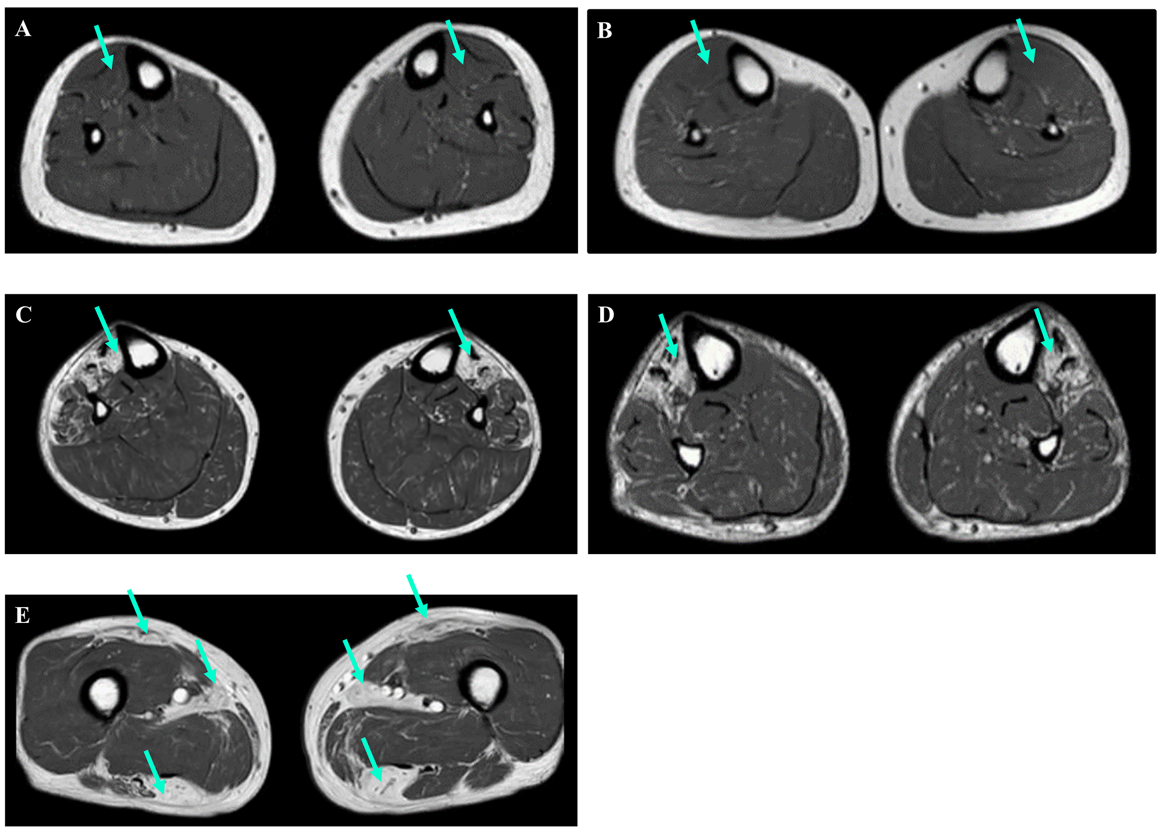
Figure 2. Muscle MRI images of the lower limbs (axial T1-weighted images). (A and B) Individuals 5 and 6, both under the age of 50, showed no signs of degeneration in their tibialis anterior muscles (green arrows). (C and D) In contrast, Individuals 7 and 12, both over the age of 50, displayed changes in their tibialis anterior muscles (green arrows). (E) Additionally, MRI results for Individual 12 revealed changes in the rectus femoris, adductor longus, and hamstring muscles (green arrows).


