


fig4
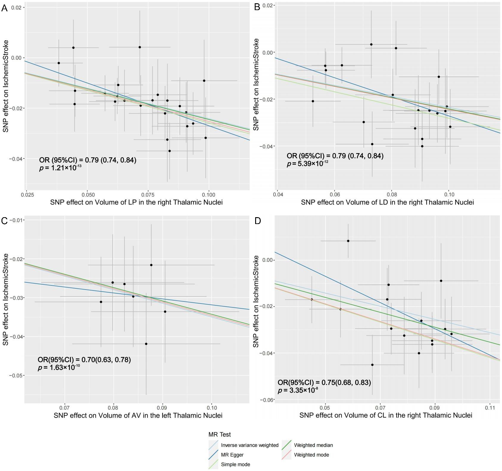
Figure 4. Scatter plots of individual SNP effects and estimates from different MR methods. (A) Volume of the right LP thalamic nucleus and risk of ischemic stroke; (B) Volume of the right LD thalamic nucleus and risk of ischemic stroke; (C) Volume of the left AV thalamic nucleus and risk of ischemic stroke; (D) Volume of the left CL thalamic nucleus and risk of ischemic stroke. LP: Lateral posterior; LD: lateral dorsal; AV: anteroventral; CL: central lateral; MR: mendelian randomization.


