

Mitochondrial homeostasis in heart failure with preserved ejection fraction: from metabolic remodeling to multi-organ crosstalk
 0
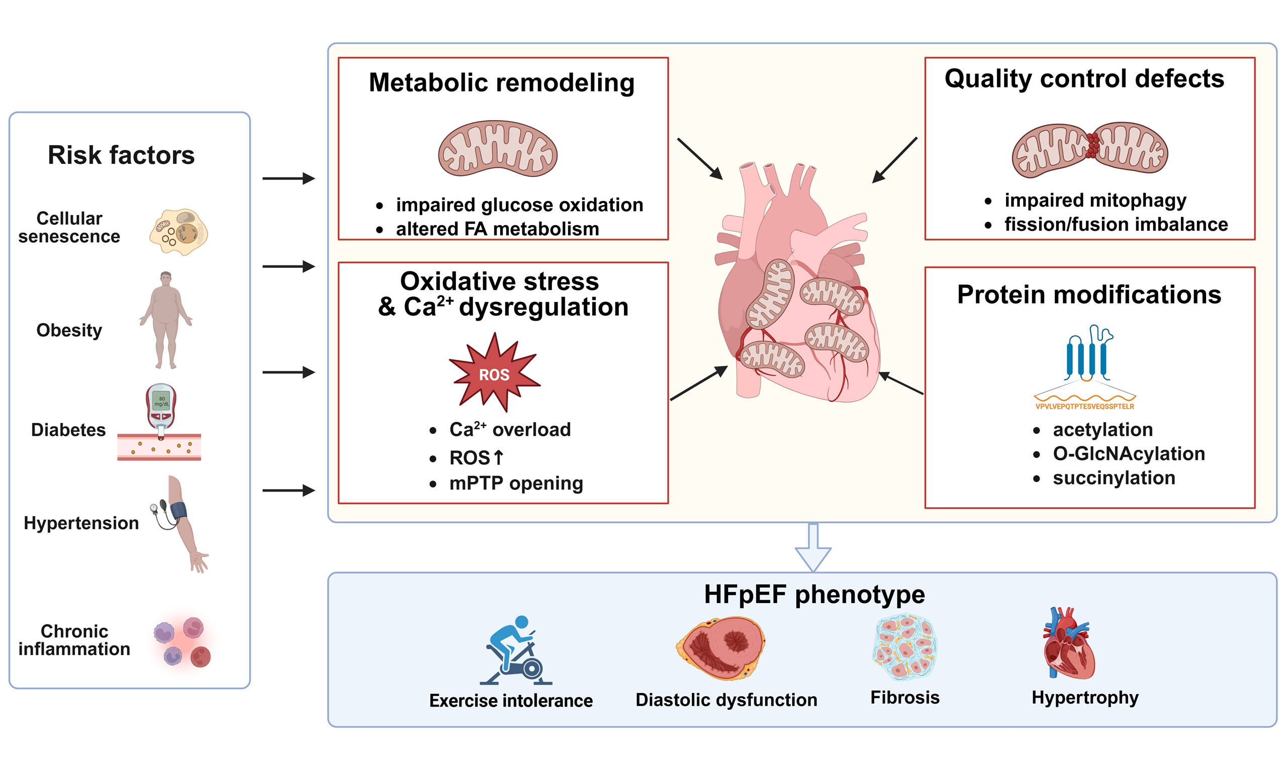
0 Graphical Abstract
Abstract
Heart failure with preserved ejection fraction (HFpEF) represents a predominant heart failure phenotype characterized by intricate pathophysiology and an escalating global prevalence. Beyond classical hemodynamic disturbances, emerging evidence identifies mitochondrial dysfunction as a central mechanism in HFpEF pathogenesis. This review synthesizes the pivotal mitochondrial aberrations driving HFpEF, including impaired bioenergetics, disrupted mitochondrial quality control, oxidative stress, and Ca2+ dyshomeostasis, particularly under the burden of cardiometabolic stressors. Importantly, we highlight that HFpEF represents a multisystem disorder wherein the mitochondria of adipose tissue, the vasculature, the immune system, and skeletal muscle actively contribute to the pathological process. By reclassifying HFpEF as a bioenergetic failure, we hope to provide a conceptual framework for the development of integrated, mitochondria-targeted therapeutic strategies.
Keywords


 Download PDF
INTRODUCTION
Heart failure with preserved ejection fraction (HFpEF) represents a predominant clinical phenotype of heart failure, impacting around 32 million individuals worldwide. While it makes up roughly half of all heart failure cases globally, regional estimates fluctuate between 30% and 75% due to varying diagnostic standards[1]. HFpEF is characterized by the presence of classic symptoms and signs despite a left ventricular ejection fraction (LVEF) ≥ 50%, alongside objective evidence of diastolic dysfunction or elevated filling pressure. Unlike heart failure with reduced ejection fraction (HFrEF), the pathophysiological mechanism of HFpEF is primarily driven by ventricular diastolic dysfunction and increased chamber stiffness, resulting in a clinically highly heterogeneous syndrome[2]. In recent years, with the rise in the elderly population and related chronic diseases such as obesity, hypertension, type 2 diabetes mellitus, chronic kidney disease and atrial fibrillation, the prevalence of HFpEF has steadily risen and is now considered a serious problem for public health. HFpEF is particularly prevalent among elderly individuals and accounts for approximately half of all heart failure cases, with its prevalence increasing progressively with advancing age[3]. Although patients with HFpEF appear to retain preserved systolic function, they still have a high risk of hospitalization and death, and long-term outcomes are relatively poor[4]. Most patients with HFpEF are associated with several other severe cardiovascular and metabolic diseases, such as exercise-induced dyspnea, fatigue and reduced exercise tolerance. However, these clinical features are often not specific, and therefore early diagnosis still faces a persistent diagnostic problem[5].
HFpEF is now perceived as a whole-body disease of older people that can be caused by many factors concurrently, including aging, obesity, diabetes, hypertension, and systemic inflammation. Pathobiology includes mild-to-moderate inflammation, endothelial dysfunction and coronary microvascular damage, myocardial hypertrophy and fibrosis. All of these routes ultimately contribute to the onset and development of diastolic dysfunction and increased filling pressures[6]. Among these mechanisms, mitochondrial injury has emerged as a key pathophysiological hallmark of HFpEF, encompassing impaired bioenergetics, altered substrate utilization, oxidative stress, dysregulated dynamics, and defective quality control[7]. Given mitochondria’s essential role in cardiac energetics and homeostasis, disrupted mitochondrial integrity may represent a fundamental mechanism driving HFpEF. In this context, a comprehensive understanding of mitochondrial dysfunction in HFpEF may refine our understanding of disease pathobiology and inform the development of mechanism-based therapies.
Moreover, HFpEF should not be viewed as a single disease entity but rather as a clinically heterogeneous illness. Obesity, diabetes, hypertension, atrial fibrillation, aging, and renal failure all have a significant impact on its phenotype in clinical practice, and these comorbidities aid in identifying the predominant pathophysiological factors in individual patients[8]. Since no single marker is adequate to make the diagnosis in every situation, modern diagnostic approaches place an emphasis on a systematic approach that incorporates symptoms, natriuretic peptides, echocardiography, and functional testing. Given that the essential contributions of oxidative stress, metabolic rigidity, poor bioenergetics, and peripheral muscle dysfunction may vary significantly within HFpEF subgroups, this clinical heterogeneity is extremely relevant to mitochondrial biology[9].
Although mitochondrial dysfunction is present in both HFpEF and HFrEF, the extent and pattern of mitochondrial remodeling drastically differ between these two phenotypes. Human myocardial studies have identified mitochondrial fragmentation and cristae disruption in both settings, although HFrEF generally displays more prominent loss of mitochondrial content, biogenesis, and oxidative capacity[10]. Distinct metabolic profiles further differentiate these entities. Although glucose oxidation and mitochondrial oxidative capacity are diminished across both phenotypes, fatty acid oxidation is frequently preserved or upregulated in HFpEF, whereas it is blunted in HFrEF. Conversely, enhanced ketone utilization and impaired branched-chain amino acid catabolism are more characteristic of HFrEF[11]. Notably, these metabolic discrepancies may attenuate over time as HFpEF advances and incurs widespread impairments in substrate utilization and electron transport chain activity[12]. Consequently, no single mitochondrial abnormality is unique to HFpEF; rather, HFpEF is defined by impaired metabolic flexibility and compromised mitochondrial reserve under persistent cardiometabolic stress, which precede the pervasive energetic collapse typical of end-stage heart failure.
RISK FACTOR-ASSOCIATED MITOCHONDRIAL DYSFUNCTION IN HFPEF
HFpEF develops gradually under the combined burden of global aging and chronic metabolic stress. Cellular senescence has emerged as a central mechanism in HFpEF pathogenesis. Animal studies further link telomere shortening to HFpEF development and enhanced p53 activation[13]. Shorter leukocyte telomere length is associated with a higher prevalence of HFpEF incidence and adverse cardiovascular events in high-risk hypertensive individuals, and also reflects accelerated age-related molecular remodeling[14]. In early-stage HFpEF, the myocardium appears to mount a compensatory mitochondrial response: basal respiration remains preserved, whereas maximal respiratory capacity is already reduced. Consistent with this temporal pattern, cardiomyocyte-specific p53 inhibition delays HFpEF progression[15]. Together, these findings suggest that age-related stress primes the heart for HFpEF by eroding mitochondrial reserve capacity[14,15]. Pressure-overload models further support this staged transition, where progressive metabolic and mitochondrial remodeling parallels the shift from compensatory hypertrophy to overt diastolic dysfunction[16].
Beyond aging, major cardiometabolic risk factors, including obesity, diabetes, hypertension, and chronic inflammation, impair mitochondrial homeostasis through overlapping inflammatory and nitric oxide (NO)-dependent pathways. In metabolically stressed HFpEF models, inducible nitric oxide synthase (iNOS) activation promotes oxidative stress, mitochondrial dysfunction, and impaired insulin-mediated glucose uptake via protein kinase B (Akt) S-nitrosylation, whereas genetic or pharmacological iNOS inhibition alleviates mitochondrial injury and improves the HFpEF phenotype[17]. In the “two-hit” HFpEF model, reduced cardiac and circulating nitrite levels reflect impaired NO bioavailability, accompanied by blunted mitochondrial respiration, vascular dysfunction, and reduced exercise capacity. Restoration of NO bioavailability alleviates oxidative and nitrosative stress while improving mitochondrial respiration and exercise performance and attenuating myocardial fibrosis[18]. Smoking is also an environmental stressor that can affect mitochondrial homeostasis; long-term exposure to cigarettes increases the E/e’ ratio [the ratio of peak early mitral inflow velocity (E) to the early diastolic mitral annular velocity (e’)], which is vital for assessment of left ventricular (LV) filling pressures and diastolic dysfunction in the presence of preserved systolic function. This diastolic deterioration is associated with impaired mitochondrial respiration and reduced myocardial sirtuin 1 (SIRT1) expression. Therefore, smoking-related diastolic dysfunction may be triggered by mitochondrial damage due to SIRT1 downregulation[19]. Thus, diverse upstream stressors appear to converge on oxidative stress, inflammatory cytokine activation, and disrupted NO signaling, creating a sustained mitochondrial burden in the HFpEF myocardium.
Chronic risk factor exposure impairs mitochondrial bioenergetics early in HFpEF, involving not only respiratory chain dysfunction but also defective biogenesis, altered substrate metabolism, and disrupted quality control[20]. Restoration of NO-soluble guanylate cyclase-cyclic guanosine monophosphate (NO-sGC-cGMP) signaling improves diastolic function, reduces hypertrophy and fibrosis, and normalizes mitochondrial ultrastructure, linking impaired NO signaling to mitochondrial dysfunction[21]. Similarly, activation of AMP-activated protein kinase (AMPK) ameliorates the HFpEF phenotype by increasing mitochondrial abundance, improving mitochondrial quality, and enhancing respiratory function[22]. The AMPK/peroxisome proliferator-activated receptor gamma coactivator 1-alpha (PGC-1α) axis further promotes mitochondrial biogenesis through upregulation of mitochondrial transcription factor A (TFAM) and nuclear respiratory factor 1 (NRF1). Activation of this pathway alleviates mitochondrial injury, limits mitochondrial reactive oxygen species (mtROS) production, and suppresses cardiomyocyte apoptosis[23]. Beyond transcriptional control, myocardial mRNA/microRNA profiling has identified distinct post-transcriptional regulatory networks in HFpEF, although their direct contribution to mitochondrial remodeling remains incompletely understood[24]. At a broader systems level, large-animal studies and human transcriptomic analyses have revealed an HFpEF-associated molecular signature characterized by downregulation of mitochondrial programs and progressive metabolic remodeling[25,26]. Taken together, mitochondrial dysfunction in HFpEF encompasses impaired respiration, defective biogenesis, and widespread metabolic dysregulation.
HFpEF exhibits sex-specific dimorphism closely linked to mitochondrial biology. In the “two-hit” model, female mice develop greater diastolic deterioration despite milder mtDNA loss and respiratory dysfunction, whereas male human cardiac tissue generally shows higher expression of mitochondrial and electron transport chain genes. Integrative mouse and human analyses identified ACSL6 as a sex-biased mitochondrial regulator of diastolic function. In mice, cardiac Acsl6 expression was lower in females than in males and decreased further after HFpEF induction. Cardiac Acsl6 overexpression improved diastolic function and exercise capacity in both sexes, although its effects on mitochondrial function were more pronounced in males, supporting a sex-dependent role for ACSL6 in HFpEF[27] [Figure 1].
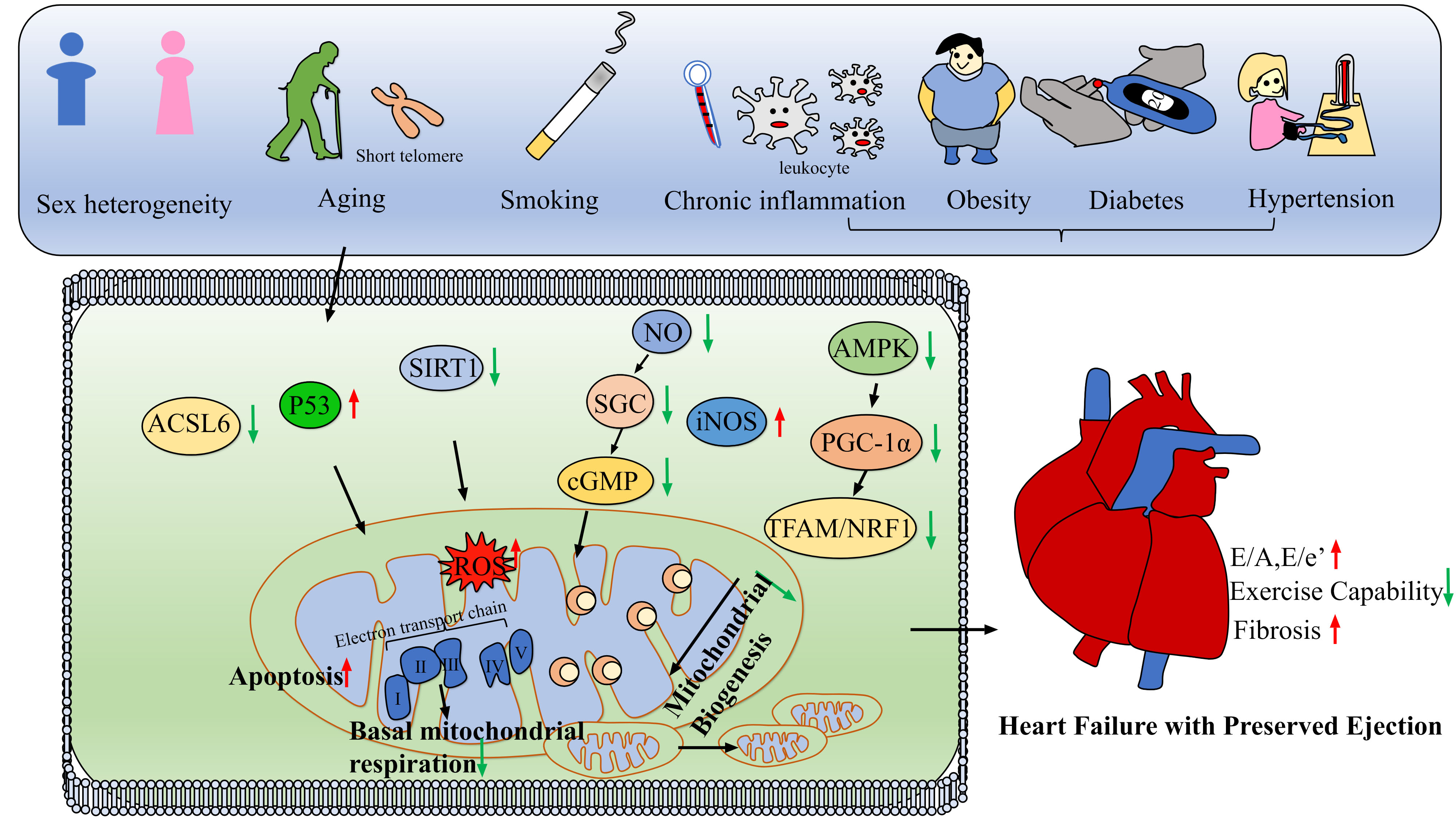
Figure 1. Pathogenesis and susceptibility of Heart Failure with Preserved Ejection Fraction (HFpEF). Schematic overview of how systemic risk factors converge on mitochondrial pathways to drive HFpEF pathogenesis. Aging, metabolic stress, inflammation/smoking, and female susceptibility disrupt key molecular hubs (p53, AMPK/PGC-1α, SIRT1, ACSL6, and NO/iNOS signaling), increase mtROS production, impair mitochondrial respiration and biogenesis, and promote apoptosis. The above mitochondrial damage is a cause of left ventricular hypertrophy and fibrosis in HFpEF, leading to diastolic dysfunction and reduced exercise capacity. In the schematic, red upward and green downward arrows indicate increased and decreased expression or activity, respectively. Black arrows indicate regulatory relationships or signaling direction. NO: Nitric oxide; mtROS: mitochondrial reactive oxygen species; iNOS: inducible nitric oxide synthase; AMPK: AMP-activated protein kinase; PGC-1α: peroxisome proliferator-activated receptor gamma coactivator 1-alpha; SIRT1: sirtuin 1; ACSL6: acyl-CoA synthetase long-chain family member 6; TFAM: mitochondrial transcription factor A; NRF1: nuclear respiratory factor 1; ROS: reactive oxygen species; SGC: soluble guanylate cyclase; cGMP: cyclic guanosine monophosphate.
Aging, cardiometabolic stress, smoking, impaired NO signaling, and sex-related susceptibility all place sustained pressure on myocardial mitochondria in HFpEF. Loss of respiratory reserve and metabolic flexibility may occur early, before overt energetic failure, leaving the myocardium increasingly vulnerable to further injury.
IMBALANCED MITOCHONDRIAL QUALITY CONTROL IN HFPEF
HFpEF involves not only impaired bioenergetics but also disrupted mitochondrial quality control. In cardiomyocytes, fusion, fission, and mitophagy work in concert to maintain mitochondrial integrity and metabolic adaptability. In HFpEF, dysregulation of this network promotes dysfunctional mitochondrial accumulation and metabolic-structural injury. Human myocardial biopsies and electron microscopy studies have revealed early mitochondrial abnormalities, including fragmentation, cristae disruption, reduced mitochondrial area, swelling, and lipid droplet accumulation, particularly in obesity-associated HFpEF[10,28]. These findings indicate that mitochondrial ultrastructural remodeling occurs early in HFpEF and is not limited to end-stage systolic heart failure. At the level of mitochondrial dynamics, impaired fission-fusion balance, particularly excessive dynamin-related protein 1 (Drp1)-mediated fission, contributes to HFpEF pathogenesis. F-box and leucine-rich repeat protein 4 (FBXL4) restrains pathological fission by promoting Drp1 ubiquitination and degradation. Accordingly, FBXL4 overexpression improves diastolic function, attenuates cardiac remodeling, preserves mitochondrial integrity, and restores Ca2+ handling, including sarcoplasmic/endoplasmic reticulum Ca2+-ATPase 2a (SERCA2a), in HFpEF models[29]. However, the role of mitochondrial fission in HFpEF is not uniformly detrimental. A controlled degree of fission is required for mitochondrial renewal and Adenosine Triphosphate (ATP) production. For instance, in high-salt-induced hypertensive HFpEF models, PTEN-induced kinase 1 (PINK1)-mediated phosphorylation of Drp1 at S616 promotes mitochondrial fission to preserve mitochondrial function and delay HFpEF progression[30]. Conversely, partial downregulation of Drp1 in obesity-related HFpEF phenotypes may worsen diastolic dysfunction, myocardial fibrosis, cell death and inflammation by inhibiting mitophagy and increasing oxidative stress. Therefore, mitochondrial quality-control dysfunction in HFpEF likely reflects an imbalance between fusion and fission rather than excessive fission alone[31]. Inflammatory-metabolic crosstalk also promotes mitochondrial fragmentation in HFpEF. Macrophage-derived S100A9 induces SPI1-dependent pyruvate dehydrogenase kinase 4 (PDK4) expression, leading to Drp1 phosphorylation, fission protein 1 (Fis1) upregulation, membrane potential loss, and ATP depletion; these effects are reversed by PDK4 or SPI1 silencing[32].
In addition to abnormalities in mitochondrial dynamics, defective mitophagy is another major driver of HFpEF progression. In a high-fat diet plus Nω-nitro-L-arginine methyl ester (HFD+L-NAME)-induced HFpEF model, genistein binds BCL2/adenovirus E1B 19-kDa interacting protein 3-like (BNIP3L) and stabilizes its dimeric conformation, thereby enhancing BNIP3L-mediated mitophagy, improving diastolic dysfunction, attenuating cardiac remodeling, and increasing exercise capacity[33]. In the same model, PINK1 and its downstream antioxidant partner peroxiredoxin 2 (Prdx2) are markedly downregulated, whereas PINK1 deficiency further reduces Prdx2 expression and exacerbates lipotoxicity-induced reactive oxygen species (ROS) accumulation, cardiomyocyte apoptosis, and cardiac dysfunction. By contrast, cardiac-specific overexpression of PINK1 restores Prdx2 levels, inhibits activation of the c-Jun N-terminal kinase (JNK) and p38 signaling pathways, and alleviates myocardial lipotoxicity and diastolic dysfunction[34]. Recently, the matricellular protein thrombospondin 1 (Thbs1) was identified as a new upstream regulator of mitophagy in HFpEF. In the high-fat diet + L-NAME model, Thbs1 is significantly upregulated in the myocardium, and knockdown of cardiac-specific Thbs1 improves diastolic function and reduces myocardial hypertrophy, fibrosis, inflammation and oxidative stress. Thbs1 inhibits mitophagy by activating the phosphoinositide 3-kinase (PI3K)/Akt/mammalian target of rapamycin (mTOR) signaling pathway, resulting in reduced clearance of damaged mitochondria and loss of mitochondrial homeostasis; thus, the inhibition of mitophagy by Thbs1 may be a new way of advancing HFpEF[35].
In SAUNA (salty drinking water/unilateral nephrectomy/aldosterone)-induced HFpEF, an increase in the expression of vasohibin 1 (VASH1) promotes α-tubulin dehydroxylation, impairs mitochondrial respiration, and suppresses mitophagy by inhibiting Parkin recruitment and preventing polyubiquitination of voltage-dependent anion channel 1 (VDAC1). As a result, mitochondrial quality control collapses and damaged mitochondria accumulate within the myocardium. Cardiac-specific VASH1 overexpression promotes HFpEF-like diastolic dysfunction and exercise intolerance[36]. Direct evidence also shows that although HFpEF hearts are reminiscent of those with diet-induced obesity and rely mainly on fatty acid oxidation for energy supply, they have a reduced myocardial phosphocreatine (PCr)/ATP ratio, impaired mitochondrial respiration, and increased ROS accumulation; thus, there is a significant disruption of mitochondrial energy homeostasis. Further analysis showed that, in the presence of an isolated high-fat condition, fatty acid overload can trigger mitophagy for the preservation of mitochondrial quality control as an adaptive mechanism. Nonetheless, this compensatory mechanism is significantly compromised in HFpEF hearts. Enhancing fatty acid oxidation may trigger mitophagy and improve mitochondrial function in HFpEF[37]. Therefore, a significant cause of the progression of HFpEF is impaired mitochondrial quality control.
In HFpEF, mitochondrial quality-control defects arise from disrupted fission-fusion balance together with insufficient clearance of damaged mitochondria. The resulting accumulation of dysfunctional mitochondria further compromises cellular homeostasis and contributes to progressive myocardial injury.
MITOCHONDRIAL OXIDATIVE STRESS, CA2+ DYSHOMEOSTASIS, AND CELL DEATH IN HFPEF
As bioenergetic impairment and defective quality control progress, redox imbalance, Ca2+ dysregulation, and altered membrane permeability further amplify mitochondrial injury, promoting cell death and myocardial remodeling in HFpEF. Mitochondria-targeted NADPH oxidase 4 (NOX4) overexpression is sufficient to evoke diastolic dysfunction, accompanied by mitochondrial fragmentation, increased Drp1 phosphorylation, mitofusin 2 (MFN2) downregulation, reduced respiratory reserve, cardiomyocyte apoptosis, and interstitial fibrosis. Conversely, NOX1/4 inhibition or mitochondrial H2O2 scavenging reduces fibrosis and prevents diastolic deterioration[38]. Mitochondrial redox homeostasis is thus essential for cardiac health in HFpEF. The model of mitochondrial DNA mutation that lacks reverse electron transport-derived ROS also induces diastolic dysfunction, cardiac hypertrophy and exercise intolerance due to disruption of mitochondrial redox signaling; therefore, maintaining the balance of mitochondrial ROS is essential for preserving the health of the heart in HFpEF[39]. Genetic evidence points to mitochondrial redox dysfunction as a cause. For example, intact nicotinamide nucleotide transhydrogenase (NNT) worsens cardiometabolic HFpEF by exacerbating nicotinamide adenine dinucleotide (NAD+) depletion and glutathione redox imbalance, while its loss-of-function is protective[40]. Environmental stressors such as fine particulate matter (PM2.5) can further worsen this situation by increasing mitochondrial ROS production, lowering mitochondrial membrane potential, fostering the opening of mitochondrial permeability transition pore (mPTP), and thus inducing programmed necrosis that exacerbates cardiac dysfunction[41]. Together, these results indicate that mitochondrial damage in HFpEF causes excessive oxidative stress, leads to cellular injury, and thus advances the disease.
Disrupted intracellular Ca2+ homeostasis represents an important component of mitochondrial injury in HFpEF. In the Zucker fatty/spontaneously hypertensive heart failure F1 hybrid (ZSF1)-HFpEF rat model, mitochondrial Ca2+ levels are elevated both at baseline and under stimulated conditions, accompanied by delayed cytosolic Ca2+ clearance, a reduced SERCA2a-to-phospholamban ratio, impaired mitochondrial respiration, and increased mitochondrial swelling[42]. Improving mitochondrial Ca2+ buffering capacity, reducing mitochondrial swelling, and lowering ROS generation alleviates left atrial remodeling in a metabolic syndrome-associated HFpEF model[43]. Moreover, transient receptor potential canonical 6 (TRPC6) deficiency reduces mitochondrial ROS production and preserves mitochondrial oxygen consumption in HFD+L-NAME-induced HFpEF mice, thereby maintaining diastolic function and exercise capacity[44]. These findings suggest that mitochondrial Ca2+ overload in HFpEF may represent both a short-term adaptive response and a critical inflection point at which compensation gives way to overt injury.
Chronic oxidative stress and cytosolic Ca2+ overload can activate cardiomyocyte death programs in HFpEF. In an aging-related high-fat diet plus L-NAME model, mice develop diastolic dysfunction, myocardial hypertrophy, interstitial fibrosis, and reduced exercise capacity, accompanied by ATP accumulation, activation of the P2X7/NOD-like receptor family pyrin domain-containing 3 (P2X7/NLRP3) inflammasome signaling, and pronounced pyroptosis. Renal denervation, P2X7 antagonism with A438079, or NLRP3 inhibition using MCC950 blocks the ATP-P2X7-NLRP3 axis, thereby reducing fibrosis and hypertrophy while improving E/e’ ratio and exercise tolerance[45]. The oxidative stress-responsive forkhead box O3a-BCL2/adenovirus E1B 19-kDa interacting protein 3 (FOXO3a-BNIP3) axis bridges mitochondrial Ca2+ dysregulation and cardiomyocyte injury. Specifically, FOXO3a activation upregulates BNIP3 expression, driving mitochondrial Ca2+ accumulation, dissipation of mitochondrial membrane potential, mitochondrial fragmentation, and apoptosis, a cascade that culminates in diastolic dysfunction. Conversely, targeted inhibition of cardiac FOXO3a signaling restores mitochondrial function and blunts adverse cardiac remodeling in experimental HFpEF models[46]. In the two-hit HFpEF model, levosimendan improves diastolic function and attenuates myocardial hypertrophy and pulmonary congestion. Mechanistically, it restores mitochondrial structure and function, reduces ROS, NOX4 expression, and cytochrome c release, increases the reduced glutathione/oxidized glutathione (GSH/GSSG) ratio, upregulates glutathione peroxidase 4 (GPX4), xCT, and ferroptosis suppressor protein 1 (FSP1), and lowers Fe2+, malondialdehyde (MDA), and 4-hydroxynonenal (4-HNE), thereby suppressing myocardial ferroptosis[47]. Ferritinophagy is also activated in hypertensive HFpEF rats, marked by increased nuclear receptor coactivator 4 (NCOA4), microtubule-associated protein 1 light chain 3 (LC3), Beclin-1, p62, and sideroflexin 1 (SFXN1), along with elevated ROS and reduced ferritin heavy chain 1 (FTH1). Canagliflozin reverses these abnormalities and improves diastolic function, implicating iron-dependent mitochondrial injury and ferroptosis-related signaling in HFpEF progression[48]. Collectively, mitochondrial injury in HFpEF is translated into myocardial hypertrophy, fibrosis, and worsening diastolic dysfunction through multiple forms of regulated cell death, including pyroptosis, ferroptosis, regulated necrosis, and apoptosis [Figure 2]. Despite substantial preclinical evidence implicating mitochondrial dysfunction in HFpEF pathogenesis, effective mitochondria-targeted therapies remain limited. Few therapeutic trials have demonstrated durable improvements in integrated exercise capacity, quality of life, or clinical outcomes, although the majority of available therapies in preclinical models enhance isolated pathways like redox balance, Ca2+ management, or mitophagy. This disparity probably indicates that HFpEF is caused by a network of interrelated disorders involving cardiomyocytes, fibroblasts, endothelium, immune cells, skeletal muscle, and the circulation rather than a single mitochondrial abnormality. Future mitochondria-targeted therapies should be tailored to the distinct phenotypes of cardiometabolic disease[49] [Table 1].
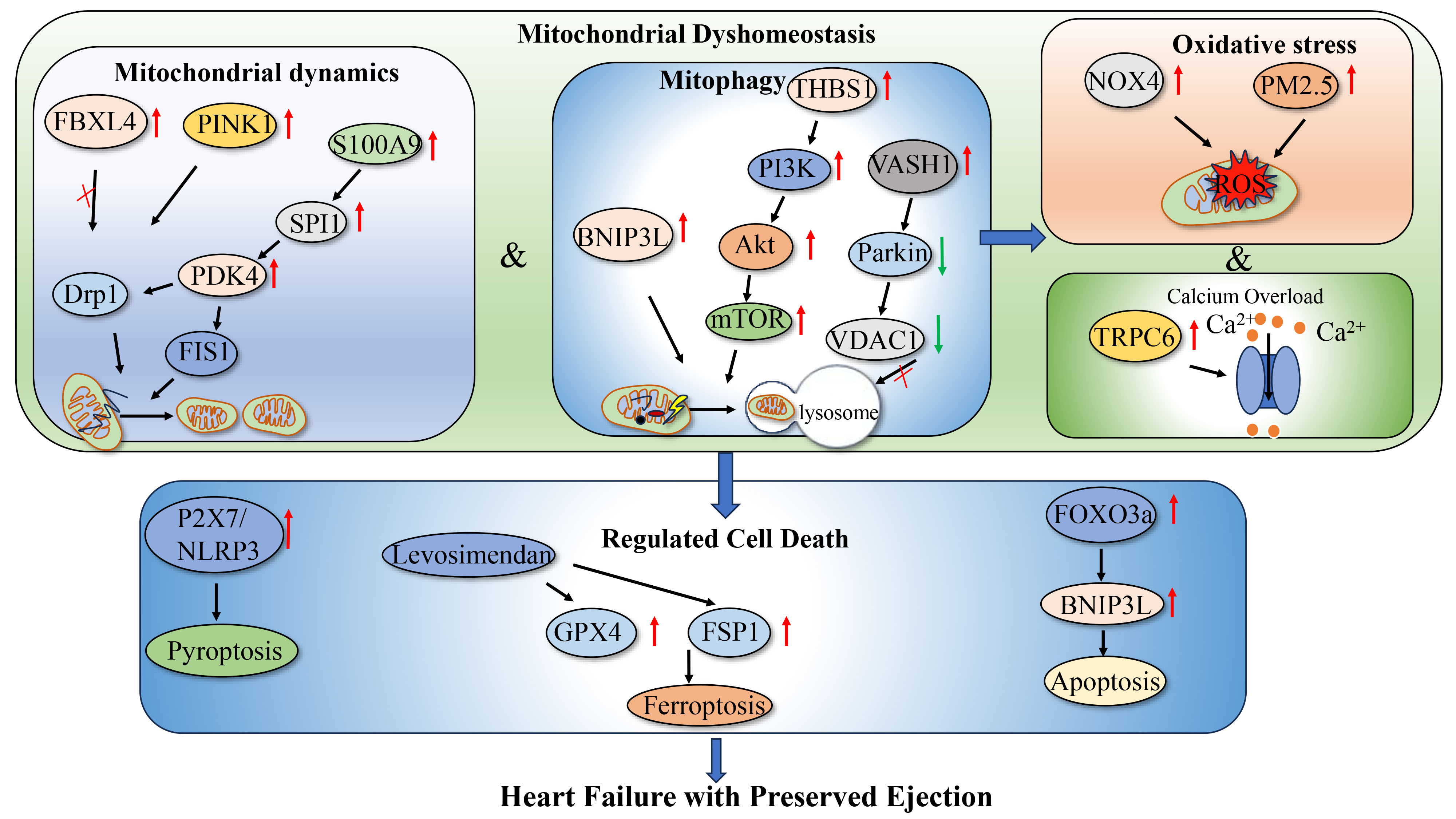
Figure 2. Mitochondrial Dyshomeostasis in HFpEF: Mechanisms and Pathogenesis. Schematic illustrating the major mechanisms linking mitochondrial dyshomeostasis to myocardial injury in HFpEF. Disrupted mitochondrial dynamics and defective mitophagy promote the accumulation of damaged mitochondria, while oxidative stress and Ca2+ overload further impair mitochondrial function and activate regulated cell death, including pyroptosis, ferroptosis, and apoptosis. These interconnected processes contribute to myocardial remodeling and diastolic dysfunction. In the schematic, red upward and green downward arrows indicate increased and decreased expression or activity, respectively. Solid arrows indicate regulatory relationships or signaling direction. Drp1: Dynamin-related protein 1; FBXL4: F-box and leucine-rich repeat protein 4; PINK1: PTEN-induced kinase 1; BNIP3L: BCL2/adenovirus E1B 19-kDa interacting protein 3-like; VASH1: vasohibin 1; VDAC1: voltage-dependent anion channel 1; NOX4: NADPH oxidase 4; ROS: reactive oxygen species; TRPC6: transient receptor potential canonical 6; mPTP: mitochondrial permeability transition pore; NLRP3: NOD-like receptor family pyrin domain-containing 3; GPX4: glutathione peroxidase 4; FSP1: ferroptosis suppressor protein 1; FOXO3a: forkhead box O3a; BNIP3: BCL2/adenovirus E1B 19-kDa interacting protein 3; HFpEF: heart failure with preserved ejection fraction; PDK4: pyruvate dehydrogenase kinase 4; PI3K: phosphoinositide 3-kinase; mTOR: mammalian target of rapamycin; NOX4: NADPH oxidase 4; SPI1: spi-1 proto-oncogene; FIS1: mitochondrial fission 1 protein; THBS1: thrombospondin 1; Akt: protein kinase B.
Mitochondrial oxidative stress, Ca2+ dyshomeostasis, and cell death in HFpEF
| Pathological process | Disease/phenotype | Experimental model | Main findings | References |
| Mitochondrial oxidative stress | HFpEF/diastolic dysfunction | Mitochondria-targeted NOX4 overexpression model | Mitochondrial NOX4 overexpression induced: Mitochondrial fragmentation ↑ Drp1 phosphorylation ↑ MFN2 ↓ Respiratory reserve ↓ Cardiomyocyte apoptosis ↑ Interstitial fibrosis ↑ | [38] |
| Mitochondrial oxidative stress/redox imbalance | HFpEF/diastolic dysfunction, cardiac hypertrophy, exercise intolerance | Mitochondrial DNA mutation model | Disruption of mitochondrial redox signaling induced: Diastolic dysfunction ↑ Cardiac hypertrophy ↑ Exercise tolerance ↓ | [39] |
| Mitochondrial oxidative stress/redox imbalance | Cardiometabolic HFpEF | Cardiometabolic HFpEF model with intact or loss-of-function nicotinamide nucleotide transhydrogenase (NNT) | Intact NNT exacerbated cardiometabolic HFpEF: NAD+ depletion ↑ Glutathione redox imbalance ↑ | [40] |
| Mitochondrial oxidative stress/mPTP opening/programmed necrosis | HFpEF/cardiac dysfunction | Model with fine particulate matter (PM2.5) exposure | Mitochondrial ROS production ↑ Mitochondrial membrane potential ↓ mPTP opening ↑ Programmed necrosis ↑ | [41] |
| Mitochondrial Ca2+ dyshomeostasis | HFpEF/diastolic dysfunction | ZSF1-HFpEF rat model | Mitochondrial Ca2+ levels ↑ Cytosolic Ca2+ clearance ↓ SERCA2a-to-phospholamban ratio ↓ Mitochondrial respiration ↓ Mitochondrial swelling ↑ | [42] |
| Mitochondrial Ca2+ overload | Metabolic syndrome-associated HFpEF/left atrial remodeling | Metabolic syndrome-associated HFpEF model | Mitochondrial Ca2+ buffering capacity ↑ Mitochondrial swelling ↓ ROS generation ↓ Left atrial remodeling ↓ | [43] |
| Mitochondrial Ca2+ dyshomeostasis/oxidative stress | HFpEF/diastolic dysfunction, reduced exercise capacity | HFD + L-NAME-induced HFpEF mouse model | TRPC6 deficiency induced: Mitochondrial ROS production ↓ Mitochondrial oxygen consumption ↑ Diastolic function ↑ Exercise capacity ↑ | [44] |
| Cardiomyocyte death/pyroptosis/inflammatory activation | HFpEF/diastolic dysfunction, myocardial hypertrophy, interstitial fibrosis, reduced exercise capacity | Aging-related high-fat diet plus L-NAME HFpEF mouse model | Pyroptosis ↑ Diastolic dysfunction ↑ Myocardial hypertrophy ↑ Interstitial fibrosis ↑ ATP accumulation ↑ Exercise capacity ↓ P2X7/NLRP3 activation ↑ | [45] |
| Cardiomyocyte death/apoptosis/mitochondrial Ca2+ dyshomeostasis | HFpEF/diastolic dysfunction, adverse myocardial remodeling | Experimental HFpEF model with FOXO3a-BNIP3 axis activation | FOXO3a induced: BNIP3 expression ↑ Mitochondrial Ca2+ accumulation ↑ Mitochondrial membrane potential ↓ Mitochondrial fragmentation ↑ and apoptosis ↑ Diastolic function ↓ | [46] |
| Cardiomyocyte death/ferroptosis/mitochondrial oxidative stress | HFpEF/diastolic dysfunction, myocardial hypertrophy, pulmonary congestion | Two-hit HFpEF mouse model | Levosimendan resulted in: ROS production ↓NOX4 expression ↓Cytochrome c release ↓GSH/GSSG ratio ↑GPX4, xCT, and FSP1 ↑Fe2+, MDA, and 4-HNE ↓Ferroptosis ↓Diastolic function ↑ | [47] |
| Cardiomyocyte death/ferroptosis/iron-dependent mitochondrial injury | Hypertensive HFpEF/diastolic dysfunction | Hypertensive HFpEF rat model with ferritinophagy activation | Ferritinophagy activation was marked by: NCOA4, LC3, Beclin-1, p62, SFXN1, ROS ↑ FTH1 ↓ | [48] |
Redox imbalance and Ca2+ dysregulation provide a direct link between mitochondrial injury and myocardial remodeling. Once mitochondrial buffering capacity is exceeded, cell death, inflammation, and fibrosis further impair relaxation and accelerate HFpEF progression.
METABOLIC REMODELING, POST-TRANSLATIONAL MODIFICATIONS, AND LIPID SIGNALING IN HFPEF
In addition to altered mitochondrial abundance and quality control, HFpEF is characterized by broad metabolic remodeling, aberrant post-translational regulation, and disrupted membrane lipid homeostasis, all of which converge on mitochondrial dysfunction.
HFpEF is characterized by disordered substrate utilization rather than a simple energy deficit. Insulin-stimulated glucose oxidation is markedly suppressed, whereas ATP production becomes increasingly dependent on fatty acid oxidation, with relatively limited changes in ketone oxidation and glycolysis[50]. Human myocardial metabolomics show increased glucose and GLUT1 but reduced glycolytic intermediates, pyruvate accumulation, and downregulated MPC1, indicating impaired glycolytic flux and limited mitochondrial substrate entry[51]. Earlier metabolic studies showed that increased glycolysis may precede the decline in fatty acid oxidation during HFpEF development, without a corresponding increase in glucose oxidation. This glycolysis-glucose oxidation mismatch increases proton production and may contribute to diastolic dysfunction by reducing contractile efficiency and increasing metabolic stress[52]. Impaired pyruvate oxidation may be an early metabolic defect in HFpEF. In an aged high-fat diet mouse model of diastolic dysfunction, Thapa and colleagues showed that general control of amino acid synthesis 5-like 1 (GCN5L1) promotes pyruvate dehydrogenase E1 subunit alpha 1 (PDHA1) acetylation, suppresses pyruvate dehydrogenase activity, and limits myocardial pyruvate utilization. Cardiomyocyte-specific GCN5L1 deletion reverses these effects and improves diastolic function. Thus, mitochondrial protein acetylation may impair metabolic flexibility by suppressing pyruvate oxidation, contributing to early diastolic dysfunction in HFpEF[53]. Consistently, multi-omics analyses in experimental HFpEF models highlight mitochondrial dysfunction, metabolic derangement, and inflammatory signaling as convergent pathways underlying myocardial stiffening and contractile impairment[54]. HFpEF also displays a prominent pattern of mitochondrial protein hyperacetylation, with abnormally acetylated proteins enriched in key metabolic pathways, including fatty acid oxidation, the tricarboxylic acid cycle, and oxidative phosphorylation[55,56]. Among these, dihydrolipoamide S-acetyltransferase (DLAT)-mediated acetylation of specific sites on hydroxyacyl-CoA dehydrogenase trifunctional multienzyme complex subunit alpha (HADHA) suppresses its enzymatic activity, limits fatty acid oxidation, and aggravates the HFpEF phenotype[55]. By contrast, restoration of the NAD+/nicotinamide adenine dinucleotide reduced form (NADH) ratio, together with reduced mitochondrial protein acetylation, improves mitochondrial function and alleviates HFpEF phenotypes[56]. Together, these findings indicate that metabolic derangements in HFpEF are not limited to shifts in myocardial fuel preference but also involve broad disturbances across multiple intramitochondrial metabolic pathways.
Beyond substrate utilization and protein acetylation, dysregulated lipid handling also contributes to metabolic remodeling in HFpEF. PCSK9 deficiency promotes cardiac lipid overload, impairs mitochondrial oxidative phosphorylation, and induces a diastolic dysfunction phenotype despite preserved ejection fraction[57]. Integrated systems biology analyses in cardiometabolic HFpEF further reveal marked mitochondrial ultrastructural remodeling, impaired respiration, energetic deficit, and lipid droplet accumulation adjacent to mitochondria[58]. Consistent with this, human myocardial proteomics demonstrates reduced oxidative phosphorylation, fuel metabolism, and protein translation, together with increased inflammatory and ROS-related signaling in HFpEF, particularly in severe obesity[59].
Osteopontin (OPN) contributes to chronic kidney disease (CKD)-associated HFpEF by disrupting mitochondrial metabolism. Col4a3 encodes the α3 chain of type IV collagen, an essential basement membrane component, and Col4a3-/- mice are widely used as a model of Alport syndrome-associated CKD. In this model, renal and circulating OPN levels are increased, whereas cardiac OPN is not markedly upregulated, suggesting that kidney-derived or systemic OPN signaling may contribute to cardiac remodeling. Col4a3-/- mice develop a HFpEF-like phenotype characterized by diastolic dysfunction, myocardial hypertrophy, fibrosis, pulmonary congestion, and impaired mitochondrial function, whereas OPN deficiency alleviates these abnormalities. Mechanistically, OPN is associated with dysregulation of oxoglutarate dehydrogenase-like (OGDHL), and cardiac-specific OGDHL restoration improves mitochondrial respiration and cardiac energetic status. Thus, the OPN/OGDHL axis may link CKD-related inflammatory signaling to mitochondrial metabolic dysfunction in HFpEF[60]. Dysregulated mitochondrial biogenesis may also drive HFpEF progression. Loss of microRNA-29 (miR-29) abnormally upregulates its target gene PGC-1α, causing imbalanced mitochondrial biogenesis, pathological accumulation of small mitochondria, and an HFpEF-like phenotype characterized by systemic hypertension, myocardial fibrosis, and diastolic dysfunction. Reducing PGC-1α gene dosage partially reverses these abnormalities, suggesting that the miR-29/PGC-1α axis links disordered metabolic regulation to mitochondrial remodeling[61].
Mitochondrial dysfunction in HFpEF is therefore not merely the consequence of impaired energy metabolism or structural disruption; aberrant post-translational modifications and altered membrane lipid metabolism also contribute substantially. As noted above, mitochondrial protein hyperacetylation is one of the hallmark metabolic features of HFpEF and can directly affect the activity of enzymes involved in fatty acid oxidation, the tricarboxylic acid cycle, and oxidative phosphorylation[55,56]. Dysregulated deacetylation has also been implicated in HFpEF progression. Histone deacetylase 6 (HDAC6) expression is increased in HFpEF mouse hearts, whereas treatment with the selective HDAC6 inhibitor TYA-018 reverses the established HFpEF phenotype. Mechanistically, HDAC6 inhibition not only restores expression of genes related to mitochondrial energy production and oxidative phosphorylation, but also enhances mitochondrial respiratory capacity in cardiomyocytes and suppresses fibroblast activation[62]. Similarly, leucine supplementation improves diastolic function at an early stage in ZSF1-HFpEF rats and attenuates left ventricular stiffness, fibrosis, and hypertrophy, effects accompanied by enhanced myocardial mitochondrial respiration and reduced HDAC4 expression and nuclear translocation[63]. These findings suggest that acetylation- and deacetylation-dependent pathways influence not only mitochondrial energetics but also fibrosis, stiffness, and diastolic dysfunction. Other metabolically sensitive post-translational modifications also contribute to HFpEF pathobiology. iNOS-mediated S-nitrosylation of Akt suppresses insulin-dependent glucose utilization, thereby aggravating mitochondrial metabolic dysfunction[17]. In addition, dissociation of hexokinase 1 (HK1) from mitochondria in endothelial cells enhances O-linked β-N-acetylglucosamine transferase (OGT) activity and increases protein O-linked β-N-acetylglucosamine (O-GlcNAc) modification, ultimately leading to endothelial dysfunction and an HFpEF phenotype; correspondingly, OGT inhibition or O-GlcNAcase overexpression reverses angiogenic defects and improves HFpEF manifestations[64]. Together, these studies indicate that mitochondrial abnormalities in HFpEF are not limited to impaired energy metabolism, but are also closely linked to altered signal transduction and abnormal protein modification arising from disturbed metabolic processes.
Another form of damage to mitochondrial homeostasis in HFpEF is disruption of membrane lipid metabolism due to post-translational modifications. Empagliflozin has been shown to improve diastolic function and hemodynamics in an established ZSF1 rat model of HFpEF, likely associated with restoration of mitochondrial respiratory chain activity (particularly complex IV activity), myocardial cardiolipin content, and normalization of mitochondrial volume[65]. The ability of SGLT2 inhibitors to maintain the integrity of mitochondrial cristae and stabilize the supramolecular assembly of electron transport chain supercomplexes, which maximizes electron flux and minimizes off-pathway electron leak that produces ROS, may be the mechanism underlying their positive effects on mitochondrial respiratory efficiency. Another study shows that the amount of myocardial mitochondrial phosphatidylethanolamine (PE) in older mice is reduced, and this has been linked to a decrease in membrane potential, an increase in ROS generation, and severe diastolic dysfunction. Lysophosphatidylethanolamine (LPE) can increase the amount of PE in the mitochondria, optimize the structure and membrane potential of the organelles, reduce ROS production, and thus alleviate diastolic dysfunction in old mice[66]. Therefore, the reasons for mitochondrial damage in HFpEF include dysregulation of enzymes and metabolism, as well as changes in the structure of lipid membranes that affect mitochondrial stability and respiratory chain function.
Inflammation and myocardial stiffening also contribute to HFpEF-like pathophysiology. In patients with diabetes mellitus and aortic stenosis, left ventricular myocardium shows increased inflammation and oxidative stress, marked by upregulation of high mobility group box 1 (HMGB1), calprotectin, Toll-like receptor 2/4 (TLR2/4), receptor for advanced glycation end products (RAGE), NLRP3, and interleukin-1 (IL-1), IL-6, and IL-18. These changes impair NO-sGC-cGMP-PKG (nitric oxide-soluble guanylate cyclase-cyclic guanosine monophosphate-cGMP-dependent protein kinase) signaling, reduce titin phosphorylation, and increase cardiomyocyte passive stiffness. The modest reduction in passive myocardial tension after anti-inflammatory, antioxidant, PKG-targeted, or empagliflozin-based interventions suggests that inflammation- and oxidative stress-driven myocardial stiffening is difficult to fully reverse and may sustain HFpEF-related diastolic dysfunction[67] [Table 2].
Metabolic remodeling, post-translational modifications, and lipid signaling in HFpEF
| Pathological process | Disease/phenotype | Experimental model | Main findings | References |
| Glycolytic flux impairment | HFpEF | Human myocardial metabolomics | Glucose ↑ / GLUT1 ↑ Glycolytic intermediates ↓ Pyruvate accumulation ↑ / MPC1 ↓ | [51] |
| Pyruvate oxidation impairment | HFpEF diastolic dysfunction (early metabolic defect) | Aged HFD mouse; CM-specific GCN5L1 KO | GCN5L1 ↑ → PDHA1 acetylation ↑ → PDH activity ↓ Pyruvate oxidation ↓ CM-specific GCN5L1 deletion reverses diastolic dysfunction | [53] |
| Multi-omics metabolic derangement | HFpEF myocardial stiffening & contractile impairment | Experimental HFpEF multi-omics | Mitochondrial dysfunction Metabolic derangement Inflammatory signaling | [54] |
| Mitochondrial protein hyperacetylation | HFpEF | HFpEF myocardial acetyl-proteomics | DLAT acetylates HADHA → HADHA activity ↓ / FAO ↓ NAD+/NADH restoration + acetylation ↓ → mitochondrial function ↑ | [55,56] |
| Cardiac lipid overload | Diastolic dysfunction with preserved EF | Pcsk9 KO mouse | Cardiac lipid overload Mitochondrial OXPHOS ↓ | [57] |
| OPN/OGDHL axis (CKD → HFpEF) | CKD-associated HFpEF | Col4a3-/- Alport CKD mouse; OPN-/- rescue; cardiac OGDHL restoration | Renal/circulating OPN ↑ OGDHL dysregulation Mitochondrial respiration ↓ OPN deletion & OGDHL restoration rescue phenotype | [60] |
| Mitochondrial biogenesis dysregulation | HFpEF | miR-29 loss-of-function mouse | miR-29 ↓ / PGC-1α ↑ Imbalanced mitochondrial biogenesis Small mitochondria accumulation | [61] |
| HDAC6-mediated deacetylation dysregulation | HFpEF | HFpEF mouse; HDAC6 inhibitor TYA-018 | HDAC6 ↑ Mitochondrial energy gene expression ↓ / fibroblast activation ↑ TYA-018 reverses HFpEF phenotype | [62] |
| HDAC4-mediated deacetylation dysregulation | ZSF1-HFpEF (early stage) | ZSF1-HFpEF rat | HDAC4 expression & nuclear translocation ↑ Leucine → HDAC4 ↓ / mitochondrial respiration ↑ LV stiffness, fibrosis, hypertrophy ↓ / diastolic function ↑ | [63] |
| Akt S-nitrosylation | HFpEF metabolic dysfunction | HFpEF models | iNOS ↑ → Akt S-nitrosylation ↑ Insulin-dependent glucose utilization ↓ Mitochondrial metabolic dysfunction aggravated | [17] |
| Endothelial O-GlcNAc modification | HFpEF endothelial dysfunction | Endothelial cell models | HK1 dissociates from mitochondria OGT activity ↑ / O-GlcNAc modification ↑ Endothelial dysfunction ↑/angiogenesis ↓ | [64] |
| Inflammation-driven myocardial stiffening | HFpEF (Diabetes Mellitus + aortic stenosis) | Human LV myocardium | HMGB1, calprotectin, TLR2/4, RAGE, NLRP3, IL-1/6/18 ↑ NO-sGC-cGMP-PKG signaling ↓ Titin phosphorylation ↓ Cardiomyocyte passive stiffness ↑ | [67] |
Mitochondrial dysfunction can also directly impair active myocardial relaxation. Depleted energetic reserve and unfavorable ATP/adenosine diphosphate (ADP) ratio constrain SERCA2a-dependent Ca2+ reuptake and delay actin-myosin cross-bridge detachment. Simultaneously, mitochondrial Ca2+ overload and excessive ROS production further disrupt cytosolic Ca2+ clearance[38,68]. Coupled with the inflammation- and titin-mediated increases in passive stiffness, these cellular deficits impair ventricular relaxation and elevate left ventricular filling pressures in HFpEF. Importantly, a preserved LVEF does not necessarily equate to intact systolic mechanics. In early-stage HFpEF, basal mitochondrial respiration may appear preserved even when maximal respiratory capacity and energetic reserve are significantly compromised[15]. Moreover, adaptive concentric remodeling alongside compensatory radial and circumferential shortening may mask underlying impairments in longitudinal strain and systolic reserve, thereby sustaining LVEF within normal limits[69,70]. Consequently, mitochondrial dysfunction often manifests first as impaired relaxation and diminished functional reserve before any detectable decline in resting LVEF occurs.
Metabolic remodeling in HFpEF extends beyond changes in fuel preference and includes impaired substrate entry, abnormal post-translational modifications, and disruption of mitochondrial membrane lipids. These abnormalities compromise ATP-dependent relaxation and redox balance, while inflammation and reduced titin phosphorylation increase passive myocardial stiffness and filling pressures despite preserved LVEF.
MULTICELLULAR AND MULTI-ORGAN MITOCHONDRIAL DYSREGULATION IN HFPEF
Accumulating evidence indicates that mitochondrial homeostasis in HFpEF is disrupted beyond cardiomyocytes, involving adipose tissue, vascular endothelium, immune cells, and skeletal muscle. Accordingly, HFpEF is increasingly viewed not as a heart-limited myocardial disorder, but as a systemic immunometabolic-mitochondrial syndrome. Consistent with this multicellular view, recent transcriptomic and single-cell analyses identified three mitochondrial hub genes, Decr1 (2,4-Dienoyl-CoA Reductase 1), Mthfd2 (Methylenetetrahydrofolate dehydrogenase 2), and Vwa8 (Von Willebrand Factor A domain-containing 8), in HFpEF. Decr1 regulates mitochondrial fatty acid metabolism, Mthfd2 is involved in mitochondrial one-carbon metabolism and redox regulation, and Vwa8 contributes to mitochondrial homeostasis. Fibroblasts showed high expression of these genes, linking mitochondrial dysfunction to fibroblast-driven extracellular matrix remodeling and intercellular communication[71]. Consistent with this systemic metabolic perspective, alterations in circulating acylcarnitine metabolism have also been linked to HFpEF severity. An increased acylcarnitine-to-free carnitine ratio, reflecting impaired mitochondrial fatty acid oxidation, predicts adverse clinical outcomes specifically in patients with HFpEF, underscoring the clinical relevance of mitochondrial metabolic dysfunction beyond the myocardium[72]. Circulating mitochondrial and metabolic biomarkers also vary across different clinical states: decompensated HFpEF exhibits higher levels of mitochondrial superoxide, mitochondrial mass, and lactate, alongside reduced mitochondrial DNA copy number, compared with stable disease[73].
Adipocyte stress may impair cardiac mitochondrial function through long-distance intercellular communication. In type 2 diabetes mellitus exhibiting HFpEF phenotype, injured adipocytes generate extracellular vesicles carrying oxidatively modified mitochondrial proteins. After uptake by cardiomyocytes, these vesicles increase ROS, reduce mitochondrial membrane potential, promote mitochondrial fragmentation, suppress oxygen consumption and ATP synthesis, and activate the intrinsic apoptotic pathway[74]. These findings suggest that adipose tissue stress can transmit mitochondrial injury signals to the heart through adipose-cardiac vesicular communication, thereby contributing to diabetes mellitus-associated HFpEF progression.
Vascular endothelial cells likewise participate in the network of mitochondrial injury in HFpEF. In db/db diabetic mice, coronary endothelial cell-specific upregulation of aldehyde dehydrogenase 2 (ALDH2) increases cardiac ALDH2 activity, reduces 4-HNE adduct formation, and improves diastolic function[75]. In another study of type 2 diabetes presenting HFpEF phenotype, elevated 12-hydroxyeicosatetraenoic acid (12-HETE) levels were linked to vascular dysfunction and disease progression, whereas inhibition of 12/15-lipoxygenase improved endothelial function, capillary density, and mitochondrial function, while attenuating the HFpEF phenotype[76]. Collectively, these results show that mitochondrial oxidative damage and metabolic dysregulation in HFpEF are not confined to cardiomyocytes; microvascular endothelial cells are also affected. Endothelial mitochondrial dysfunction, in turn, can worsen diastolic impairment by disrupting blood flow, angiogenesis, and local inflammatory signaling.
The immune-inflammatory system represents another major amplifying axis in HFpEF pathophysiology. Cross-species single-cell transcriptomic analyses of circulating immune cells show that HFpEF has a distinct inflammatory profile compared with HFrEF, marked by obesity-related cytokine signaling, including C-C motif chemokine ligand 2 (CCL2) and tumor necrosis factor (TNF), as well as increased mitochondrial and metabolic gene expression. Nitro-oleic acid (NO2-OA) partially reverses these immune abnormalities in mouse models and improves diastolic function[77]. High-fat diet-induced diastolic dysfunction depends on the accumulation of inflammatory macrophages and their secretion of IL-1β; blockade of IL-1β signaling, depletion of macrophages, or inhibition of inflammatory macrophage polarization all markedly improve diastolic function, while scavenging mtROS confers similar protection[78]. In early HFpEF, circulating Ly6Chi monocytes and C-C motif chemokine receptor 2-positive (CCR2+) monocyte-derived macrophages are increased, whereas protective T-cell immunoglobulin and mucin domain-containing protein 4-positive (TIMD4+) resident macrophages are reduced, indicating marked remodeling of the cardiac immune microenvironment. CCR2 deletion reduces inflammatory macrophage recruitment and attenuates left ventricular hypertrophy, but has limited effects on myocardial fibrosis and diastolic dysfunction[79]. Taken together, these experimental results indicate that systemic immune inflammation in HFpEF is closely linked to mitochondrial metabolic dysfunction, and together they may promote the progression of the disease; thus, a rationale for immunometabolic therapy has been provided.
Emerging evidence suggests that immunometabolic remodeling in HFpEF also involves the peripheral hematopoietic system, beyond expansion of inflammatory immune cells. Patients with cardiometabolic HFpEF show increased circulating hematopoietic stem cells, a finding recapitulated in mice fed a high-fat diet plus L-NAME. In this setting, enhanced mitochondrial fatty acid metabolism in splenic red pulp macrophages upregulates VCAM-1, remodels the hematopoietic stem cell niche, and promotes hematopoietic stem cell proliferation and systemic inflammation, thereby contributing to diastolic dysfunction. SGLT2 inhibitors partially suppress this process, highlighting a link between immune-inflammatory activation and myeloid metabolic reprogramming in HFpEF[80].
Skeletal muscle mitochondrial dysfunction contributes to exercise intolerance in HFpEF. Compared with skeletal muscle biopsies from patients with HFrEF and sedentary controls, HFpEF biopsies show increased muscle RING-finger protein 1 (MuRF1) expression, global ubiquitination, and proteasome activity, indicating enhanced protein degradation. Mitochondrial complex I and malate dehydrogenase (MDH) activities are also reduced, consistent with impaired skeletal muscle energy metabolism. Proteasome activity correlates with IL-6 levels and worse exercise performance, linking inflammation-driven protein degradation and mitochondrial dysfunction to reduced exercise capacity in HFpEF[81]. Alterations in skeletal muscle contractile proteins, such as titin hyperphosphorylation, are associated with impaired muscle function and mitochondrial activity in HFpEF[82]. The small-molecule MuRF1 inhibitor MyoMed-205 improves key HFpEF features, including elevated E/e’, increased left ventricular end-diastolic pressure, and myocardial fibrosis, while also attenuating skeletal muscle atrophy and enhancing contractile function. In skeletal muscle, MyoMed-205 reduces MuRF1 expression and global protein ubiquitination, increases mitochondrial metabolic protein expression, and enhances respiratory chain complex I/II and citrate synthase activities. In the myocardium, it reduces perivascular fibrosis, matrix metalloproteinase 2 activity, advanced glycation end product (AGE) modification, and titin hypophosphorylation, thereby improving left ventricular compliance and alleviating diastolic dysfunction[83]. These findings suggest that targeting MuRF1 may simultaneously improve cardiac diastolic abnormalities and peripheral skeletal muscle dysfunction in HFpEF.
Likewise, in HFpEF rats, empagliflozin improves cardiac diastolic function while also boosting skeletal muscle contractility, lowering intramuscular lipid buildup, and enhancing muscle mitochondrial function, particularly by upregulating Complex IV activity[84]. In cardiometabolic HFpEF, skeletal muscle shows impaired mitochondrial substrate oxidation, particularly defective fatty acid oxidation and branched-chain amino acid catabolism. Voluntary exercise training improves exercise capacity without substantially altering resting cardiac hemodynamics, while restoring skeletal muscle fatty acid and branched-chain amino acid utilization and upregulating β-oxidation, branched-chain amino acid catabolism, and mitochondrial electron transport chain pathways[85]. Based on the above studies, metabolic dysfunction in skeletal muscles is likely to be one of the reasons for exercise intolerance in HFpEF, and thus, improving peripheral skeletal muscle metabolism could offer a new treatment approach. Therefore, exercise intolerance in HFpEF is not only due to heart disease, but also significantly caused by mitochondrial dysfunction in skeletal muscles.
HFpEF-related mitochondrial dysfunction extends beyond the myocardium to adipose tissue, the vascular endothelium, immune and hematopoietic cells, and skeletal muscle. Crosstalk among these tissues amplifies metabolic stress, inflammation, and exercise intolerance, supporting the view of HFpEF as a systemic disorder rather than a disease confined to cardiomyocytes.
MITOCHONDRIA-TARGETED THERAPEUTIC STRATEGIES IN HFPEF
Based on the mechanistic insights outlined above, interventions targeting disrupted mitochondrial homeostasis, metabolic abnormalities, and associated inflammatory amplification pathways have emerged as an important therapeutic strategy in HFpEF. While these approaches target distinct molecular pathways, they ultimately aim for the same core goals: restoring mitochondrial function, limiting myocardial remodeling, and easing diastolic dysfunction [Figure 3].
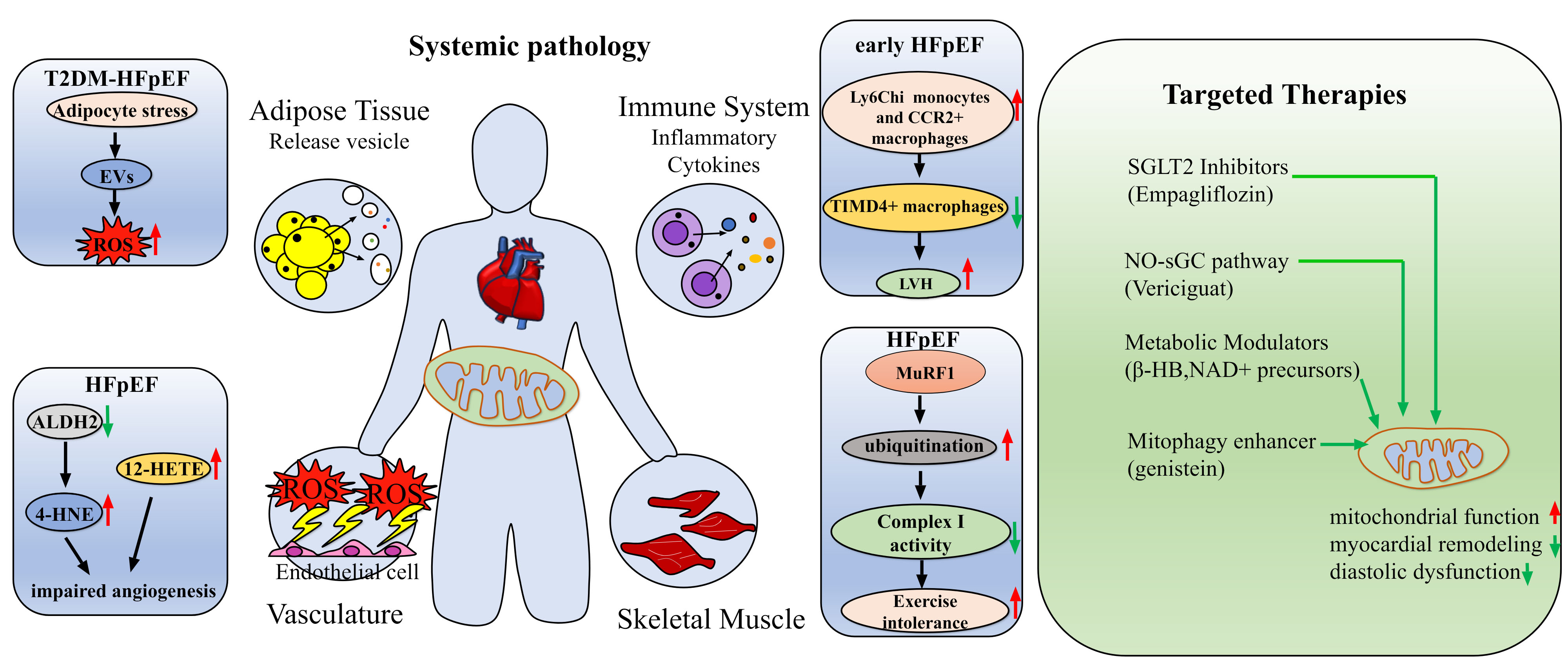
Figure 3. Multiorgan crosstalk and mitochondria-targeted therapeutic strategies in HFpEF. The left panel illustrates interactions among adipose tissue, the vasculature, the immune system, skeletal muscle, and the heart, highlighting mitochondrial dysfunction, oxidative stress, inflammation, and exercise intolerance. The right panel summarizes therapeutic strategies that restore mitochondrial homeostasis and alleviate myocardial remodeling and diastolic dysfunction. In the schematic, red upward and green downward arrows indicate increased and decreased expression or activity, respectively. Black arrows indicate regulatory relationships, orange arrows illustrate inter-organ crosstalk, and green arrows indicate therapeutic effects on mitochondrial function. HFpEF: Heart failure with preserved ejection fraction; ROS: reactive oxygen species; SGLT2: sodium-glucose cotransporter 2; NO-sGC-cGMP: nitric oxide-soluble guanylate cyclase-cyclic guanosine monophosphate; NAD+: nicotinamide adenine dinucleotide; AMPK: AMP-activated protein kinase; PGC-1α: peroxisome proliferator-activated receptor gamma coactivator 1-alpha; β-HB: Β-hydroxybutyrate; ALDH2: aldehyde dehydrogenase 2; TIMD4+: T-cell immunoglobulin and mucin domain-containing protein 4-positive; LVH: left ventricular hypertrophy; EVs: extracellular vesicles; 4-HNE: 4-hydroxynonenal; 12-HETE: 12-hydroxyeicosatetraenoic acid.
Vericiguat and NO2-OA primarily improve mitochondrial respiration and energy metabolism through restoration of NO-related signaling. As a soluble guanylate cyclase stimulator, vericiguat enhances NO-sGC-cGMP signaling and, in HFpEF models, increases basal mitochondrial respiration, upregulates electron transport chain-related proteins, and improves mitochondrial ultrastructure, thereby attenuating diastolic dysfunction[21]. NO2-OA, a nitrated fatty acid with anti-inflammatory and metabolic regulatory properties, not only improves immune-inflammatory status but also enhances mitochondrial abundance and expression of functional mitochondrial proteins through AMPK activation, thereby improving mitochondrial respiration, myocardial energy metabolism, and the HFpEF phenotype[22]. Several metabolic regulators and non-pharmacological interventions have also shown the capacity to restore mitochondrial metabolic homeostasis. Fibroblast growth factor 21, a key metabolic regulator with beneficial effects on lipid metabolism and energy balance, enhances mitochondrial bioenergetics in HFpEF via activation of the adiponectin (APN)-PI3K-Akt-PDK4 axis, thereby attenuating myocardial injury[86]. Berberine, a natural bioactive with blood glucose-lowering, lipid-lowering, and antioxidant properties, promotes mitochondrial biogenesis by upregulating TFAM and NRF1 through the AMPK/PGC-1α pathway, thereby alleviating mitochondrial injury and oxidative stress[23]. Astragaloside IV (AS-IV) may protect against HFpEF by attenuating inflammation and metabolic dysfunction. In high-fat diet plus L-NAME mice, AS-IV improves cardiac function and myocardial remodeling, reduces inflammatory markers including growth differentiation factor 15, C-reactive protein, monocyte chemoattractant protein-1, myocardial NLRP3, IL-1β, caspase-1, and IL-6, and restores myocardial glucose and fatty acid metabolism. These effects are accompanied by increased complex I activity, ATP production, plasma NAD+ levels, and pyruvate dehydrogenase activity, together with reduced pyruvate and lactate accumulation[87]. As a cornerstone non-pharmacological strategy, exercise training improves systemic metabolic health and functional capacity in HFpEF. Notably, recent human skeletal muscle biopsy studies indicate that these improvements are driven substantially by peripheral skeletal muscle adaptations, including the restoration of mitochondrial oxidative phosphorylation and substrate utilization[88]. In parallel, exercise may confer direct cardioprotective effects by augmenting mitochondrial function and mitigating metabolic stress through the inhibition of TLR4-mediated necroptosis[89].
At the level of mitochondrial quality control, genistein, a phytoestrogen-derived natural compound with antioxidant and metabolic regulatory properties, directly binds BNIP3L and stabilizes its dimeric conformation in HFpEF models, thereby enhancing BNIP3L-mediated mitophagy, promoting clearance of damaged mitochondria, improving diastolic dysfunction, and attenuating myocardial remodeling[33]. These findings suggest that restoring mitophagy may help slow HFpEF progression. Reducing mitochondrial oxidative injury and preserving membrane integrity have also become therapeutic priorities. In HFpEF models, empagliflozin improves diastolic function and hemodynamics while restoring mitochondrial respiratory chain function, enhancing complex IV activity, and correcting reduced myocardial cardiolipin content and mitochondrial volume[65]. Empagliflozin reduces ICAM-1, VCAM-1, TNF-α, and IL-6 levels in human HFpEF myocardium and animal models, while attenuating cytosolic and mitochondrial oxidative stress and restoring NO-sGC-cGMP-PKG signaling. By reducing protein kinase G 1α (PKG1α) oxidation and abnormal oligomerization, empagliflozin improves phosphorylation of titin, cardiac troponin I (cTnI), and vasodilator-stimulated phosphoprotein (VASP), thereby lowering passive cardiomyocyte stiffness and improving diastolic function[90]. Its ability to improve mitochondrial function in skeletal muscle further suggests that this class of agents may boost overall exercise capacity through coordinated effects on both the heart and peripheral tissues[84]. Catestatin, an endogenous peptide with anti-inflammatory and antioxidant properties, exerts cardioprotective effects by suppressing mitochondrial ROS production and improving mitochondrial ultrastructure[91]. In contrast, LPE functions more directly as a membrane lipid stabilizer. It replenishes key mitochondrial membrane lipids, preserves membrane potential and ultrastructural integrity, and ultimately ameliorates diastolic dysfunction[66].
Correcting metabolic intermediate accumulation and aberrant post-translational modifications represents another therapeutic strategy. Β-hydroxybutyrate (β-HB), a ketone body with metabolic and signaling functions, may improve mitochondrial dysfunction and myocardial remodeling in HFpEF by reducing the acetyl-coenzyme A (acetyl-CoA) pool, limiting mitochondrial hyperacetylation, and suppressing NLRP3 inflammasome activation[92]. Nicotinamide riboside, a precursor of NAD+, is mainly used to restore cellular redox balance and deacetylation capacity; in HFpEF, it improves mitochondrial function and myocardial remodeling by restoring the NAD+/NADH ratio and reducing mitochondrial protein hyperacetylation[56]. More recent work indicates that, in HFpEF, the benefit of NAD+ repletion is contingent on cardiomyocyte HMGCS2, linking restored ketogenesis to improved lipid metabolism, mitochondrial function, and cardiac performance[93]. Beyond effects on oxidative stress and membrane integrity, metabolic reprogramming may also contribute to the benefits of SGLT2 inhibition in HFpEF. In the “two-hit” HFpEF model, dapagliflozin mitigates myocardial hypertrophy, inflammation, oxidative stress, and fibrosis, while improving both diastolic dysfunction and subsequent systolic impairment. Mechanistically, it elevates circulating β-HB, boosts citrate synthase activity, and lowers acetyl-CoA buildup. These changes enhance the expression of oxidative phosphorylation complex proteins, increase ATP generation, and curb aberrant fatty acid uptake, collectively resolving the myocardial metabolic disturbances characteristic of HFpEF[94].
Notably, in patients with HFpEF receiving dapagliflozin, circulating β-HB and citrate synthase levels also tend to increase, further supporting the possibility that this agent improves myocardial energy metabolism through a “β-HB-citrate synthase” axis[94]. Mitochondrial abnormalities in HFpEF extend beyond impaired respiration to include metabolic intermediate accumulation and aberrant post-translational modifications, supporting metabolic reprogramming as a therapeutic strategy. Epigallocatechin gallate shows preventive effects in age-related diastolic dysfunction, improving diastolic function and exercise tolerance while attenuating cardiomyocyte apoptosis and mitochondrial injury. These effects are accompanied by increased cTnI expression and reduced HDAC1 expression and activity, suggesting that targeting age-related epigenetic remodeling and mitochondrial injury may protect against pre-HFpEF progression[95].
Current evidence also suggests that mitochondrial abnormalities in HFpEF are unlikely to be fully reversed through manipulation of a single pathway. Mitochondrial uncoupling strategies enhance substrate oxidation while reducing ROS production, representing a potential approach to improving metabolic efficiency in HFpEF[96]. However, clinical trials in HFpEF have shown that the mitochondrial uncoupling agent HU6 leads to significant reductions in body weight, total fat mass, and visceral adiposity. Importantly, these benefits do not translate into meaningful improvements in peak oxygen consumption, 6-minute walk distance, or diastolic function[97]. Similarly, elamipretide, a peptide developed to target mitochondria by stabilizing the inner membrane and enhancing respiratory chain activity, has demonstrated modest enhancements in complex I/II respiration in experimental HFpEF models, yet fails to improve diastolic function, myocardial remodeling, or inflammation, and may even be associated with mild deterioration in systolic performance[98]. Together, these observations indicate that correction of selected mitochondrial metabolic indices alone is insufficient to reverse the established HFpEF phenotype. More promising future strategies will likely need to address metabolic remodeling, oxidative stress, mitochondrial dynamics and quality control, inflammatory signaling, and multi-organ dysfunction in an integrated manner [Table 3].
Mitochondria-targeted therapeutic strategy for HFpEF
| Therapeutic Strategies | Model | Key effects | References |
| Vericiguat | HFpEF model | NO-sGC-cGMP signaling ↑ Basal mitochondrial respiration ↑ ETC-related proteins ↑ Diastolic dysfunction ↓ | [21] |
| NO2-OA | HFD + L-NAME-induced HFpEF mouse model | AMPK signaling ↑ Mitochondrial abundance ↑ Functional mitochondrial proteins ↑ Mitochondrial respiration and myocardial energy metabolism ↑ Inflammation ↓ | [22] |
| Fibroblast growth factor 21 (FGF21) | HFpEF model | APN-PI3K-Akt signaling ↑ PDK4 ↓ Mitochondrial bioenergetics ↑ Myocardial injury ↓ | [86] |
| Berberine | HFD + L-NAME-induced HFpEF mouse model | AMPK/PGC-1α pathway ↑ TFAM and NRF1 ↑ Mitochondrial biogenesis ↑ Mitochondrial injury and oxidative stress ↓ Apoptosis ↓ | [23] |
| Astragaloside IV (AS-IV) | HFD + L-NAME-induced HFpEF mouse model | Myocardial remodeling ↓ Inflammatory markers ↓ Complex I activity, ATP production, plasma NAD+, and PDH activity ↑ Pyruvate and lactate accumulation ↓ | [87] |
| Exercise training | HFpEF model | Myocardial metabolic stress ↓ TLR4-related necroptosis ↓ | [89] |
| Genistein | HFD + L-NAME-induced HFpEF model | BNIP3L-mediated mitophagy ↑ Diastolic dysfunction and myocardial remodeling ↓ | [33] |
| Empagliflozin | HFpEF models/human HFpEF myocardium | Complex IV activity ↑ Inflammatory markers ↓ Cytosolic and mitochondrial oxidative stress ↓ PKG1α oxidation and abnormal oligomerization ↓ | [65,90,84] |
| Catestatin | HFpEF model | Mitochondrial ETC-derived ROS ↓ Diastolic dysfunction ↓ | [91] |
| LPE | Aged mouse model of diastolic dysfunction | Mitochondrial PE content ↑ Mitochondrial ROS ↓ Diastolic dysfunction ↓ | [66] |
| β-hydroxybutyrate | HFpEF model | Acetyl-CoA pool ↓ Mitochondrial hyperacetylation ↓ NLRP3 inflammasome activation ↓ | [92] |
| Nicotinamide riboside | HFpEF model | NAD+/NADH ratio restored Cellular redox balance ↑ Deacetylation capacity ↑ Mitochondrial protein hyperacetylation ↓ | [56] |
| Dapagliflozin | Two-hit HFpEF model/patients with HFpEF | Circulating β-hydroxybutyrate ↑ Citrate synthase activity ↑ Acetyl-CoA buildup ↓ OXPHOS complex proteins ↑ ATP generation ↑ Aberrant fatty acid uptake ↓ Diastolic dysfunction and myocardial remodeling ↓ | [94] |
| Epigallocatechin gallate | Aged mouse model of age-related diastolic dysfunction | Exercise intolerance ↓ Cardiomyocyte apoptosis ↓ Mitochondrial injury ↓ Diastolic dysfunction ↓ HDAC1 expression and activity ↓ | [95] |
| Elamipretide | ZSF1 HFpEF model | Complex I/II respiration modestly ↑ No improvement in diastolic function, myocardial remodeling, or inflammation | [98] |
While existing interventions can optimize selected mitochondrial measures, improvements in respiration, redox balance, or body composition have not reliably translated into functional gains in exercise tolerance or cardiac performance. This translational disconnect highlights a major hurdle in HFpEF management, demonstrating that therapeutic efficacy will necessitate phenotype-specific approaches that concurrently mitigate intrinsic myocardial dysfunction and systemic metabolic and inflammatory drivers.
Strengths and limitations
While this review provides a comprehensive framework positioning mitochondrial dysfunction as a central node connecting metabolic remodeling, cellular stress, and multi-organ crosstalk in HFpEF, several limitations persist. First, much of the mechanistic insight relies on preclinical models, necessitating rigorous validation in human cohorts. Second, given the pronounced heterogeneity of HFpEF, it remains uncertain whether these mitochondrial deficits serve as a universal hallmark across all disease phenotypes. Third, although inter-organ networks are increasingly recognized, the precise circulating mediators and spatiotemporal dynamics governing these interactions remain poorly defined. Finally, the translational potential, long-term safety, and clinical efficacy of mitochondria-targeted therapeutics warrant further investigation.
CONCLUSIONS
In short, the problems in mitochondria in HFpEF are not just about a reduced amount of energy production. HFpEF is a type of serious illness that can result from all sorts of reasons, including metabolic diseases, damage to the quality control system of mitochondria, oxidative stress, Ca2+ dysregulation, and damage to multiple organs. Mitochondria show defective substrate utilization, impaired oxidative phosphorylation, aberrant post-translational modifications, and altered membrane lipid metabolism; thus, both energy efficiency and adaptability are compromised. In addition to the above changes in the mitochondria, disturbed mitochondrial dynamics, impaired mitophagy, redox imbalance, Ca2+ overload and activation of various cell death pathways have all contributed to myocardial hypertrophy, fibrosis and diastolic dysfunction. Other sites of the disease include fat and blood vessels. HFpEF is a form of heart failure with preserved ejection fraction that also affects other organs in the body and is associated with mitochondrial dysfunction. Given the complex and phenotypic heterogeneity of HFpEF, targeting an isolated mitochondrial defect is unlikely to yield sufficient therapeutic efficacy. Future strategies will necessitate coordinated, multi-target interventions that simultaneously address mitochondrial dysfunction, metabolic dysregulation, systemic inflammation, and adverse fibrosis, tailored to patient-specific pathophysiological profiles. Furthermore, identifying circulating biomarkers of mitochondrial injury could refine early diagnostic strategies and enable precise longitudinal monitoring of therapeutic responses.
DECLARATIONS
Acknowledgments
The Graphical Abstract was created with BioRender.com (https://BioRender.com/o0yfu63).
Authors’ contributions
Original draft preparation: An Y
Figure preparation using Microsoft PowerPoint: Shang C
Critical review and revision of the manuscript: Abd El-Aty AM, Reiter RJ, Tuomilehto J, Qin X, Dhanasekaran M, Uversky VN, Haidara MA, Zhao L
Manuscript revision: Zhang Y, Ren J
All authors reviewed and approved the final version of the manuscript.
Availability of data and materials
Not applicable.
AI and AI-assisted tools statement
During the preparation of this manuscript, the AI tool Grammarly (version Windows v.1.2.281.1928, released 2026-07-27) was used solely for language editing. The tool did not influence the study design, data collection, analysis, interpretation, or the scientific content of the work. All authors take full responsibility for the accuracy, integrity, and final content of the manuscript.
Financial support and sponsorship
This study was funded by the National Key R&D Program of China (2021YFC2500500) and the National Natural Science Foundation of China (T2288101).
Conflicts of interest
Ren J is an Editorial Board Member of The Journal of Cardiovascular Aging. Ren J was not involved in any steps of editorial processing, notably including reviewers’ selection, manuscript handling, or decision-making. The other authors declare that there are no conflicts of interest.
Ethical approval and consent to participate
Not applicable.
Consent for publication
Not applicable.
Copyright
© The Author(s) 2026.
REFERENCES
1. Cho DH, Yoo BS. Current prevalence, incidence, and outcomes of heart failure with preserved ejection fraction. Heart Fail Clin. 2021;17:315-26.
2. Borlaug BA. Evaluation and management of heart failure with preserved ejection fraction. Nat Rev Cardiol. 2020;17:559-73.
3. Gharagozloo K, Mehdizadeh M, Heckman G, et al. Heart failure with preserved ejection fraction in the elderly population: basic mechanisms and clinical considerations. Can J Cardiol. 2024;40:1424-44.
4. Dunlay SM, Roger VL, Redfield MM. Epidemiology of heart failure with preserved ejection fraction. Nat Rev Cardiol. 2017;14:591-602.
5. Campbell P, Rutten FH, Lee MM, Hawkins NM, Petrie MC. Heart failure with preserved ejection fraction: everything the clinician needs to know. Lancet. 2024;403:1083-92.
6. Omote K, Verbrugge FH, Borlaug BA. Heart failure with preserved ejection fraction: mechanisms and treatment strategies. Annu Rev Med. 2022;73:321-37.
7. Kumar AA, Kelly DP, Chirinos JA. Mitochondrial dysfunction in heart failure with preserved ejection fraction. Circulation. 2019;139:1435-50.
8. Abudureyimu M, Luo X, Wang X, et al. Heart failure with preserved ejection fraction (HFpEF) in type 2 diabetes mellitus: from pathophysiology to therapeutics. J Mol Cell Biol. 2022:14.
9. Pieske B, Tschöpe C, de Boer RA, et al. How to diagnose heart failure with preserved ejection fraction: the HFA-PEFF diagnostic algorithm: a consensus recommendation from the Heart Failure Association (HFA) of the European Society of Cardiology (ESC). Eur Heart J. 2019;40:3297-317.
10. Chaanine AH, Joyce LD, Stulak JM, et al. Mitochondrial morphology, dynamics, and function in human pressure overload or ischemic heart disease with preserved or reduced ejection fraction. Circ Heart Fail. 2019;12:e005131.
11. De Jong KA, Lopaschuk GD. Complex energy metabolic changes in heart failure with preserved ejection fraction and heart failure with reduced ejection fraction. Can J Cardiol. 2017;33:860-71.
12. Ferro F, Spelat R, Valente C, Contessotto P. Understanding how heart metabolic derangement shows differential stage specificity for heart failure with preserved and reduced ejection fraction. Biomolecules. 2022;12:969.
13. Leri AS, Franco A, Zacheo A, et al. Ablation of telomerase and telomere loss leads to cardiac dilatation and heart failure associated with p53 upregulation. EMBO J. 2003;22:131-9.
14. Zhang A, Tang Z, Chang ACY, et al. Leukocyte telomere length and risk of heart failure with preserved ejection fraction in high-risk Chinese patients with hypertension under 65 years. Geroscience. 2025.
15. Chen X, Lin H, Xiong W, et al. p53-dependent mitochondrial compensation in heart failure with preserved ejection fraction. J Am Heart Assoc. 2022;11:e024582.
16. Li X, Tan W, Zheng S, et al. Differential mRNA Expression and circular RNA-based competitive endogenous RNA networks in the three stages of heart failure in transverse aortic constriction mice. Front Physiol. 2022;13:777284.
17. Guo Y, Wen J, He A, et al. iNOS contributes to heart failure with preserved ejection fraction through mitochondrial dysfunction and Akt S-nitrosylation. J Adv Res. 2023;43:175-86.
18. LaPenna KB, Li Z, Doiron JE, et al. Combination sodium nitrite and hydralazine therapy attenuates heart failure with preserved ejection fraction severity in a “2-Hit” murine model. J Am Heart Assoc. 2023;12:e028480.
19. Slotabec L, Wang H, Seale B, Wen C, Filho F, Li J. Cardiac diastolic dysfunction by cigarette smoking is associated with mitochondrial integrity in the heart. FASEB J. 2024;38:e23826.
20. Ren J, Pulakat L, Whaley-Connell A, Sowers JR. Mitochondrial biogenesis in the metabolic syndrome and cardiovascular disease. J Mol Med. 2010;88:993-1001.
21. Vázquez-Abuín X, Moraña-Fernández S, Aragón-Herrera A, et al. Soluble guanylate cyclase stimulation improves cardiac function and mitochondrial activity in a rat model of early-stage heart failure with preserved ejection fraction. Biomed Pharmacother. 2025;191:118439.
22. Müller M, Schubert T, Welke C, et al. Nitro-oleic acid enhances mitochondrial metabolism and ameliorates heart failure with preserved ejection fraction in mice. Nat Commun. 2025;16:3933.
23. Hu Y, Chen X, Zhao Q, et al. Berberine improves cardiac insufficiency through AMPK/PGC-1α signaling-mediated mitochondrial homeostasis and apoptosis in HFpEF mice. Int Immunopharmacol. 2025;155:114613.
24. Inácio JM, Cristo F, Pinheiro M, et al. Myocardial RNA sequencing reveals new potential therapeutic targets in heart failure with preserved ejection fraction. Biomedicines. 2023;11:2131.
25. Gibb AA, Murray EK, Eaton DM, et al. Molecular signature of HFpEF: systems biology in a cardiac-centric large animal model. JACC Basic Transl Sci. 2021;6:650-72.
26. Hahn VS, Knutsdottir H, Luo X, et al. Myocardial gene expression signatures in human heart failure with preserved ejection fraction. Circulation. 2021;143:120-34.
27. Cao Y, Vergnes L, Wang YC, et al. Sex differences in heart mitochondria regulate diastolic dysfunction. Nat Commun. 2022;13:3850.
28. Meddeb M, Koleini N, Binek A, et al. Myocardial ultrastructure of human heart failure with preserved ejection fraction. Nat Cardiovasc Res. 2024;3:907-14.
29. Abudureyimu M, Luo X, Jiang L, et al. FBXL4 protects against HFpEF through Drp1-Mediated regulation of mitochondrial dynamics and the downstream SERCA2a. Redox Biol. 2024;70:103081.
30. Shou J, Huo Y. PINK1 Phosphorylates Drp1(S616) to improve mitochondrial fission and inhibit the progression of hypertension-induced HFpEF. Int J Mol Sci. 2022;23:11934.
31. Wu D, Hu Q, Li H, Yin Y, Wang P, Wang W. Drp1 knockdown aggravates obesity-induced cardiac dysfunction and remodeling. Mitochondrion. 2025;83:102023.
32. Wang M, Ren B, Wu X, et al. S100A9 inhibition ameliorates HFpEF by modulating mitochondrial fission and oxidative stress. Int Immunopharmacol. 2025;163:115280.
33. Dou Z, Sun D, Huang Q, et al. Genistein improves heart failure with preserved ejection fraction through BNIP3L-mediated mitophagy. Biochem Pharmacol. 2026;245:117680.
34. Zhang H, Xu T, Mei X, et al. PINK1 modulates Prdx2 to reduce lipotoxicity-induced apoptosis and attenuate cardiac dysfunction in heart failure mice with a preserved ejection fraction. Clin Transl Med. 2025;15:e70166.
35. Bu X, Sha S, Zhang Z, et al. Thrombospondin 1 aggravates cardiac remodeling in heart failure with preserved ejection fraction by inhibiting mitophagy. iScience. 2026;29:114639.
36. Miura S, Misaka T, Sekine T, et al. Detyrosinated α-tubulin mediates mitochondrial dysfunction and diastolic impairment in heart failure with preserved ejection fraction. FEBS Lett. 2025;599:2474-90.
37. Yoshii A, McMillen TS, Wang Y, et al. Blunted cardiac mitophagy in response to metabolic stress contributes to HFpEF. Circ Res. 2024;135:1004-17.
38. Lozhkin A, Vendrov AE, Ramos-Mondragón R, et al. Mitochondrial oxidative stress contributes to diastolic dysfunction through impaired mitochondrial dynamics. Redox Biol. 2022;57:102474.
39. Vujic A, Koo A, Bidault G, et al. Loss of RET-ROS at complex I induces diastolic dysfunction in mice that is reversed by aerobic exercise. Am J Physiol Heart Circ Physiol. 2025;329:H154-68.
40. Pepin ME, Konrad PJM, Nazir S, et al. Mitochondrial NNT promotes diastolic dysfunction in cardiometabolic HFpEF. Circ Res. 2025;136:1564-78.
41. Wu T, Tong M, Chu A, Wu K, Niu X, Zhang Z. PM2.5-induced programmed myocardial cell death via mPTP opening results in deteriorated cardiac function in HFpEF mice. Cardiovasc Toxicol. 2022;22:746-62.
42. Miranda-Silva D, Wüst RCI, Conceição G, et al. Disturbed cardiac mitochondrial and cytosolic calcium handling in a metabolic risk-related rat model of heart failure with preserved ejection fraction. Acta Physiol. 2020;228:e13378.
43. Bode D, Semmler L, Wakula P, et al. Dual SGLT-1 and SGLT-2 inhibition improves left atrial dysfunction in HFpEF. Cardiovasc Diabetol. 2021;20:7.
44. Li X, Fu Y, Dai X, et al. TRPC6 deficiency attenuates mitochondrial and cardiac dysfunction in heart failure with preserved ejection fraction induced by high-fat diet plus L-NAME. Int J Mol Sci. 2025;26:9383.
45. Li Z, Sun X, Wang Y, et al. Renal denervation attenuates cardiac dysfunction in HFpEF by inhibiting the ATP-P2X7-NLRP3 inflammasome axis. Basic Res Cardiol. 2025;120:1225-47.
46. Chaanine AH, Kohlbrenner E, Gamb SI, et al. FOXO3a regulates BNIP3 and modulates mitochondrial calcium, dynamics, and function in cardiac stress. Am J Physiol Heart Circ Physiol. 2016;311:H1540-59.
47. Zhang LL, Chen GH, Tang RJ, et al. Levosimendan reverses cardiac malfunction and cardiomyocyte ferroptosis during heart failure with preserved ejection fraction via connexin 43 signaling activation. Cardiovasc Drugs Ther. 2024;38:705-18.
48. Ma S, Zuo QJ, He LL, et al. Canagliflozin ameliorates ferritinophagy in HFpEF rats. J Geriatr Cardiol. 2025;22:178-89.
49. Paulus WJ, Tschöpe C. A novel paradigm for heart failure with preserved ejection fraction: comorbidities drive myocardial dysfunction and remodeling through coronary microvascular endothelial inflammation. J Am Coll Cardiol. 2013;62:263-71.
50. Sun Q, Güven B, Wagg CS, et al. Mitochondrial fatty acid oxidation is the major source of cardiac adenosine triphosphate production in heart failure with preserved ejection fraction. Cardiovasc Res. 2024;120:360-71.
51. Koleini N, Meddeb M, Zhao L, et al. Landscape of glycolytic metabolites and their regulating proteins in myocardium from human heart failure with preserved ejection fraction. Eur J Heart Fail. 2024;26:1941-51.
52. Fillmore N, Levasseur JL, Fukushima A, et al. Uncoupling of glycolysis from glucose oxidation accompanies the development of heart failure with preserved ejection fraction. Mol Med. 2018;24:3.
53. Thapa D, Bugga P, Mushala BAS, et al. GCN5L1 impairs diastolic function in mice exposed to a high fat diet by restricting cardiac pyruvate oxidation. Physiol Rep. 2022;10:e15415.
54. Zhang W, Zhang H, Yao W, et al. Morphometric, hemodynamic, and multi-omics analyses in heart failure rats with preserved ejection fraction. Int J Mol Sci. 2020;21:3362.
55. Wang Y, Guo D, Zhu J, et al. Pyruvate metabolism enzyme Dlat induces mitochondria protein hyperacetylation to limit fatty acid oxidation in the HFpEF heart. Nat Commun. 2026:17.
56. Liu X, Zhang Y, Deng Y, et al. Mitochondrial protein hyperacetylation underpins heart failure with preserved ejection fraction in mice. J Mol Cell Cardiol. 2022;165:76-85.
57. Da Dalt L, Castiglioni L, Baragetti A, et al. PCSK9 deficiency rewires heart metabolism and drives heart failure with preserved ejection fraction. Eur Heart J. 2021;42:3078-90.
58. Gibb AA, LaPenna K, Gaspar RB, et al. Integrated systems biology identifies disruptions in mitochondrial function and metabolism as key contributors to HFpEF. JACC Basic Transl Sci. 2025;10:101334.
59. Jani VP, Yoo EJ, Binek A, et al. Myocardial proteome in human heart failure with preserved ejection fraction. J Am Heart Assoc. 2025;14:e038945.
60. Yousefi K, Irion CI, Takeuchi LM, et al. Osteopontin promotes left ventricular diastolic dysfunction through a mitochondrial pathway. J Am Coll Cardiol. 2019;73:2705-18.
61. Caravia XM, Fanjul V, Oliver E, et al. The microRNA-29/PGC1α regulatory axis is critical for metabolic control of cardiac function. PLoS Biol. 2018;16:e2006247.
62. Ranjbarvaziri S, Zeng A, Wu I, et al. Targeting HDAC6 to treat heart failure with preserved ejection fraction in mice. Nat Commun. 2024;15:1352.
63. Alves PKN, Schauer A, Augstein A, et al. Leucine supplementation improves diastolic function in HFpEF by HDAC4 inhibition. Cells. 2023;12:2561.
64. Tatekoshi Y, Mahmoodzadeh A, Shapiro JS, et al. Protein O-GlcNAcylation and hexokinase mitochondrial dissociation drive heart failure with preserved ejection fraction. Cell Metab. 2025;37:1584-1600.e10.
65. Schauer A, Adams V, Kämmerer S, et al. Empagliflozin improves diastolic function in HFpEF by restabilizing the mitochondrial respiratory chain. Circ Heart Fail. 2024;17:e011107.
66. Xu G, Xiao W, Sun P, et al. Lysophosphatidylethanolamine improves diastolic dysfunction by alleviating mitochondrial injury in the aging heart. J Lipid Res. 2025;66:100713.
67. Herwig M, Sieme M, Kovács A, et al. Diabetes mellitus aggravates myocardial inflammation and oxidative stress in aortic stenosis: a mechanistic link to HFpEF features. Cardiovasc Diabetol. 2025;24:203.
68. McCandless MG, Altara R, Booz GW, Kurdi M. What role do mitochondria have in diastolic dysfunction? Implications for diabetic cardiomyopathy and heart failure with preserved ejection function. J Cardiovasc Pharmacol. 2022;79:399-406.
69. MacIver DH. Is remodeling the dominant compensatory mechanism in both chronic heart failure with preserved and reduced left ventricular ejection fraction? Basic Res Cardiol. 2010;105:227-34.
70. Morris DA, Ma XX, Belyavskiy E, et al. Left ventricular longitudinal systolic function analysed by 2D speckle-tracking echocardiography in heart failure with preserved ejection fraction: a meta-analysis. Open Heart. 2017;4:e000630.
71. You H, Fan X, Diao J, Wu F. Integrative transcriptomic and single-cell analysis reveals mitochondrial-related gene biomarkers in heart failure with preserved ejection fraction. Sci Rep. 2025;15:44241.
72. Yoshihisa A, Watanabe S, Yokokawa T, et al. Associations between acylcarnitine to free carnitine ratio and adverse prognosis in heart failure patients with reduced or preserved ejection fraction. ESC Heart Fail. 2017;4:360-4.
73. Anguita E, Chaparro A, Candel FJ, et al. Biomarkers of stable and decompensated phases of heart failure with preserved ejection fraction. Int J Cardiol. 2022;361:91-100.
74. Ji H, Cao T, Tan Z, et al. Adipocyte extracellular vesicle mitochondrial cargo is linked to cardiomyocyte dysfunction in type 2 diabetes-related heart failure with preserved ejection fraction. Free Radic Biol Med. 2026;244:435-51.
75. Pan G, Roy B, Yeboah EO, et al. Targeted overexpression of mitochondrial ALDH2 in coronary endothelial cells mitigates HFpEF in a diabetic mouse model. Biomolecules. 2025;15:1029.
76. Otto M, Brabenec L, Müller M, et al. Development of heart failure with preserved ejection fraction in type 2 diabetic mice is ameliorated by preserving vascular function. Life Sci. 2021;284:119925.
77. Kneuer JM, Müller M, Erbe S, et al. Circulating immune cell signature analysis in HFpEF across species. Circ Res. 2025;137:682-98.
78. Liu H, Huang Y, Zhao Y, et al. Inflammatory macrophage interleukin-1β mediates high-fat diet-induced heart failure with preserved ejection fraction. JACC Basic Transl Sci. 2023;8:174-85.
79. Raman J, Simmonds S, Caluwé E, et al. CCR2+ monocyte-derived macrophages drive cardiac hypertrophy in early HFpEF. Am J Physiol Heart Circ Physiol. 2025;329:H109-23.
80. Filipp M, Ge ZD, DeBerge M, et al. Myeloid fatty acid metabolism activates neighboring hematopoietic stem cells to promote heart failure with preserved ejection fraction. Circulation. 2025;151:1451-66.
81. Adams V, Wunderlich S, Mangner N, et al. Ubiquitin-proteasome-system and enzymes of energy metabolism in skeletal muscle of patients with HFpEF and HFrEF. ESC Heart Fail. 2021;8:2556-68.
82. Vahle B, Schauer A, Augstein A, et al. MyoMed205 counteracts titin hyperphosphorylation and the expression of contraction-regulating proteins in a rat model of HFpEF. J Cachexia Sarcopenia Muscle. 2025;16:e13843.
83. Adams V, Schauer A, Augstein A, et al. Targeting MuRF1 by small molecules in a HFpEF rat model improves myocardial diastolic function and skeletal muscle contractility. J Cachexia Sarcopenia Muscle. 2022;13:1565-81.
84. Winzer EB, Schauer A, Langner E, et al. Empagliflozin preserves skeletal muscle function in a HFpEF rat model. Int J Mol Sci. 2022;23:10989.
85. Quiriarte H, Noland RC, Stampley JE, et al. Exercise therapy rescues skeletal muscle dysfunction and exercise intolerance in cardiometabolic HFpEF. JACC Basic Transl Sci. 2024;9:1409-25.
86. Zhang K, Gan J, Wang B, et al. FGF21 protects against HFpEF by improving cardiac mitochondrial bioenergetics in mice. Nat Commun. 2025;16:1661.
87. Wang X, Chen X, Wang Y, et al. Astragaloside IV alleviates inflammation and improves myocardial metabolism in heart failure mice with preserved ejection fraction. Front Pharmacol. 2024;15:1467132.
88. Winzer EB, Augstein A, Schauer A, et al. Impact of different training modalities on molecular alterations in skeletal muscle of patients with heart failure with preserved ejection fraction: a substudy of the OptimEx trial. Circ Heart Fail. 2022;15:e009124.
89. Dong J, Wang H, Feng Y, et al. Exercise training alleviates myocardial injury in mice with heart failure with preserved ejection fraction via the TLR4-mediated necroptosis pathway. Int Immunopharmacol. 2026;176:116513.
90. Kolijn D, Pabel S, Tian Y, et al. Empagliflozin improves endothelial and cardiomyocyte function in human heart failure with preserved ejection fraction via reduced pro-inflammatory-oxidative pathways and protein kinase Gα oxidation. Cardiovasc Res. 2021;117:495-507.
91. Qiu Z, Fan Y, Wang Z, et al. Catestatin protects against diastolic dysfunction by attenuating mitochondrial reactive oxygen species generation. J Am Heart Assoc. 2023;12:e029470.
92. Deng Y, Xie M, Li Q, et al. Targeting mitochondria-inflammation circuit by β-hydroxybutyrate mitigates HFpEF. Circ Res. 2021;128:232-45.
93. Koay YC, McIntosh B, Ng YH, et al. The heart has intrinsic ketogenic capacity that mediates NAD+ therapy in HFpEF. Circ Res. 2025;136:1113-30.
94. Zhang X, Wang N, Fu P, et al. Dapagliflozin attenuates heart failure with preserved ejection fraction remodeling and dysfunction by elevating β-hydroxybutyrate-activated citrate synthase. J Cardiovasc Pharmacol. 2023;82:375-88.
95. Quan J, Jia Z, Liu L, Tian J. The effect of long-term administration of green tea catechins on aging-related cardiac diastolic dysfunction and decline of troponin I. Genes Dis. 2025;12:101284.
96. Kitzman DW, Lewis GD, Pandey A, et al. A novel controlled metabolic accelerator for the treatment of obesity-related heart failure with preserved ejection fraction: rationale and design of the phase 2a HuMAIN trial. Eur J Heart Fail. 2024;26:2013-24.
97. Pandey A, Lewis GD, Borlaug BA, et al. Novel controlled metabolic accelerator for obesity-related HFpEF: the HuMAIN-HFpEF randomized clinical trial. JAMA Cardiol. 2025;10:609-16.
Cite This Article
 Open Access
How to Cite
Download Citation
If you have the appropriate software installed, you can download article citation data to the citation manager of your choice. Simply select your manager software from the list below and click on download.
Export Citation File
Type of Import
Tips on Downloading Citation
Citation Manager File Format
Type of Import
Direct Import: When the Direct Import option is selected (the default state), a dialogue box will give you the option to Save or Open the downloaded citation data. Choosing Open will either launch your citation manager or give you a choice of applications with which to use the metadata. The Save option saves the file locally for later use.
Indirect Import: When the Indirect Import option is selected, the metadata is displayed and may be copied and pasted as needed.
About This Article
Special Topic
Copyright

Data & Comments
Data
0











Comments
Comments must be written in English. Spam, offensive content, impersonation, and private information will not be permitted. If any comment is reported and identified as inappropriate content by OAE staff, the comment will be removed without notice. If you have any queries or need any help, please contact us at [email protected].