

A cardio-hematopoietic signaling axis after myocardial infarction: platelet extracellular vesicles drive inflammatory remodeling
 0
0 


 Download PDF
Cardiovascular disease remains the leading cause of mortality worldwide, with myocardial infarction (MI) representing a major manifestation of ischemic heart disease. Although timely reperfusion therapies and advances in antithrombotic treatment have substantially improved survival after acute coronary syndromes, many patients continue to develop adverse cardiac remodeling and heart failure following MI[1,2]. Increasing evidence over the past two decades has established that these complications are not solely driven by the initial ischemic insult, but also profoundly influenced by post-infarction inflammatory responses. Inflammation has therefore emerged as a central regulator of cardiac repair, ventricular remodeling, and long-term cardiovascular outcomes. Clinical studies involving anti-inflammatory therapies, including interleukin-1β inhibition and colchicine treatment, have further validated inflammation as a therapeutically actionable pathway in cardiovascular disease[2].
Although activation of bone marrow myelopoiesis after MI is increasingly recognized[3,4], the mechanisms by which injury-derived signals are transmitted from the ischemic heart to distant hematopoietic niches remain poorly understood. In particular, how circulating mediators generated during cardiac injury regulate hematopoietic stem and progenitor cell (HSPC) fate and coordinate systemic inflammatory remodeling remains a major unresolved question. Platelet-derived extracellular vesicles (pEVs) have recently emerged as potential mediators of interorgan communication, providing a new framework for understanding the heart-bone marrow signaling axis. Despite their abundance and established roles in vascular inflammation and thrombosis[5], whether pEVs actively regulate distant hematopoietic remodeling remains unclear. Specifically, whether pEVs serve as molecular messengers linking myocardial injury to bone marrow reprogramming, and which cargo components mediate this process, represent important unanswered questions in cardiovascular immunology.
A recent study by Ohayon-Steckel and colleagues provides a mechanistic framework linking ischemic cardiac injury to systemic hematopoietic reprogramming through pEV-mediated signaling[6]. The authors identify circulating pEVs as key mediators of emergency myelopoiesis following MI, establishing a cardio-hematopoietic signaling axis that connects platelet activation to bone marrow remodeling and subsequent amplification of systemic inflammation. As summarized in Figure 1, the proposed axis involves five steps. First, MI initiates the signaling cascade through acute cardiac injury. Second, cardiac injury induces platelet activation and the release of abundant submicron pEVs into the circulation. Third, circulating pEVs traffic to the bone marrow, where they are internalized by quiescent HSPCs. Fourth, pEV-derived miR-499 and miR-184 suppress lactoferrin expression in HSPCs, thereby relieving quiescence and promoting cell-cycle entry. Fifth, this transition drives emergency myelopoiesis, characterized by the expansion of myeloid progenitors and mature myeloid populations that sustain and exacerbate post-MI inflammation.
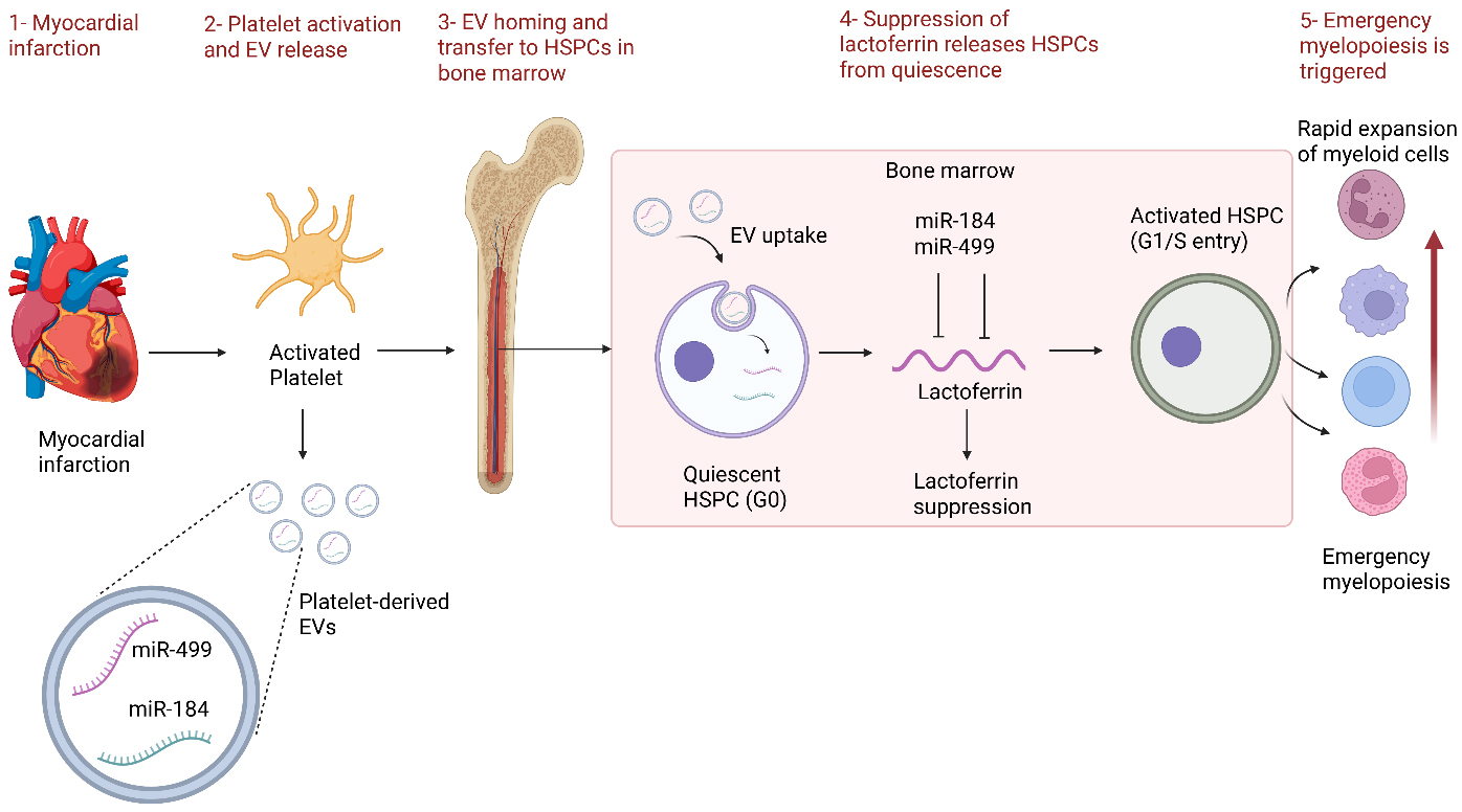
Figure 1. Platelet-derived EVs suppress lactoferrin in HSPCs and trigger emergency myelopoiesis during myocardial infarction. Figure created with BioRender.com. HSPCs: Hematopoietic stem and progenitor cells; EVs: extracellular vesicles. Figure 1 was created using BioRender.com.
A major conceptual advance of this work is the expanded view of platelet function in MI. Beyond their established roles in thrombosis and hemostasis, platelets are revealed as active regulators of systemic immune signaling. Following acute MI, activated platelets release abundant submicron pEVs into the circulation. The authors identify the damage-associated alarmin S100A8/9 as a key upstream driver of this response, consistent with accumulating evidence implicating S100A8/9 in platelet activation, neutrophil-mediated inflammation, and adverse cardiac remodeling after MI[7]. Rather than functioning as inert cellular byproducts, these vesicles serve as intercellular signaling mediators that deliver regulatory cargo to distant HSPCs, ultimately reshaping stem cell fate and promoting inflammatory myelopoiesis.
The authors validate this model using complementary approaches that integrate human observational data with rigorous mechanistic studies in murine systems. In both patients and murine models of MI, circulating pEV levels are markedly increased and correlate with leukocytosis and expansion of activated HSPCs. Parabiosis experiments, in which MI mice are surgically connected to healthy partners, demonstrate that circulating factors from infarcted mice are sufficient to activate HSPCs and expand myeloid progenitors in uninjured recipients, although this approach does not distinguish pEVs from other circulating mediators. The specific contribution of pEVs is established through direct transfer experiments, in which injection of pEVs isolated from miR-499+/+ versus miR-499-/- mice into sham animals respectively recapitulates or attenuates the hematopoietic activation phenotype. Together, these findings identify pEVs as important mediators within the circulating cardio-hematopoietic signaling pathway activated following MI.
Mechanistically, MI-derived pEVs are enriched in miR-499 and miR-184, with miR-499 being particularly notable due to its high expression in the heart and its established role as a biomarker of myocardial injury[8]. Unbiased transcriptomic profiling combined with luciferase 3’-untranslated region reporter assays confirmed direct binding of both miR-499 and miR-184 to the lactoferrin transcript, identifying lactoferrin, a protein classically involved in tumor suppression[9], as a shared downstream target suppressed by both miRNAs. Lactoferrin silencing increased expression of the proliferation marker MKI67 and other cell-cycle-associated genes and promoted expansion of myeloid cells and progenitors, suggesting that lactoferrin negatively regulates HSPC activation. Following MI, pEV-delivered miR-499 and miR-184 suppress lactoferrin expression in the bone marrow, thereby releasing HSPCs from quiescence and triggering emergency myelopoiesis. These findings reveal a previously underappreciated role for lactoferrin in regulating stem cell activity and immune responses after cardiac injury.
The functional importance of this miRNA-lactoferrin axis is confirmed through gain- and loss-of-function studies. pEVs deficient in miR-499 fail to induce maladaptive hematopoiesis, while lentiviral restoration of lactoferrin attenuates inflammatory myelopoiesis and preserves cardiac function after MI. Consistently, adoptive transfer of MI-derived pEVs into healthy mice enhances HSPC proliferation and emergency myelopoiesis. In disease models, these pEVs destabilize atherosclerotic plaques in ApoE-/- mice and worsen cardiac dysfunction in MI-injured recipients. Conversely, pEVs lacking miR-499 reduce fibrosis and mitigate these deleterious effects.
Beyond its mechanistic insights, this study has important therapeutic implications. It highlights several potentially targetable nodes within the cardio-hematopoietic axis, including platelet activation, pEV biogenesis and uptake, and the miR-499/miR-184-lactoferrin pathway. Notably, the authors show that ticagrelor reduces the miR-499 and miR-184 content of circulating pEVs without significantly affecting overall vesicle abundance[6]. This finding raises the exciting possibility that commonly used antiplatelet agents may exert previously unrecognized immunomodulatory effects through alteration of extracellular vesicle (EV) cargo composition rather than solely through inhibition of platelet aggregation.
Despite its many strengths, the study leaves several important questions unresolved that warrant further investigation. One notable limitation, acknowledged by the authors, concerns the precise origin and trafficking of miR-499 within pEVs. Although cardiomyocytes are the most likely source given the cardiac enrichment of miR-499, it remains unclear how this miRNA is incorporated into pEVs following MI. Specifically, do activated platelets endocytose circulating cardiomyocyte-derived miRNAs or cardiac EVs, or do they instead upregulate and package miR-499 de novo in response to systemic injury signals? Clarifying this mechanism will be critical for defining the temporal dynamics and cellular coordination of post-MI interorgan communication. In addition, the small size of the clinical cohorts limits statistical power and warrants cautious interpretation of the associations between circulating pEV levels, leukocytosis, and HSPC expansion in patients. Larger independent cohorts will be required to validate the clinical relevance and generalizability of these findings.
Additionally, while the study convincingly demonstrates effects on HSPCs, the broader systemic consequences of pEV-mediated signaling remain incompletely explored. EVs are known to interact with multiple cardiovascular and immune cell types, including endothelial cells, fibroblasts, tissue-resident macrophages, and adaptive immune cells, all of which play critical roles in post-infarction inflammation and ventricular remodeling[10]. It remains unclear how pEV-derived cargo influences these diverse cell types or whether it coordinately reshapes the cardiac and vascular microenvironments alongside its effects on the bone marrow. Future studies dissecting cell type-specific uptake and functional responses to pEVs will be essential to fully define their role in post-MI pathophysiology and translational potential.
These findings also raise broader questions regarding the role of pEV-mediated signaling as a general mechanism of inflammatory regulation across diverse diseases. Emergency myelopoiesis is not unique to MI but also occurs in sepsis, cancer, autoimmune disorders, and chronic metabolic disease[4]. Consistent with this concept, the authors show that pathological states beyond MI, such as hindlimb ischemia and sepsis, similarly induce pEV production. In the case of sepsis, pEVs are also enriched for miR-499 and miR-184[6]. This observation suggests that pEV signaling may represent a conserved systemic danger-response pathway that links peripheral tissue injury to bone marrow activation. An important question moving forward is whether distinct pathological conditions generate distinct pEV cargo signatures that selectively program hematopoietic responses, or whether diverse inflammatory insults converge on a common EV-mediated activation pathway. Defining how different disease states shape pEV composition and downstream HSPC behavior will therefore be critical for understanding the broader role of EV-mediated communication in systemic inflammation and immune regulation.
The identification of a pEV-mediated cardio-hematopoietic axis highlights several potential therapeutic opportunities, including modulation of platelet activity, inhibition of EV release, targeting of pathogenic microRNAs, and restoration of lactoferrin signaling. However, successful clinical translation will require careful consideration of specificity, safety, and patient selection. P2Y12 receptor antagonists, such as ticagrelor, represent the most immediately translatable strategy because they are already widely used after MI. The finding that ticagrelor reduces miR-499 and miR-184 enrichment in circulating pEVs suggests potential immunomodulatory effects beyond platelet inhibition[6]. However, whether these effects contribute to improved clinical outcomes, are shared among different P2Y12 inhibitors, or depend on treatment timing remains unclear. EV secretion inhibitors could directly block pathological pEV-mediated signaling but may also interfere with physiological EV functions, creating challenges in achieving platelet-selective inhibition without adverse effects[11]. MicroRNA inhibitors targeting miR-499 or miR-184 provide greater molecular specificity but face limitations related to delivery to bone marrow HSPCs, off-target effects, and long-term safety[12]. Alternatively, lactoferrin-based therapies may restore HSPC quiescence downstream of pEV signaling; however, their efficacy and optimal dosing after MI remain to be established. A major translational barrier shared by these approaches is the lack of validated biomarkers to identify patients with excessive pEV-mediated hematopoietic activation. Future studies integrating pEV cargo profiling with hematopoietic and cardiovascular outcomes will be essential to define responsive patient populations and determine the therapeutic window for intervention.
In conclusion, this study represents a major advance in the field of cardio-immunology by elucidating the molecular and cellular mechanisms linking MI to systemic inflammatory remodeling. Through sophisticated translational experimentation, the authors identify pEVs as potent regulators of emergency myelopoiesis and establish the miRNA-lactoferrin axis as a central pathway driving inflammatory propagation after ischemic injury. The work is mechanistically rigorous, conceptually innovative, and therapeutically relevant. Beyond its implications for cardiovascular disease, this study provides a broader framework for understanding inflammatory regulation, stem cell activation, and EV-mediated interorgan communication. Thus, this work represents an important contribution to the evolving fields of cardio-immunology and EV biology.
DECLARATIONS
Authors’ contributions
Performed the literature review, drafted the manuscript, and prepared the figure: Bahreyni A
Conceived and designed the commentary, critically revised the manuscript for important intellectual content, and supervised the work: Luo H
Availability of data and materials
Not applicable.
AI and AI-assisted tools statement
Not applicable.
Financial support and sponsorship
This work was supported by the Canadian Institutes of Health Research (PJT-173318 and PJT-186101), the Heart and Stroke Foundation of Canada (G-26-0047343), and the Natural Sciences and Engineering Research Council of Canada (RGPIN-2022-02979).
Conflicts of interest
Luo H is the Guest Editor of the Special Issue “Inflammation and Infection in Cardiac Injury and Remodeling” in The Journal of Cardiovascular Aging, and also serves as an Editorial Board Member of this journal. Luo H was not involved in any steps of the editorial process for this manuscript, including reviewer selection, manuscript handling, or decision-making. Bahreyni A declares that there are no conflicts of interest.
Ethical approval and consent to participate
Not applicable.
Consent for publication
Not applicable.
Copyright
© The Author(s) 2026.
REFERENCES
1. Heusch G. Myocardial ischaemia-reperfusion injury and cardioprotection in perspective. Nat Rev Cardiol. 2020;17:773-89.
2. Imbesi A, Greco A, Spagnolo M, et al. Targeting inflammation after acute myocardial infarction. J Am Coll Cardiol. 2025;86:1146-69.
3. Nahrendorf M, Swirski FK. Innate immune cells in ischaemic heart disease: does myocardial infarction beget myocardial infarction? Eur Heart J. 2016;37:868-72.
4. Swann JW, Olson OC, Passegué E. Made to order: emergency myelopoiesis and demand-adapted innate immune cell production. Nat Rev Immunol. 2024;24:596-613.
5. Buntsma N, van der Pol E, Nieuwland R, Gąsecka A. Extracellular Vesicles in Coronary Artery Disease. In: Xiao J, Editor. Extracellular vesicles in cardiovascular and metabolic diseases. Singapore: Springer Nature; 2023. pp. 81-103.
6. Ohayon-Steckel L, Zhang X, Haque S, et al. miR-499 in platelet-derived extracellular vesicles augments inflammatory cell generation and cardiac remodeling after myocardial infarction. Circulation. 2026;153:243-59.
7. Cai Z, Xie Q, Hu T, et al. S100A8/A9 in myocardial infarction: a promising biomarker and therapeutic target. Front Cell Dev Biol. 2020;8:603902.
8. Adachi T, Nakanishi M, Otsuka Y, et al. Plasma microRNA 499 as a biomarker of acute myocardial infarction. Clin Chem. 2010;56:1183-5.
9. Hu Z, Li W, Wei L, Ma J. Lactoferrin in cancer: focus on mechanisms and translational medicine. Biochim Biophys Acta Rev Cancer. 2025;1880:189330.
10. Boulanger CM, Loyer X, Rautou PE, Amabile N. Extracellular vesicles in coronary artery disease. Nat Rev Cardiol. 2017;14:259-72.
11. Ma Y, Dong S, Grippin AJ, et al. Engineering therapeutical extracellular vesicles for clinical translation. Trends Biotechnol. 2025;43:61-82.
Cite This Article
 Open Access
How to Cite
Download Citation
If you have the appropriate software installed, you can download article citation data to the citation manager of your choice. Simply select your manager software from the list below and click on download.
Export Citation File
Type of Import
Tips on Downloading Citation
Citation Manager File Format
Type of Import
Direct Import: When the Direct Import option is selected (the default state), a dialogue box will give you the option to Save or Open the downloaded citation data. Choosing Open will either launch your citation manager or give you a choice of applications with which to use the metadata. The Save option saves the file locally for later use.
Indirect Import: When the Indirect Import option is selected, the metadata is displayed and may be copied and pasted as needed.
About This Article
Special Topic
Copyright

Data & Comments
Data
0











Comments
Comments must be written in English. Spam, offensive content, impersonation, and private information will not be permitted. If any comment is reported and identified as inappropriate content by OAE staff, the comment will be removed without notice. If you have any queries or need any help, please contact us at [email protected].