

Bacterial extracellular vesicles in the microbiota-gut-brain axis: biogenesis, cargo, delivery, and neuromodulatory functions
 0
Graphical Abstract
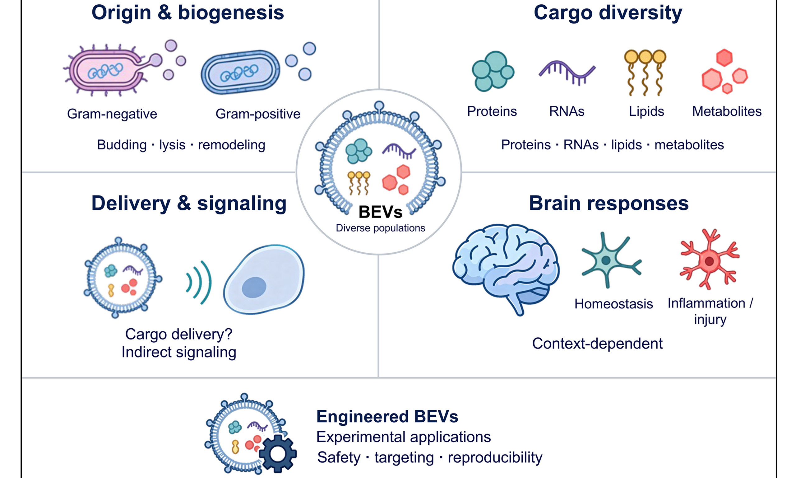
Abstract
Bacterial extracellular vesicles (BEVs) are potential mediators of the microbiota-gut-brain axis (MGBA). These membrane-enclosed nanoparticles protect their molecular cargo from gastrointestinal and systemic degradation, allowing bioactive signals to act at distant sites and potentially influence the central nervous system (CNS). This review covers the biogenesis of BEVs and their environmental regulation, the composition of their neuroactive cargo, the putative routes through which they reach the CNS, and their opposing roles in supporting homeostasis or driving neurodegeneration. We also discuss the prospects and current limitations of engineered BEVs platforms as CNS-targeted therapeutics. Overall, BEVs act as cargo-defined, context-dependent messengers in gut-brain communication.
Keywords


 Download PDF
INTRODUCTION
Communication between the gut microbiota and the central nervous system (CNS) occurs through immune, vagal, neuroendocrine, and metabolic pathways, collectively termed the microbiota-gut-brain axis (MGBA)[1,2]. Animal studies indicate that the microbiota contributes to blood-brain barrier (BBB) development and microglial homeostasis[3,4]. Experimental alterations of the microbiota have also been associated with behavioral and metabolic changes in models of neurological and psychiatric disorders[5-8]. Human studies and systematic reviews have linked intestinal dysbiosis to Alzheimer’s disease (AD), Parkinson’s disease (PD), autism spectrum disorder (ASD), and major depressive disorder (MDD)[9-15]. Although the biological importance of gut-brain crosstalk is no longer seriously doubted, the molecular vehicles that relay microbial signals to the CNS remain only partially characterized.
Most investigations of microbial effectors in gut-brain signaling have focused on soluble metabolites, including short-chain fatty acids[16], indole derivatives[17], and bile acid conjugates[18], or on intact microbial cells. Bacterial extracellular vesicles (BEVs) provide another potential route for this communication[19,20]. BEVs are membrane-enclosed particles released by both Gram-negative and Gram-positive bacteria; reported sizes commonly span tens to several hundred nanometers[21,22]. Their lipid bilayers can protect associated cargo, although stability depends on vesicle composition and environmental conditions[23]. Biological activity depends on the producing strain, cargo, exposure, and host state. Selected commensal-derived preparations have supported barrier integrity and immune regulation in experimental models[24]. Other preparations contain proinflammatory lipopolysaccharide (LPS), virulence factors, or cargo associated with tissue injury[25]. These findings support a role for microbiota-derived extracellular vesicles (EVs) in interkingdom communication[26]. Existing reviews have examined BEVs in gut-brain communication and therapeutic applications[19,20,27,28]. However, many studies establish cargo composition, delivery, and CNS outcomes separately, without connecting them into a causal pathway.
Here, we apply a cargo-delivery-function framework, with vesicle biogenesis as a source of heterogeneity, to critically assess these connections. We examine direct CNS delivery alongside peripheral immune, metabolic, and vagal signaling, while distinguishing evidence from gut-resident bacteria from findings involving oral pathobionts or mammalian EVs. We also assess whether experimental administration informs endogenous gut-derived BEVs activity. By identifying supported links and gaps in causal evidence, this review defines priorities for mechanistic validation and the development of CNS-directed BEVs therapeutics.
CLASSIFICATION AND BIOGENESIS OF BEVs
BEVs biogenesis is shaped by the bacterial cell envelope. Commonly described categories include outer membrane vesicles (OMVs), explosive outer membrane vesicles (EOMVs), and outer-inner membrane vesicles (OIMVs) from Gram-negative bacteria, and cytoplasmic membrane vesicles (CMVs) from Gram-positive bacteria[22,29]. These categories describe membrane origin, topology, and aspects of biogenesis. Table 1 summarizes representative features.
Classification and representative features of bacterial extracellular vesicles
| Vesicle type | Reported size (nm) | Membrane topology | Biogenesis | Representative cargo | Representative bacteria | References |
| OMV | 20-250 | Single outer-membrane bilayer | Outer-membrane budding after local envelope remodeling or stress | LPS, outer-membrane and periplasmic proteins; selected RNAs | Escherichia coli; Salmonella spp.; Porphyromonas gingivalis; Helicobacter pylori; Bacteroides thetaiotaomicron | [22,29-31] |
| EOMV | 50-250 | Membrane fragments reassemble after lysis; composition can be heterogeneous | Endolysin-associated explosive lysis and membrane reassembly | Membrane components and released cytoplasmic material | Pseudomonas aeruginosa | [22,32] |
| OIMV | 40-260 | Outer and inner membrane bilayers enclose cytoplasmic material | Double-membrane protrusion; structural class is not uniquely diagnostic of a lytic origin | DNA, cytoplasmic proteins, and ribosome-like material | P. aeruginosa; Neisseria gonorrhoeae; Acinetobacter baumannii | [21,22] |
| CMV | 20-400 | Single cytoplasmic-membrane bilayer | Wall remodeling, membrane-active modulins, or prophage-associated wall damage | Lipoteichoic acid, lipoproteins, cytoplasmic proteins, nucleic acids | Staphylococcus aureus; Bacillus subtilis; Lacticaseibacillus casei | [25,29,33-38] |
OMV biogenesis in Gram-negative bacteria
Classical OMVs form by outward budding of the outer membrane through several non-mutually exclusive mechanisms. These include localized weakening of Braun’s lipoprotein (Lpp)-peptidoglycan (PG) links[30], accumulation of PG fragments or misfolded proteins in the periplasm[39], and altered phospholipid transport through the VacJ/Yrb ATP-binding cassette (ABC) system[40]. Lipid A remodeling and reduced outer membrane protein A (OmpA)-PG attachment can also favor membrane curvature and release[31]. In Escherichia coli, YdcZ interacts with YdiH and BssR to regulate vesicle formation and selective protein loading[41]. In Vibrio fischeri, rotation of sheathed flagella promotes OMV release, providing evidence for a mechanical contribution to biogenesis[42]. Different pathways can coexist within a bacterial population. This heterogeneity complicates attribution of specific cargo and functions to individual vesicle subpopulations.
Explosive lysis and double-bilayer vesicles
Beyond orderly blebbing, phage-derived endolysins can trigger explosive lysis, as demonstrated in Pseudomonas aeruginosa. Released membrane fragments can reassemble into vesicles[32]. OIMVs contain both outer and inner membranes and can enclose cytoplasmic material. OIMVs have been observed in Neisseria gonorrhoeae, P. aeruginosa, and Acinetobacter baumannii; double-membrane protrusion was directly visualized in N. gonorrhoeae[21]. Double-membrane topology therefore does not by itself establish an explosive-lysis origin. The relative contribution of these pathways to the intestinal vesicle pool remains unknown. Density-gradient and size-exclusion purification alone do not reliably assign biogenesis subtypes. The Minimal Information for Studies of Extracellular Vesicles 2023 (MISEV2023) recommendations include bacterial EVs, but subtype assignment requires organism- and preparation-specific structural evidence[43].
CMV biogenesis in Gram-positive bacteria
The thick PG layer of Gram-positive bacteria was once thought to prevent vesicle release[33]. However, CMVs have been isolated from Staphylococcus aureus, Bacillus subtilis, and Lacticaseibacillus casei[34-36]. Autolysin-mediated PG hydrolysis facilitates passage through the cell wall, as demonstrated for Sle1 in S. aureus[34]. Phenol-soluble modulins (PSMs) promote membrane remodeling and vesicle release; PSM-deficient mutants show reduced production in the studied conditions[34,37]. Prophage-encoded holin-endolysin systems can also promote vesiculation through cell-wall damage[44]. In B. subtilis, endolysin-generated openings permit cytoplasmic membrane protrusion and vesicle release, followed by membrane failure and cell death[35]. The relative contribution of each mechanism depends on the strain and growth conditions.
Environmental regulation of vesicle production in the gut niche
Vesicle production is not a fixed constitutive process, but rather, it responds dynamically to environmental cues, a property that is directly relevant to the gut ecosystem. Antibiotic exposure, nutrient limitations, oxidative and envelope stress, temperature shifts, and host immune pressure can alter OMV production rates, size distributions, and cargo loading preferences[39,45-49]. For example, cysteine or other essential amino acid deprivation stimulates vesicle production in Neisseria meningitidis and Francisella spp., respectively[45,48], whereas hydrogen peroxide and elevated oxygen tension promote OMV release in P. aeruginosa and
Temperature also affects vesicle production and cargo loading; however, the direction and magnitude of these effects vary among bacterial species. In S. aureus, temperature variation alters EV abundance, size, and protein and RNA cargo, emphasizing the context-dependent nature of thermal regulation[49]. Subinhibitory exposure to the β-lactam antibiotic ceftazidime increases membrane-vesicle formation in A. baumannii[53], while gentamicin exposure increases OMV abundance and alters vesicular protein composition in susceptible extraintestinal pathogenic E. coli[54].
These observations may have implications for patients receiving long-term antibiotic therapy, although their relevance to BEVs production in the human gut remains to be established. Within the gastrointestinal tract, the local chemical environment varies dramatically along the cephalocaudal axis: the pH, bile salt concentration, oxygen tension, and antimicrobial peptide gradients each impose selective pressures on the resident microbiota. Host-like changes in pH, divalent-cation availability, and antimicrobial exposure have been shown to affect OMV production, size, and lipid composition in Salmonella[47]. However, the ways in which these gradients shape the vesiculation behavior of individual gut commensals and pathobionts in situ remain largely unexplored. Single-species in vitro assays capture only a fraction of the regulatory complexity; future work employing microfluidic gut-on-chip models and in vivo vesicle-tracking reporters will be essential for mapping the spatiotemporal dynamics of BEVs production across intestinal niches.
NEUROACTIVE CARGO REPERTOIRE OF BEVs
BEVs preparations contain diverse cargo classes[29], including pattern recognition receptor (PRR) ligands[38,55,56], neuroactive metabolites[57], regulatory RNAs[58], and catalytically active enzymes[59]. Detection in bulk preparations does not establish that these components coexist within individual vesicles. We therefore distinguish cargo diversity from demonstrated co-packaging and evaluate the functional evidence for each class separately [Figure 1].
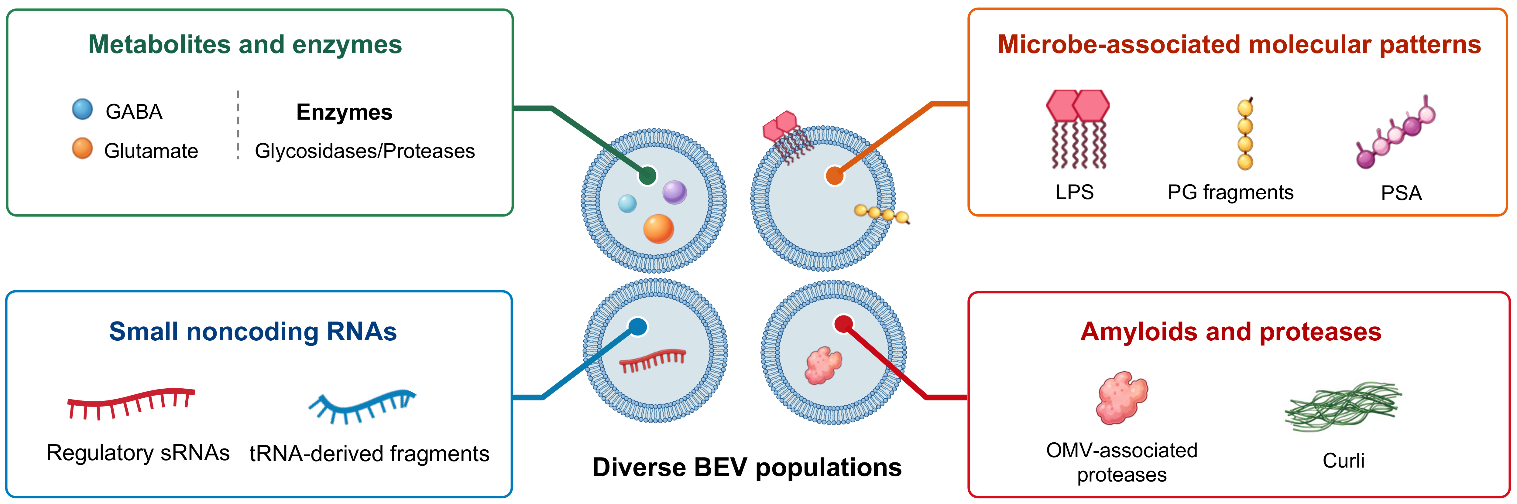
Figure 1. Cargo diversity across bacterial extracellular vesicle populations. Representative cargo classes include metabolites and enzymes, microbe-associated molecular patterns, sRNAs, and amyloids and proteases. Separate vesicles illustrate population-level diversity and do not imply universal co-packaging. The contribution of vesicular delivery to curli-mediated effects remains unresolved. BEVs: Bacterial extracellular vesicles; GABA: γ-aminobutyric acid; LPS: lipopolysaccharide; OMVs: outer membrane vesicles; PG: peptidoglycan; PSA: polysaccharide A; sRNAs: small noncoding RNAs; tRNA: transfer RNA.
Neuroactive metabolites and biosynthetic enzymes
BEVs preparations contain neuroactive metabolites and associated metabolic enzymes, suggesting potential vesicle-associated metabolic activity. A metabolomic and metaproteomic study of stool-derived and ex vivo microbiota-derived vesicles reported neuroactive metabolites and enzymes associated with glutamine-glutamate-γ-aminobutyric acid (GABA) metabolism[57]. GABA was also detected in vesicles from a cultured Bacteroides finegoldii isolate. These findings establish cargo association but do not demonstrate neurotransmitter synthesis within circulating vesicles. Metabolite annotations, particularly xenobiotic-like signals such as gabapentin, require confirmation before being interpreted as products of bacterial biosynthesis. Functional enzyme activity has been demonstrated in Bacteroides-derived OMVs enriched in acidic glycosidases and proteases[59]. Separately, Bacteroides species synthesize sphingolipids that contribute to intestinal homeostasis and host immune regulation[60], while microbial tryptophan metabolites can influence CNS-relevant aryl hydrocarbon receptor (AhR) signaling[17]. Vesicular packaging and sustained production of these metabolites at distant sites require direct validation.
Microbe-associated molecular patterns
Microbe-associated molecular patterns (MAMPs) are an important class of immunologically active BEVs cargo. Gram-negative OMVs contain LPS and can deliver PG fragments to host cells. These components engage distinct receptors, including Toll-like receptor 4 (TLR4) and nucleotide-binding oligomerization domain-containing protein 1 (NOD1); the cited epithelial-cell study directly demonstrated NOD1 activation[61]. Gram-positive vesicles can contain lipoteichoic acid and lipoproteins that engage Toll-like receptor 2 (TLR2)[38,62]. Vesicle-associated presentation can alter cellular delivery and receptor activation, but its potency depends on the preparation, dose metric, and target cell[62]. The number of intact BEVs reaching brain parenchyma under physiological conditions remains unknown.
LPS preparations differ in their biological activity. Lipid A acylation and phosphate position influence TLR4 responses, so LPS from different bacteria should not be treated as functionally interchangeable[63]. Bulk endotoxin measurements alone do not resolve lipid A structure or predict all host responses. Structural analysis and functional receptor assays can therefore complement measurements of circulating LPS-positive BEVs[64]. Capsular polysaccharide A (PSA) from Bacteroides fragilis provides another example of context-dependent immune signaling. PSA-containing OMVs engage dendritic-cell TLR2 and promote regulatory T-cell responses and interleukin-10 (IL-10) production[55]. This finding complements earlier evidence that PSA contributes to host immune maturation[65]. It does not establish that PSA activity is exclusive to vesicular presentation. Ligand identity, molecular structure, and delivery context should all be considered when evaluating BEV-mediated neuroimmune signaling.
Small noncoding RNAs
BEVs carry small noncoding RNAs (sRNAs), including regulatory RNAs and transfer RNA-derived fragments. Selected RNAs can retain biological activity after uptake by host cells, although cargo selection and function vary among species and preparations[66]. Enterotoxigenic and nontoxigenic B. fragilis OMVs differ in their molecular composition and metabolic activity[67]. P. aeruginosa OMVs deliver an sRNA to human airway epithelial cells and reduce interleukin-8 (IL-8) secretion in the studied system[58]. OMV-associated extracellular RNAs from the oral pathogen Aggregatibacter actinomycetemcomitans promote tumor necrosis factor alpha (TNF-α) production in human macrophages, and brain-associated RNA signal has been reported after administration in mice[68]. These findings should not be interpreted as proof of intact gut-derived vesicle entry into brain parenchyma. The abundance, host targets, and intracellular mechanism of transferred bacterial RNAs require further investigation, including whether particular effects depend on host Argonaute proteins[58,66].
Amyloidogenic and proteolytic cargo
Bacterial amyloids and proteolytic virulence factors have potential relevance to neurodegeneration, but evidence for their vesicular delivery differs. Curli produced by E. coli and Salmonella species form β-sheet-rich fibrils and can promote aggregation of host amyloidogenic proteins. Gut bacterial curli accelerated
Proteolytic virulence factors provide more direct examples of biologically active vesicle-associated cargo. Porphyromonas gingivalis produces the lysine-specific gingipain Kgp and arginine-specific gingipains RgpA/RgpB in soluble and OMV-associated forms[70]. Gingipains detected in postmortem AD brain tissue correlate with tau and ubiquitin pathology. In P. gingivalis-challenged BALB/c mice, gingipain inhibition reduced cerebral bacterial burden and neurodegenerative changes[71]. The human findings are associative, whereas the mouse experiment supports a mechanistic contribution in that model. Neither establishes a causal role for gingipains in human AD or their delivery into the brain by intact vesicles[72]. Outside the CNS, Fusobacterium nucleatum OMVs containing Fusobacterium adhesin A (FadA) aggravated rheumatoid arthritis in an experimental study[73]. S. aureus vesicles also induced atopic dermatitis-like inflammation[74]. These peripheral examples justify investigating cargo-specific effects but do not establish corresponding CNS mechanisms.
Current limitations and the emerging role of multiomics approaches
Current cargo inventories derive mainly from bulk preparations and a limited range of culturable species. Important questions remain about the in vivo proteome, metabolome, and RNA content of gut BEVs, and whether particular cargo molecules coexist within individual vesicles. Molecular heterogeneity, including lipid A acylation, RNA modifications, and amyloid conformations, may further influence biological activity. Single-vesicle methods can help resolve selected cargo combinations, but each platform has limits in sensitivity, specificity, and throughput[75]. Combining vesicle analysis with single-cell and spatial profiling of recipient host cells could connect cargo exposure to cellular responses. Such measurements must be paired with perturbation and transport experiments to test causality.
ROUTES LINKING GUT-DERIVED BEVs TO CNS RESPONSES
Gut-derived BEVs may influence CNS function through direct delivery of vesicles or cargo, or indirectly through peripheral immune, metabolic, and neural signaling[19]. Delivery from the gut lumen to brain parenchyma through the circulation requires passage across the intestinal barrier and BBB[76]. Vagal signaling is another potential link between intestinal BEVs exposure and CNS responses[27,77]. The proposed routes and their different levels of supporting evidence are summarized in Figure 2 and Table 2. We distinguish vesicle-associated signals from demonstrated transport of intact vesicles.
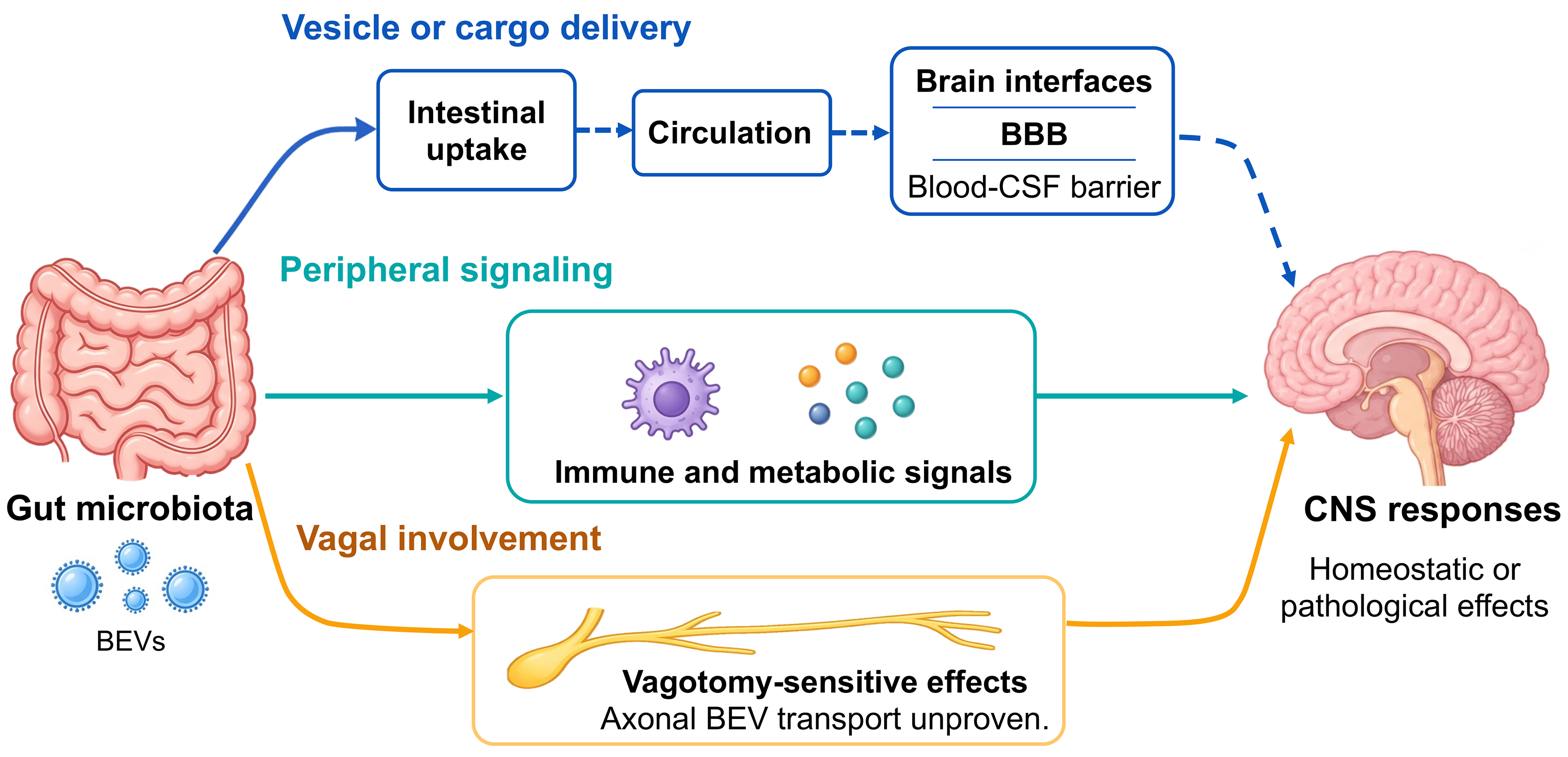
Figure 2. Proposed routes of BEV-mediated gut-brain communication. Vesicle or cargo delivery, peripheral immune and metabolic signaling, and vagal involvement may contribute to homeostatic or pathological CNS responses. The BBB and blood-CSF barrier represent distinct candidate interfaces. Solid arrows indicate observations supported in selected experimental models; dashed arrows indicate proposed delivery steps. Intact-vesicle entry into brain tissue remains unresolved, and vagotomy-sensitive effects do not establish axonal BEVs transport. BEVs: Bacterial extracellular vesicles; BBB: blood-brain barrier; CNS: central nervous system; CSF: cerebrospinal fluid.
Representative evidence for bacterial vesicle delivery and CNS-related effects
| Evidence or route | Representative system | Observed result | Interpretive limit | References |
| Intestinal uptake | Bacteroides thetaiotaomicron OMVs; epithelial cells and oral mouse administration | Cellular uptake and extraintestinal fluorescent signal | Fluorescence does not establish intact brain entry | [78] |
| Producer-derived distal cargo | Engineered B. thetaiotaomicron colonization in mice | Active NanoLuc reporter detected at distal sites, including brain | Cargo activity does not resolve intact-vesicle transit across the BBB | [79] |
| BBB interaction in vitro | B. thetaiotaomicron BEVs; multicellular barrier models | Barrier and cellular responses in controlled models | Does not quantify endogenous gut-to-brain vesicle flux in vivo | [80] |
| Barrier injury and inflammation | Porphyromonas gingivalis OMVs administered to mice | Glial activation, tight junction changes, tau phosphorylation | Oral pathobiont and experimental exposure; not universal gut-BEV transport | [81,82] |
| Vagal involvement | Paenalcaligenes hominis EVs; mice with or without vagotomy | Vagotomy attenuated cognitive effects | Supports neural involvement; intact axonal vesicle transport unproven | [77] |
| AD-model pathology | Germ-free AppNL-G-F mice; oral plus intrarectal fecal BEVs, three times weekly for three weeks | Increased amyloid-β plaque pathology; limited microglial effects | No behavioral assay or inflammatory-marker panel; not evidence of cognitive decline | [83] |
| AD-model cognition and inflammation | APP/PS1 mice; AD donor fecal OMV preparations, 120-day gavage | Impaired cognition; increased inflammatory markers and amyloid-β production | Prolonged exogenous dosing; cannot establish physiological exposure | [84] |
Traversal of the intestinal epithelial barrier
BEVs may cross the intestinal epithelium through paracellular passage or cellular uptake and transport. Barrier disruption can facilitate passage, although transport has also been studied in models without overt epithelial injury. Patients with intestinal barrier dysfunction have elevated circulating levels of LPS-positive BEVs[64]. B. fragilis OMVs are taken up by dendritic cells and induce immunoregulatory responses[55]; this finding does not establish microfold-cell transcytosis as their route across the epithelium. Bacteroides thetaiotaomicron OMVs undergo rapid epithelial uptake and can traverse primary intestinal epithelial monolayers[78]. A separate reporter study demonstrated transfer of microbial biomolecules to host cells
Systemic circulation and hepatic clearance
Once in the bloodstream, BEVs may be taken up by the mononuclear phagocyte system, although BEV-specific pharmacokinetic data remain limited. Intraperitoneally administered E. coli OMVs accumulated in the liver, lungs, spleen, and kidneys[85]. This experiment describes exogenous administration rather than endogenous gut-to-brain delivery. General principles established in mammalian EV studies provide a conceptual framework but should not be presented as BEV-specific pharmacokinetic measurements[86]. Plasma half-life, organ-specific uptake, and the brain-associated fraction of endogenous gut-derived BEVs remain poorly quantified. Tissue detection of vesicular components or labels does not alone establish intact-vesicle entry into brain parenchyma. Translocated OMVs can nevertheless affect distant organs; gut pathobiont-derived OMVs promote liver inflammation and fibrosis in primary sclerosing cholangitis-associated inflammatory bowel disease[87].
Crossing the BBB
The BBB contains tightly connected brain microvascular endothelial cells that restrict entry into brain parenchyma. Several candidate routes or interfaces may contribute to CNS exposure, but their anatomical location and supporting evidence differ.
Barrier disruption is one candidate route. Oral administration of P. gingivalis OMVs reduced brain tight junction gene expression and occludin protein abundance and was associated with NOD-like receptor family pyrin domain-containing 3 (NLRP3) inflammasome activation, tau phosphorylation, and memory impairment in mice[81]. These observations do not establish direct gingipain-mediated cleavage of BBB claudin-5 or occludin. A comparison of P. gingivalis and its OMVs found different inflammatory responses and tissue-associated signals after exposure[82]. Another study linked OMV exposure to BBB dysfunction and glycogen synthase kinase 3β-associated tau phosphorylation[88]. Together, these findings support barrier dysfunction as a possible contributor to CNS exposure, but do not resolve whether intact vesicles, released cargo, or secondary inflammatory mediators cross the barrier.
Receptor-mediated transcytosis is a candidate route across an intact BBB, but BEV-specific receptor mechanisms remain incompletely defined. Studies of mammalian-cell-derived EVs found that transport depended on vesicle origin and could change after systemic LPS exposure[89]. These findings are not direct evidence for BEVs transcytosis. OmpA contributes to invasion of brain microvascular endothelial cells by intact E. coli[90], but this does not establish an equivalent receptor-dependent pathway for OMVs. The relevant receptors and in vivo kinetics of gut-resident BEVs transport remain unresolved.
The choroid plexus is a candidate route across the blood-cerebrospinal fluid (CSF) barrier rather than the BBB proper. After repeated intraperitoneal administration of P. gingivalis OMVs, gingipain immunoreactivity was detected around cerebral ventricles, in the choroid plexus, and in ependymal cells[91]. This distribution does not identify the transported entity or the route taken. Entry from choroid plexus capillaries into CSF would still require passage across the choroid plexus epithelial barrier. Cathepsin B-associated stress-activated protein kinase/c-Jun N-terminal kinase (SAPK/JNK) signaling has been implicated in downstream pathology after OMV exposure[92], but does not establish a transport mechanism.
A further hypothesis is transport within migrating leukocytes, sometimes termed a Trojan horse mechanism. Postmortem detection of bacterial 16S ribosomal RNA (rRNA) gene sequences does not identify vesicles or establish this route[93]. Such signals may reflect bacterial components, intact bacteria, or contamination. BEV-associated neuroinflammatory changes in the brain and trigeminal ganglion likewise do not identify the carrier or route of entry[94]. Direct evidence that leukocytes transport native gut-derived BEVs across the BBB remains insufficient.
Mechanistic BBB studies have emphasized oral pathobionts, but relevant evidence also exists for gut commensals. B. thetaiotaomicron BEVs have been studied in multicellular intestinal and BBB models, with vesicle-associated signals detected in recipient neuronal cells[80]. Gut microbiota-derived preparations have also been tested in epithelial and brain endothelial transport models[57]. More recently, colonization with B. thetaiotaomicron engineered to produce NanoLuc-labeled BEVs yielded reporter activity in distal organs, including the brain[79]. This advances evidence for endogenous microbial cargo delivery. It does not establish the precise in vivo route or continuously visualize intact vesicles crossing the BBB. Receptor dependence, vesicle integrity during transit, and quantitative delivery to brain cell types remain unresolved.
Transport experiments also require attention to labeling, dose, and purity. Lipophilic dyes such as PKH26 and DiR can yield signals from free dye or dye-derived particles, while labeled cargo can separate from vesicles. Dye-only controls, removal of free label, independent membrane and cargo reporters, and cellular or ultrastructural localization strengthen interpretation[43]. Experimentally concentrated preparations may not reproduce endogenous exposure, which is poorly quantified. Mixed vesicle subtypes and co-isolated material further limit attribution to intact OMVs. These limitations preclude equating brain-associated signal with verified vesicle entry into parenchyma.
The vagal nerve route
The vagus nerve may transmit BEV-induced signals from the gut without requiring BBB crossing. Evidence for this mechanism rests largely on a study of Paenalcaligenes hominis EVs in mice, in which bilateral subdiaphragmatic vagotomy attenuated cognitive impairment[77]. Vagotomy also changes gut motility, immune regulation, and neuroendocrine signaling. The result therefore supports vagal involvement but does not demonstrate retrograde axonal transport of intact BEVs. Helicobacter pylori OMVs have been linked to AD-related pathology through complement component 3-C3a receptor (C3-C3aR) signaling, with hematogenous and vagal mechanisms proposed[95]. Gastric infection studies provide additional, indirect support for gut-to-brain inflammatory signaling[96]. The relative contributions of intact vesicles, released cargo, circulating mediators, and neural activation remain unresolved.
BEVs AS FUNCTIONAL MODULATORS OF CNS HOMEOSTASIS AND PATHOLOGY
The effects of BEVs on the CNS depend on their cargo composition, bacterial source, dose, route of exposure, and host physiological state[28]. BEVs from selected commensal strains have shown neuroprotective effects in experimental models, whereas those from certain pathobionts have been associated with barrier dysfunction, neuroinflammation, and neurodegenerative changes. These outcomes are context-dependent and may involve direct cargo delivery or indirect peripheral signaling. Here, we examine the evidence for protective and detrimental effects, distinguishing observed CNS outcomes from the mechanisms proposed to explain them.
Commensal BEV-mediated neuroprotection
Akkermansia muciniphila-derived extracellular vesicles (AmEVs) are among the best-studied commensal BEVs with reported neuroprotective effects. In a postoperative cognitive dysfunction model induced by intestinal ischemia-reperfusion, AmEVs suppressed microglial activation through TLR2/4-associated signaling. Pharmacological microglial depletion abolished their reported protection of BBB integrity[97]. Earlier work showed improved intestinal barrier function and increased tight junction protein expression after AmEV treatment[98]. In a colitis-associated cognitive impairment model, AmEVs improved intestinal and BBB integrity, reduced hippocampal inflammation, and improved behavioral performance. They reduced excessive colonic serotonin while restoring hippocampal serotonin levels, indicating tissue- and disease-dependent regulation[24]. Garlic exosome-like nanoparticle exposure also altered the activity of A. muciniphila-derived OMVs, which improved high-fat-diet-associated metabolic dysfunction in mice[99]. This metabolic study should not be interpreted as direct evidence of cognitive benefit. In a smoking-exposure model, treatment with AmEVs or exogenous indole-3-lactic acid improved cognitive outcomes in association with AhR-dependent microglial regulation[100]. This does not by itself establish delivery of indole-3-lactic acid within intact AmEVs.
Neuroprotective effects are not restricted to Akkermansia. Lactiplantibacillus plantarum (formerly Lactobacillus plantarum)-derived EV treatment reduced ischemic neuronal injury and increased host microRNA-101a-3p (miR-101a-3p), implicating a c-Fos/transforming growth factor beta (TGF-β) pathway[101]. These findings support modulation of host microRNA signaling; they do not demonstrate delivery of bacterial vesicle-encapsulated microRNAs.
Pathobiont- and disease-associated BEVs effects
Oral gavage of P. hominis EVs induced cognitive impairment and colitis in mice. Subdiaphragmatic vagotomy attenuated the cognitive deficits without significantly reducing colitis[77]. Hippocampal fluorescein isothiocyanate (FITC) signals were more frequently detected after labeled EVs than labeled LPS, and vagotomy reduced EV-associated signals. However, the investigators did not detect intact EVs in the hippocampus by transmission electron microscopy. Label-associated signals therefore support exposure but do not establish intact vesicle transport. FITC can label molecules that later become detached or released; it should not be conflated with the micelle-forming behavior of lipophilic dyes. A structural study identified penta- and tetra-acylated lipid A in P. hominis LPS and found weaker TLR-dependent activity than for E. coli LPS[102]. This finding motivates tests of non-LPS cargo but does not establish its contribution to the neurological phenotype.
P. gingivalis OMV administration has been associated with hippocampal and cortical signals, reduced tight junction protein expression, glial activation, increased interleukin-1 beta (IL-1β), tau phosphorylation, and NLRP3 inflammasome activation in mice[81]. A broader review summarizes the systemic effects of
Moreover, two complementary studies implicate gut-derived BEVs in AD-related pathology. In germ-free AppNL-G-F mice, fecal BEVs from specific pathogen-free (SPF)-housed donors increased amyloid-β (Aβ) plaque burden and influenced microglial Aβ uptake and synaptic parameters[83]. In amyloid precursor protein/presenilin 1 (APP/PS1) mice, prolonged intragastric administration of AD patient-derived fecal OMVs impaired cognition, enhanced neuroinflammation and Aβ production, and disrupted intestinal barrier integrity and short-chain fatty acid homeostasis[84]. These findings suggest that gut-derived BEVs preparations can influence AD-related pathological processes independently of administered live bacteria.
Open mechanistic gaps
Several gaps limit causal interpretation. Many experiments use purified, concentrated preparations, and the corresponding endogenous exposure is often unknown. Strain, cargo, isolation method, and host state can alter the outcome. Preparations may contain several vesicle subtypes, free cargo, and co-isolated bacterial material[43,105]. Evidence that BEVs administration changes CNS phenotypes does not establish intact vesicle entry into the brain. Peripheral immune responses, altered barrier function, and neural signaling may contribute. Defined producer strains, appropriate nonvesicular controls, quantitative cargo measurements, and direct tracking of intact vesicles are needed to resolve these alternatives.
ENGINEERED BEVs AS CNS-TARGETED THERAPEUTIC PLATFORMS
BEV-based strategies aim to deliver beneficial signals or inhibit harmful signaling. Engineering the producing strain can alter vesicle cargo and surface properties, as shown by lpxM/msbB deletion to reduce endotoxicity[106] and oral administration of bacteria that produce antigen-bearing OMVs in situ[107]. Nevertheless, immunogenicity, barrier transport, and CNS targeting require formulation-specific validation. The four-component meningococcal serogroup B vaccine Bexsero contains an OMV component and provides a clinical precedent for OMV-containing vaccination[108,109]. It is administered as a multidose regimen. Experience with intramuscular vaccination cannot be extrapolated directly to chronic oral or intravenous BEVs treatment. Four challenges are particularly relevant to translation.
The first challenge is residual immunogenicity. Deleting lpxM can reduce lipid A-mediated TLR4 stimulation[106], but other components, including PG fragments, lipoproteins, flagellin, and bacterial DNA, may remain immunologically active. Repeated therapeutic exposure requires separate assessment of antibody responses, complement activation, and immune-mediated clearance. Multigene engineering and synthetic vesicle formulations may help control composition. However, reducing one inflammatory pathway does not establish overall safety or therapeutic efficacy[108].
The second challenge is limited CNS selectivity. Administered BEVs can accumulate in peripheral organs, and their quantitative brain delivery remains incompletely characterized[85]. In experimental disease models, neutrophil-hitchhiking OMVs enhanced delivery to ischemic brain tissue[110], brain-targeted Limosilactobacillus reuteri (formerly Lactobacillus reuteri) EVs reduced inflammation after stroke[111], and engineered OMVs carrying doxorubicin and CD47-targeting small interfering RNA (siRNA) improved antitumor responses in glioblastoma models[112]. These findings do not establish equivalent targeting across an intact BBB or in other CNS disorders. Oral delivery of engineered bacteria that produce OMVs in situ offers a different approach[107]. Its extension to neurological therapy requires tests of dose control, sustained exposure, and safety.
The third challenge is payload compatibility. Bacteria do not reproduce the full mammalian repertoire of posttranslational processing. Efficient loading and activity of complex therapeutic proteins therefore require direct validation. Brain-derived neurotrophic factor (BDNF), glial cell line-derived neurotrophic factor (GDNF), and antibody fragments are not intrinsically excluded from bacterial platforms, but folding, disulfide formation, localization, and biological activity must be optimized for each construct. Alternative approaches include production in another expression system followed by vesicle loading, or the use of mammalian EVs or synthetic carriers. Payload selection should follow experimental compatibility rather than a categorical restriction.
The fourth challenge concerns manufacturing consistency and regulatory requirements. Production, purification, and release criteria remain product-specific. Variation in vesicle size, cargo, and residual immunostimulatory activity can complicate comparisons among preparations[29,105]. A recent fecal-vesicle study found stable measured characteristics after frozen storage and an additional freeze-thaw cycle, providing a useful example of methodological reproducibility rather than a universal preservation standard[113]. Clinical development will require reproducible processes and validated potency and safety assays. Regulatory classification should follow each product’s composition, intended use, and mode of action, with early regulatory engagement.
CONCLUSIONS AND FUTURE PERSPECTIVES
BEVs are potential mediators of gut-brain communication that complement soluble microbial metabolites and intact bacterial cells. Their biogenesis and cargo are heterogeneous, and their effects depend on bacterial source, dose, route, and host state. Selected preparations support barrier function or reduce inflammation in experimental models; others promote neuroinflammatory or neurodegenerative phenotypes. These effects should not be generalized to all commensal or pathobiont vesicles. Evidence for the proposed transport routes is unequal. In particular, changes in CNS phenotypes do not prove intact BEVs entry into brain parenchyma, and vagotomy results support neural involvement rather than direct vesicle transport.
Several limitations temper current interpretations. Most studies use isolated vesicle preparations, although producer-colonization studies now provide evidence for distal delivery of gut bacterial cargo. Preparations may contain mixed vesicle populations and nonvesicular components. Bulk cargo analyses cannot establish molecular co-occurrence within individual BEVs. BBB studies include both oral pathobionts and selected gut commensals, but generalization across gut taxa remains unsupported. Definitive real-time visualization and quantification of intact endogenous BEVs traversing the BBB or undergoing retrograde vagal transport remain major evidence gaps. For therapeutic development, immunogenicity, CNS selectivity, payload compatibility, and manufacturing consistency require product-specific assessment.
Progress requires complementary approaches. Single-vesicle analysis should establish which cargo combinations occur in relevant populations. Defined producer strains, colonization models, and independent membrane and cargo reporters should test the links between vesicle formation, delivery, and CNS responses. Finally, standardized isolation, potency assays, safety evaluation, and manufacturing controls are needed for therapeutic development. These steps will clarify when BEVs are causal mediators, associated biomarkers, or experimental delivery tools in the MGBA.
DECLARATIONS
Authors’ contributions
Conceptualised the study: Cai S, Sheng J
Contributed to literature curation: Cai S, Hu Z, Ma X
Led the drafting of the manuscript: Cai S
Contributed to visualization: Cai S, Hu Z
Contributed to writing - review and editing: Cai S, Hu Z, Ma X, Sheng J
Supervised the study: Sheng J
Contributed to funding acquisition and project administration: Sheng J
All authors have read and approved the manuscript.
Availability of data and materials
Not applicable.
AI and AI-assisted tools statement
During the preparation of this manuscript, the AI tools Claude (version Opus 4.7, released 2026-04-16) and ChatGPT (version GPT-5.5, released 2026-04-23) were used solely for language editing without unsupervised automatic generation. These tools did not influence the study design, data collection, analysis, interpretation, or the scientific content of the work. All authors take full responsibility for the accuracy, integrity, and final content of the manuscript.
Financial support and sponsorship
This work was supported by the National Natural Science Foundation of China (U22A20358) and the Zhejiang Provincial Natural Science Foundation of China (LR25H260001).
Conflicts of interest
All authors declared that there are no conflicts of interest.
Ethical approval and consent to participate
Not applicable.
Consent for publication
Not applicable.
Copyright
© The Author(s) 2026.
REFERENCES
1. Cryan JF, O’Riordan KJ, Cowan CSM, et al. The microbiota-gut-brain axis. Physiol Rev. 2019;99:1877-2013.
2. Morais LH, Schreiber HL 4th, Mazmanian SK. The gut microbiota-brain axis in behaviour and brain disorders. Nat Rev Microbiol. 2021;19:241-55.
3. Braniste V, Al-Asmakh M, Kowal C, et al. The gut microbiota influences blood-brain barrier permeability in mice. Sci Transl Med. 2014;6:263ra158.
4. Erny D, Hrabě de Angelis AL, Jaitin D, et al. Host microbiota constantly control maturation and function of microglia in the CNS. Nat Neurosci. 2015;18:965-77.
5. de Theije CG, Wopereis H, Ramadan M, et al. Altered gut microbiota and activity in a murine model of autism spectrum disorders. Brain Behav Immun. 2014;37:197-206.
6. Sampson TR, Debelius JW, Thron T, et al. Gut microbiota regulate motor deficits and neuroinflammation in a model of Parkinson’s disease. Cell. 2016;167:1469-80.e12.
7. Sharon G, Cruz NJ, Kang DW, et al. Human gut microbiota from autism spectrum disorder promote behavioral symptoms in mice. Cell. 2019;177:1600-18.e17.
8. Zheng P, Zeng B, Zhou C, et al. Gut microbiome remodeling induces depressive-like behaviors through a pathway mediated by the host’s metabolism. Mol Psychiatry. 2016;21:786-96.
9. Liu S, Gao J, Zhu M, Liu K, Zhang HL. Gut microbiota and dysbiosis in Alzheimer’s disease: implications for pathogenesis and treatment. Mol Neurobiol. 2020;57:5026-43.
10. Ferreiro AL, Choi J, Ryou J, et al. Gut microbiome composition may be an indicator of preclinical Alzheimer’s disease. Sci Transl Med. 2023;15:eabo2984.
11. Huang B, Chau SWH, Liu Y, et al. Gut microbiome dysbiosis across early Parkinson’s disease, REM sleep behavior disorder and their first-degree relatives. Nat Commun. 2023;14:2501.
12. Korteniemi J, Karlsson L, Aatsinki A. Systematic review: autism spectrum disorder and the gut microbiota. Acta Psychiatr Scand. 2023;148:242-54.
13. Zhao H, Tao L, Tang C, Cai W, Shen W. Do immune system and microbiome-gut-brain axis interactions associate with major depressive disorder? J Transl Med 2025;23:1279.
14. Nikolova VL, Smith MRB, Hall LJ, Cleare AJ, Stone JM, Young AH. Perturbations in gut microbiota composition in psychiatric disorders: a review and meta-analysis. JAMA Psychiatry. 2021;78:1343-54.
15. Valles-Colomer M, Falony G, Darzi Y, et al. The neuroactive potential of the human gut microbiota in quality of life and depression. Nat Microbiol. 2019;4:623-32.
16. Dalile B, Van Oudenhove L, Vervliet B, Verbeke K. The role of short-chain fatty acids in microbiota-gut-brain communication. Nat Rev Gastroenterol Hepatol. 2019;16:461-78.
17. Agus A, Planchais J, Sokol H. Gut microbiota regulation of tryptophan metabolism in health and disease. Cell Host Microbe. 2018;23:716-24.
18. MahmoudianDehkordi S, Arnold M, Nho K, et al. ; Alzheimer’s Disease Neuroimaging Initiative and the Alzheimer Disease Metabolomics Consortium. Altered bile acid profile associates with cognitive impairment in Alzheimer’s disease-an emerging role for gut microbiome. Alzheimers Dement. 2019;15:76-92.
19. Pirolli NH, Bentley WE, Jay SM. Bacterial extracellular vesicles and the gut-microbiota brain axis: emerging roles in communication and potential as therapeutics. Adv Biol. 2021;5:e2000540.
20. Xie J, Haesebrouck F, Van Hoecke L, Vandenbroucke RE. Bacterial extracellular vesicles: an emerging avenue to tackle diseases. Trends Microbiol. 2023;31:1206-24.
21. Pérez-Cruz C, Delgado L, López-Iglesias C, Mercade E. Outer-inner membrane vesicles naturally secreted by gram-negative pathogenic bacteria. PLoS One. 2015;10:e0116896.
22. Toyofuku M, Nomura N, Eberl L. Types and origins of bacterial membrane vesicles. Nat Rev Microbiol. 2019;17:13-24.
23. Sartorio MG, Pardue EJ, Feldman MF, Haurat MF. Bacterial outer membrane vesicles: from discovery to applications. Annu Rev Microbiol. 2021;75:609-30.
24. Chen X, Li Q, Zhang W, et al. Akkermansia muciniphila-derived extracellular vesicles alleviate colitis-related cognitive impairment via tryptophan metabolic reprogramming of the gut-brain axis. Gut Microbes. 2026;18:2611546.
25. Kim JH, Lee J, Park J, Gho YS. Gram-negative and Gram-positive bacterial extracellular vesicles. Semin Cell Dev Biol. 2015;40:97-104.
26. Díaz-Garrido N, Badia J, Baldomà L. Microbiota-derived extracellular vesicles in interkingdom communication in the gut. J Extracell Vesicles. 2021;10:e12161.
27. Kaisanlahti A, Salmi S, Kumpula S, et al. Bacterial extracellular vesicles - brain invaders? A systematic review. Front Mol Neurosci. 2023;16:1227655.
28. Arenas YM, Izquierdo-Altarejos P, Pérez-Martínez G, Felipo V, Llansola M. Bacterial extracellular vesicles in the brain: Pathological effects and therapeutic possibilities. Neural Regen Res. 2026;21:2101-9.
29. Toyofuku M, Schild S, Kaparakis-Liaskos M, Eberl L. Composition and functions of bacterial membrane vesicles. Nat Rev Microbiol. 2023;21:415-30.
30. Schwechheimer C, Kuehn MJ. Outer-membrane vesicles from Gram-negative bacteria: biogenesis and functions. Nat Rev Microbiol. 2015;13:605-19.
31. Juodeikis R, Carding SR. Outer membrane vesicles: biogenesis, functions, and issues. Microbiol Mol Biol Rev. 2022;86:e0003222.
32. Turnbull L, Toyofuku M, Hynen AL, et al. Explosive cell lysis as a mechanism for the biogenesis of bacterial membrane vesicles and biofilms. Nat Commun. 2016;7:11220.
33. Brown L, Wolf JM, Prados-Rosales R, Casadevall A. Through the wall: extracellular vesicles in Gram-positive bacteria, mycobacteria and fungi. Nat Rev Microbiol. 2015;13:620-30.
34. Wang X, Thompson CD, Weidenmaier C, Lee JC. Release of Staphylococcus aureus extracellular vesicles and their application as a vaccine platform. Nat Commun. 2018;9:1379.
35. Toyofuku M, Cárcamo-Oyarce G, Yamamoto T, et al. Prophage-triggered membrane vesicle formation through peptidoglycan damage in Bacillus subtilis. Nat Commun. 2017;8:481.
36. da Silva Barreira D, Lapaquette P, Novion Ducassou J, Couté Y, Guzzo J, Rieu A. Spontaneous prophage induction contributes to the production of membrane vesicles by the Gram-positive bacterium Lacticaseibacillus casei BL23. mBio. 2022;13:e0237522.
37. Schlatterer K, Beck C, Hanzelmann D, et al. The mechanism behind bacterial lipoprotein release: phenol-soluble modulins mediate toll-like receptor 2 activation via extracellular vesicle release from Staphylococcus aureus. mBio. 2018;9:e01851-18.
38. Bitto NJ, Cheng L, Johnston EL, et al. Staphylococcus aureus membrane vesicles contain immunostimulatory DNA, RNA and peptidoglycan that activate innate immune receptors and induce autophagy. J Extracell Vesicles. 2021;10:e12080.
39. McBroom AJ, Kuehn MJ. Release of outer membrane vesicles by Gram-negative bacteria is a novel envelope stress response. Mol Microbiol. 2007;63:545-58.
40. Roier S, Zingl FG, Cakar F, et al. A novel mechanism for the biogenesis of outer membrane vesicles in Gram-negative bacteria. Nat Commun. 2016;7:10515.
41. Li J, Li C, Han Y, et al. Unveiling a novel mechanism of enhanced secretion, cargo loading, and accelerated dynamics of bacterial extracellular vesicles following antibiotic exposure. J Extracell Vesicles. 2025;14:e70131.
42. Aschtgen MS, Lynch JB, Koch E, Schwartzman J, McFall-Ngai M, Ruby E. Rotation of vibrio fischeri flagella produces outer membrane vesicles that induce host development. J Bacteriol. 2016;198:2156-65.
43. Welsh JA, Goberdhan DCI, O’Driscoll L, et al. ; MISEV Consortium. Minimal information for studies of extracellular vesicles (MISEV2023): from basic to advanced approaches. J Extracell Vesicles. 2024;13:e12404.
44. Bołoz A, Lannoy V, Olszak T, Drulis-Kawa Z, Augustyniak D. Interplay between bacterial extracellular vesicles and phages: receptors, mechanisms, and implications. Viruses. 2025;17:1180.
45. van de Waterbeemd B, Zomer G, van den Ijssel J, et al. Cysteine depletion causes oxidative stress and triggers outer membrane vesicle release by Neisseria meningitidis; implications for vaccine development. PLoS One. 2013;8:e54314.
46. Macdonald IA, Kuehn MJ. Stress-induced outer membrane vesicle production by Pseudomonas aeruginosa. J Bacteriol. 2013;195:2971-81.
47. Bonnington KE, Kuehn MJ. Outer membrane vesicle production facilitates LPS remodeling and outer membrane maintenance in salmonella during environmental transitions. mBio. 2016;7:e01532-16.
48. Sampath V, McCaig WD, Thanassi DG. Amino acid deprivation and central carbon metabolism regulate the production of outer membrane vesicles and tubes by Francisella. Mol Microbiol. 2018;107:523-41.
49. Briaud P, Frey A, Marino EC, et al. Temperature influences the composition and cytotoxicity of extracellular vesicles in Staphylococcus aureus. mSphere. 2021;6:e0067621.
50. Gerritzen MJH, Maas RHW, van den Ijssel J, et al. High dissolved oxygen tension triggers outer membrane vesicle formation by Neisseria meningitidis. Microb Cell Fact. 2018;17:157.
51. Manning AJ, Kuehn MJ. Contribution of bacterial outer membrane vesicles to innate bacterial defense. BMC Microbiol. 2011;11:258.
52. Balhuizen MD, van Dijk A, Jansen JWA, van de Lest CHA, Veldhuizen EJA, Haagsman HP. Outer membrane vesicles protect Gram-negative bacteria against host defense peptides. mSphere. 2021;6:e0052321.
53. Koning RI, de Breij A, Oostergetel GT, Nibbering PH, Koster AJ, Dijkshoorn L. Cryo-electron tomography analysis of membrane vesicles from Acinetobacter baumannii ATCC19606T. Res Microbiol. 2013;164:397-405.
54. Chan KW, Shone C, Hesp JR. Antibiotics and iron-limiting conditions and their effect on the production and composition of outer membrane vesicles secreted from clinical isolates of extraintestinal pathogenic E. coli. Proteomics Clin Appl. 2017;11:1600091.
55. Shen Y, Giardino Torchia ML, Lawson GW, Karp CL, Ashwell JD, Mazmanian SK. Outer membrane vesicles of a human commensal mediate immune regulation and disease protection. Cell Host Microbe. 2012;12:509-20.
56. Vanaja SK, Russo AJ, Behl B, et al. Bacterial outer membrane vesicles mediate cytosolic localization of LPS and Caspase-11 activation. Cell. 2016;165:1106-19.
57. Mottawea W, Yousuf B, Sultan S, et al. Multi-level analysis of gut microbiome extracellular vesicles-host interaction reveals a connection to gut-brain axis signaling. Microbiol Spectr. 2025;13:e0136824.
58. Koeppen K, Hampton TH, Jarek M, et al. A novel mechanism of host-pathogen interaction through sRNA in bacterial outer membrane vesicles. PLoS Pathog. 2016;12:e1005672.
59. Elhenawy W, Debelyy MO, Feldman MF. Preferential packing of acidic glycosidases and proteases into Bacteroides outer membrane vesicles. mBio. 2014;5:e00909-14.
60. Brown EM, Ke X, Hitchcock D, et al. Bacteroides-derived sphingolipids are critical for maintaining intestinal homeostasis and symbiosis. Cell Host Microbe. 2019;25:668-80.e7.
61. Kaparakis M, Turnbull L, Carneiro L, et al. Bacterial membrane vesicles deliver peptidoglycan to NOD1 in epithelial cells. Cell Microbiol. 2010;12:372-85.
62. Kaparakis-Liaskos M, Ferrero RL. Immune modulation by bacterial outer membrane vesicles. Nat Rev Immunol. 2015;15:375-87.
63. Coats SR, Berezow AB, To TT, et al. The lipid A phosphate position determines differential host Toll-like receptor 4 responses to phylogenetically related symbiotic and pathogenic bacteria. Infect Immun. 2011;79:203-10.
64. Tulkens J, Vergauwen G, Van Deun J, et al. Increased levels of systemic LPS-positive bacterial extracellular vesicles in patients with intestinal barrier dysfunction. Gut. 2020;69:191-3.
65. Mazmanian SK, Liu CH, Tzianabos AO, Kasper DL. An immunomodulatory molecule of symbiotic bacteria directs maturation of the host immune system. Cell. 2005;122:107-18.
66. Dauros-Singorenko P, Blenkiron C, Phillips A, Swift S. The functional RNA cargo of bacterial membrane vesicles. FEMS Microbiol Lett. 2018;365:fny023.
67. Zakharzhevskaya NB, Vanyushkina AA, Altukhov IA, et al. Outer membrane vesicles secreted by pathogenic and nonpathogenic Bacteroides fragilis represent different metabolic activities. Sci Rep. 2017;7:5008.
68. Han EC, Choi SY, Lee Y, Park JW, Hong SH, Lee HJ. Extracellular RNAs in periodontopathogenic outer membrane vesicles promote TNF-α production in human macrophages and cross the blood-brain barrier in mice. FASEB J. 2019;33:13412-22.
69. Sampson TR, Challis C, Jain N, et al. A gut bacterial amyloid promotes α-synuclein aggregation and motor impairment in mice. Elife. 2020;9:e53111.
70. Lunar Silva I, Cascales E. Molecular strategies underlying Porphyromonas gingivalis virulence. J Mol Biol. 2021;433:166836.
71. Dominy SS, Lynch C, Ermini F, et al. Porphyromonas gingivalis in Alzheimer’s disease brains: evidence for disease causation and treatment with small-molecule inhibitors. Sci Adv. 2019;5:eaau3333.
72. Liu S, Butler CA, Ayton S, Reynolds EC, Dashper SG. Porphyromonas gingivalis and the pathogenesis of Alzheimer’s disease. Crit Rev Microbiol. 2024;50:127-37.
73. Hong M, Li Z, Liu H, et al. Fusobacterium nucleatum aggravates rheumatoid arthritis through FadA-containing outer membrane vesicles. Cell Host Microbe. 2023;31:798-810.e7.
74. Hong SW, Kim MR, Lee EY, et al. Extracellular vesicles derived from Staphylococcus aureus induce atopic dermatitis-like skin inflammation. Allergy. 2011;66:351-9.
75. Barth I, Lee H. Nanophotonic sensing and label-free imaging of extracellular vesicles. Light Sci Appl. 2025;14:177.
76. Bittel M, Reichert P, Sarfati I, et al. Visualizing transfer of microbial biomolecules by outer membrane vesicles in microbe-host-communication in vivo. J Extracell Vesicles. 2021;10:e12159.
77. Lee KE, Kim JK, Han SK, et al. The extracellular vesicle of gut microbial Paenalcaligenes hominis is a risk factor for vagus nerve-mediated cognitive impairment. Microbiome. 2020;8:107.
78. Jones EJ, Booth C, Fonseca S, et al. The uptake, trafficking, and biodistribution of Bacteroides thetaiotaomicron generated outer membrane vesicles. Front Microbiol. 2020;11:57.
79. Jones EJ, Parker A, Juodeikis R, Blackshaw LA, Brion A, Carding SR. Special delivery - extracellular vesicles released by commensal gut bacteria deliver bioactive protein to distal organs. Extracell Vesicles Circ Nucl Acids. 2025;6:791-806.
80. Modasia AA, Jones EJ, Martel LM, et al. The use of a multicellular in vitro model to investigate uptake and migration of bacterial extracellular vesicles derived from the human gut commensal Bacteroides thetaiotaomicron. J Extracell Biol. 2023;2:e93.
81. Gong T, Chen Q, Mao H, et al. Outer membrane vesicles of Porphyromonas gingivalis trigger NLRP3 inflammasome and induce neuroinflammation, tau phosphorylation, and memory dysfunction in mice. Front Cell Infect Microbiol. 2022;12:925435.
82. Qiu Y, Zhao Y, He G, Yang D. Porphyromonas gingivalis and its outer membrane vesicles induce neuroinflammation in mice through distinct mechanisms. Immun Inflamm Dis. 2025;13:e70135.
83. Xie J, Van Hoecke L, Van Wonterghem E, et al. The gut-brain axis in Alzheimer’s disease is shaped by commensal gut microbiota derived extracellular vesicles. Gut Microbes. 2025;17:2501193.
84. Wei S, Ma X, Chen Y, et al. Alzheimer’s disease-derived outer membrane vesicles exacerbate cognitive dysfunction, modulate the gut microbiome, and increase neuroinflammation and amyloid-β production. Mol Neurobiol. 2025;62:5109-32.
85. Jang SC, Kim SR, Yoon YJ, et al. In vivo kinetic biodistribution of nano-sized outer membrane vesicles derived from bacteria. Small. 2015;11:456-61.
86. Wiklander OP, Nordin JZ, O’Loughlin A, et al. Extracellular vesicle in vivo biodistribution is determined by cell source, route of administration and targeting. J Extracell Vesicles. 2015;4:26316.
87. Dorner H, Stolzer I, Mattner J, et al. Gut pathobiont-derived outer membrane vesicles drive liver inflammation and fibrosis in primary sclerosing cholangitis-associated inflammatory bowel disease. Gastroenterology. 2024;167:1183-97.e16.
88. Wei S, Peng W, Mai Y, et al. Outer membrane vesicles enhance tau phosphorylation and contribute to cognitive impairment. J Cell Physiol. 2020;235:4843-55.
89. Banks WA, Sharma P, Bullock KM, Hansen KM, Ludwig N, Whiteside TL. Transport of extracellular vesicles across the blood-brain barrier: brain pharmacokinetics and effects of inflammation. Int J Mol Sci. 2020;21:4407.
90. Prasadarao NV, Wass CA, Weiser JN, Stins MF, Huang SH, Kim KS. Outer membrane protein A of Escherichia coli contributes to invasion of brain microvascular endothelial cells. Infect Immun. 1996;64:146-53.
91. Yoshida K, Yoshida K, Seyama M, et al. Porphyromonas gingivalis outer membrane vesicles in cerebral ventricles activate microglia in mice. Oral Dis. 2023;29:3688-97.
92. Jiang M, Ge Z, Yin S, et al. Cathepsin B modulates Alzheimer’s disease pathology through SAPK/JNK signals following administration of Porphyromonas gingivalis-derived outer membrane vesicles. J Clin Periodontol. 2025;52:434-56.
93. Emery DC, Shoemark DK, Batstone TE, et al. 16S rRNA next generation sequencing analysis shows bacteria in Alzheimer’s post-mortem brain. Front Aging Neurosci. 2017;9:195.
94. Ha JY, Seok J, Kim SJ, et al. Periodontitis promotes bacterial extracellular vesicle-induced neuroinflammation in the brain and trigeminal ganglion. PLoS Pathog. 2023;19:e1011743.
95. Xie J, Cools L, Van Imschoot G, et al. Helicobacter pylori-derived outer membrane vesicles contribute to Alzheimer’s disease pathogenesis via C3-C3aR signalling. J Extracell Vesicles. 2023;12:e12306.
96. Park AM, Tsunoda I. Helicobacter pylori infection in the stomach induces neuroinflammation: the potential roles of bacterial outer membrane vesicles in an animal model of Alzheimer’s disease. Inflamm Regen. 2022;42:39.
97. Gao X, Lin C, Feng Y, et al. Akkermansia muciniphila-derived small extracellular vesicles attenuate intestinal ischemia-reperfusion-induced postoperative cognitive dysfunction by suppressing microglia activation via the TLR2/4 signaling. Biochim Biophys Acta Mol Cell Res. 2024;1871:119630.
98. Chelakkot C, Choi Y, Kim DK, et al. Akkermansia muciniphila-derived extracellular vesicles influence gut permeability through the regulation of tight junctions. Exp Mol Med. 2018;50:e450.
99. Sundaram K, Teng Y, Mu J, et al. Outer membrane vesicles released from garlic exosome-like nanoparticles (GaELNs) train gut bacteria that reverses type 2 diabetes via the gut-brain axis. Small. 2024;20:e2308680.
100. Zhu B, Huang J, Zhang H, et al. Akkermansia muciniphila vesicles attenuate smoking-induced cognitive decline via ILA-mediated AhR-dependent microglial reprogramming. J Nanobiotechnology. 2026;24:170.
101. Yang Z, Gao Z, Yang Z, et al. Lactobacillus plantarum-derived extracellular vesicles protect against ischemic brain injury via the microRNA-101a-3p/c-Fos/TGF-β axis. Pharmacol Res. 2022;182:106332.
102. Nieto-Fabregat F, Mercogliano M, Cangiano A, et al. Structure of the lipopolysaccharide from Paenalcaligenes hominis: a chemical perspective on immune recognition. JACS Au. 2025;5:3311-27.
103. Zhang Z, Liu D, Liu S, Zhang S, Pan Y. The role of Porphyromonas gingivalis outer membrane vesicles in periodontal disease and related systemic diseases. Front Cell Infect Microbiol. 2021;10:585917.
104. Koukoulis TF, Beauchamp LC, Kaparakis-Liaskos M, et al. Do bacterial outer membrane vesicles contribute to chronic inflammation in Parkinson’s disease? J Parkinsons Dis 2024;14:227-44.
105. De Langhe N, Van Dorpe S, Guilbert N, et al. Mapping bacterial extracellular vesicle research: insights, best practices and knowledge gaps. Nat Commun. 2024;15:9410.
106. Kim OY, Park HT, Dinh NTH, et al. Bacterial outer membrane vesicles suppress tumor by interferon-γ-mediated antitumor response. Nat Commun. 2017;8:626.
107. Yue Y, Xu J, Li Y, et al. Antigen-bearing outer membrane vesicles as tumour vaccines produced in situ by ingested genetically engineered bacteria. Nat Biomed Eng. 2022;6:898-909.
108. van der Pol L, Stork M, van der Ley P. Outer membrane vesicles as platform vaccine technology. Biotechnol J. 2015;10:1689-706.
109. Gerritzen MJH, Martens DE, Wijffels RH, van der Pol L, Stork M. Bioengineering bacterial outer membrane vesicles as vaccine platform. Biotechnol Adv. 2017;35:565-74.
110. Pan J, Wang Z, Huang X, et al. Bacteria-derived outer-membrane vesicles hitchhike neutrophils to enhance ischemic stroke therapy. Adv Mater. 2023;35:e2301779.
111. Sun M, Ma J, Zhang G, et al. Brain targeting bacterial extracellular vesicles enhance ischemic stroke therapy via efficient ROS elimination and suppression of immune infiltration. ACS Nano. 2025;19:15491-508.
112. You H, Zhang S, Zhang Y, et al. Engineered bacterial outer membrane vesicles-based doxorubicin and CD47-siRNA Co-delivery nanoplatform overcomes immune resistance to potentiate the immunotherapy of glioblastoma. Adv Mater. 2025;37:e2418053.
Cite This Article
 Open Access
How to Cite
Cai S, Hu Z, Ma X, Sheng J. Bacterial extracellular vesicles in the microbiota-gut-brain axis: biogenesis, cargo, delivery, and neuromodulatory functions. Extracell Vesicles Circ Nucleic Acids. 2026;7:1825-41. https://dx.doi.org/10.20517/evcna.2026.94
Download Citation
If you have the appropriate software installed, you can download article citation data to the citation manager of your choice. Simply select your manager software from the list below and click on download.
Export Citation File
Type of Import
Tips on Downloading Citation
Citation Manager File Format
Type of Import
Direct Import: When the Direct Import option is selected (the default state), a dialogue box will give you the option to Save or Open the downloaded citation data. Choosing Open will either launch your citation manager or give you a choice of applications with which to use the metadata. The Save option saves the file locally for later use.
Indirect Import: When the Indirect Import option is selected, the metadata is displayed and may be copied and pasted as needed.
About This Article
Copyright

Data & Comments
Data
0










Comments
Comments must be written in English. Spam, offensive content, impersonation, and private information will not be permitted. If any comment is reported and identified as inappropriate content by OAE staff, the comment will be removed without notice. If you have any queries or need any help, please contact us at [email protected].