





From bug to bubble: the rising prominence of Lactobacillus-derived extracellular vesicles as postbiotic wound healers
 0
0  ,
, Abstract
The therapeutic landscape of probiotics is undergoing a paradigm shift from the live bacteria “bug” to the bioactive extracellular vesicles (EVs) “bubble” they secrete. Owing to their essential defense and metabolic functions, EVs derived from Lactobacillus, a genus of anaerobic or microaerophilic probiotics well known for their ability to ferment carbohydrates into lactic acid, have garnered particular interest for wound healing. As such, the presented mini-review began by elucidating why these Lactobacillus-derived EVs (L-EVs) attract upward attention by highlighting their superior safety, stability, multifunctionality, customizability, and scalability. This was followed by elaborating how these L-EVs accelerate wound healing by detailing their multifaceted mechanisms, including removing bacteria, reprogramming the microenvironment, repairing tissue, and remodeling the matrix during the wound-healing process. Lastly, it ended by envisioning where these L-EVs head for future therapy by addressing the translational hurdles, such as the lack of isolation and purification standardizations, the existing cargo and batch variabilities, as well as the ambiguous classification and regulation of L-EVs, which must be overcome to push them from bench to bedside. Moreover, it further proposed a specialized database based on artificial intelligence and multi-omics, the combined modification based on diverse engineering approaches, as well as the advanced formulation based on the material engineering strategies of L-EVs in the future to integrate the next generation of wound management modalities with optimal treatment efficacy.
Keywords
WHY THE LACTOBACILLUS DERIVED EXTRACELLULAR VESICLES ATTRACT UPWARD ATTENTION
Lactobacillus is a genus of anaerobic or microaerophilic gram-positive bacteria with a rod shape[1]. These bacteria are the primary members of the lactic acid bacteria (LAB) group and are known for their ability to ferment carbohydrates into lactic acid[2]. They naturally exist in the human body and are important components of the microbiome in the gut and vagina[3-6]. By producing lactic acid and other short-chain fatty acids (SCFAs), these Lactobacillus species provide an acid mantle with a pH typically between 4.5 and 5.5 for the host, inhibiting the overgrowth of opportunistic pathogens such as Staphylococcus aureus and Escherichia coli, while facilitating the absorption of certain nutrients such as calcium and iron[7,8]. Besides, they are also capable of protecting the host against pathogen invasion by competing for adhesion sites and nutrients on epithelial surfaces, as well as by producing antimicrobial peptides and bacteriocins[9,10]. More importantly, they can interact with the toll-like receptors on immune cells as well, promoting immune tolerance to commensal microbes while priming defenses against actual threats[11].
Owing to these essential defense and metabolic functions, the Lactobacillus species are widely classified as probiotics and exploited as live biotherapeutics for the treatment of gastrointestinal disorders as well as vaginal infections[12]. However, even though being considered safe for healthy individuals, the live probiotics still pose risks to vulnerable populations[13]. Cases of severe infections have been reported in premature infants and immunocompromised hospital patients who were administered the live probiotics[14,15]. Furthermore, maintaining the viability of the Lactobacillus at the disease site is also technically challenging. The hostile environment of the disease site, typically characterized by high protease activities and shifted pH levels, may compromise the survival and functional activities of the Lactobacillus, leading to inconsistent therapeutic outcomes[16]. These limitations expedited the development of the Lactobacillus-derived extracellular vesicles (L-EVs), which are nonviable nanostructures containing microbial components or metabolites that confer health benefits to the host with an improved safety profile[17].
The surge of interest in L-EVs is not merely a trend but a response to their unique properties. First of all, as cell-free entities, the L-EVs do not replicate and cannot transfer pathogenic genes in the same manner as live bacteria. Therefore, they eliminate the risk of systemic infection and sepsis. Secondly, their lipid bilayer nanostructures provide inherent protection for their bioactive cargoes, ensuring the stability and potency of these cargoes in complex bodily fluids[18,19]. Their nanostructures also make them ideal for incorporation into various formulations, including injections, sprays, and gels. Thirdly, the L-EVs inherit the multifaceted biological molecules and activities of their parent Lactobacillus strains. Therefore, they are capable of exhibiting multifunctional effects, such as antimicrobial, anti-inflammatory, as well as regenerative effects simultaneously. Fourthly, the L-EVs are customizable according to the diverse therapeutic requirements. Based on biological, physical, as well as chemical engineering approaches, the cargo required can be either displayed to or loaded into the L-EVs to achieve the optimal therapeutic efficiency[20]. Last but not least, the L-EVs are more scalable compared to the mammalian cell-derived extracellular vesicles (EVs) since the Lactobacillus can grow rapidly in cost-effective fermentation media, allowing for mass production and standardized purification. All these superiorities have made the L-EVs the primary focus for both researchers and clinicians [Figure 1].
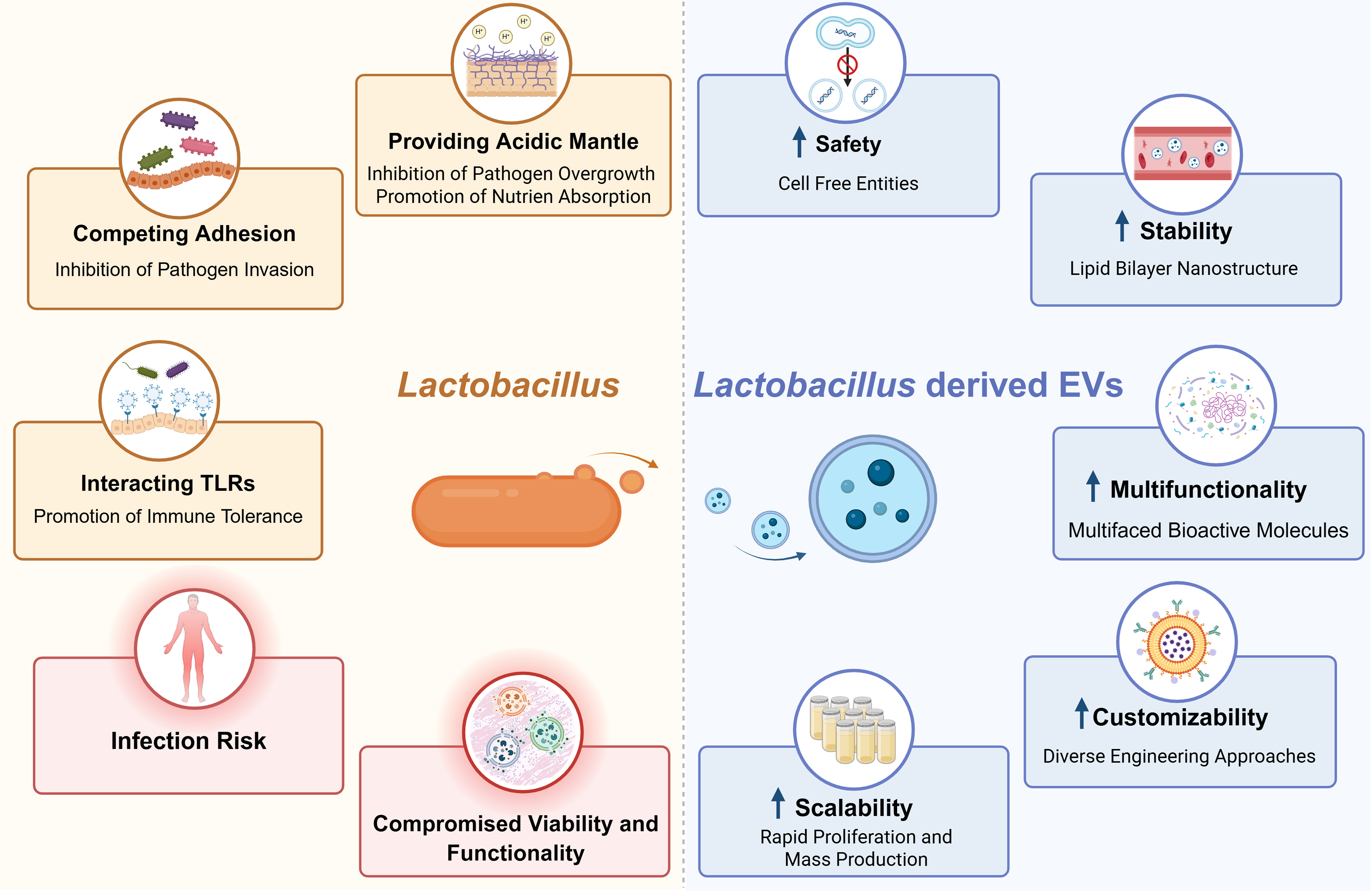
Figure 1. The advantages and shortcomings of Lactobacillus and L-EVs for wound management. Created in BioRender. Shi, Y. (2026). EV: Extracellular vesicle; L-EV: Lactobacillus-derived extracellular vesicle; TLR: Toll-like receptor.
HOW THE LACTOBACILLUS DERIVED EVS ACCELERATE WOUND HEALING
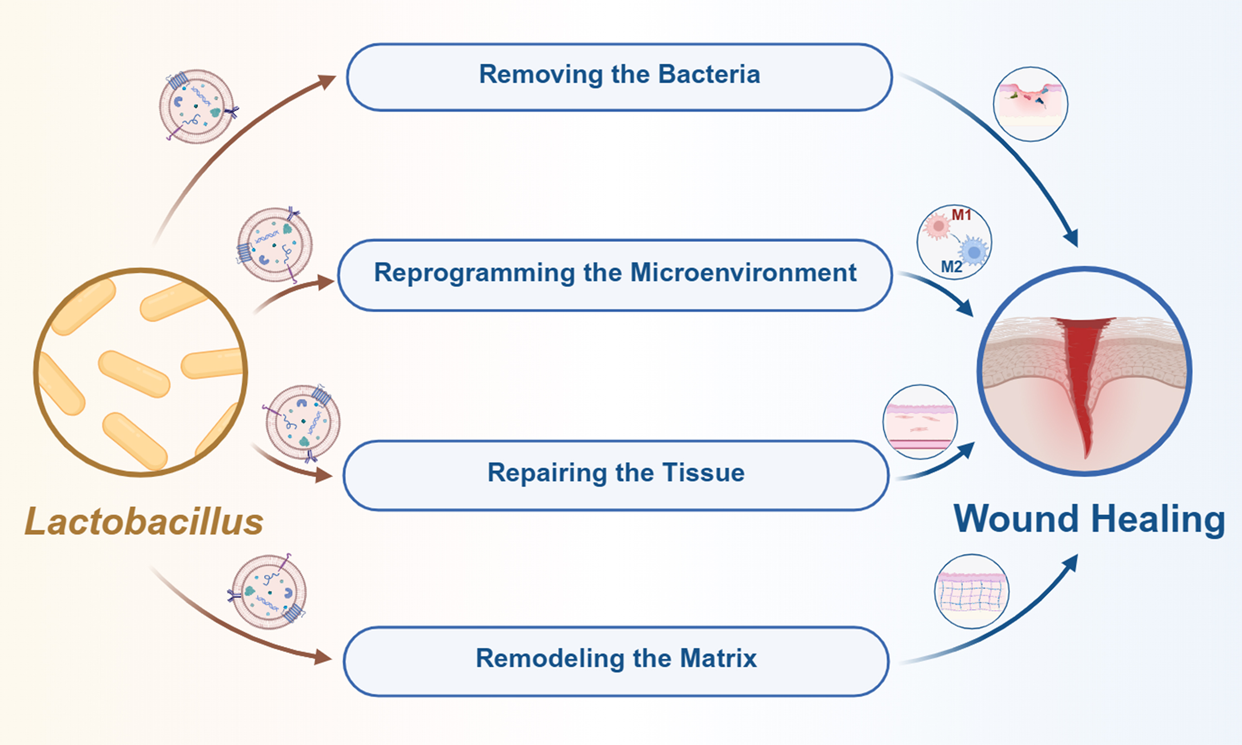
The upward attention towards the L-EVs gradually revealed their antibacterial, anti-inflammatory, as well as regenerative capacities. Therefore, these L-EVs have been considered omnipotent candidates for wound management since the wound-healing process typically involves overlapping phases of hemostasis, inflammation, proliferation, and remodeling. By removing bacteria, reprogramming the microenvironment, repairing the tissue, and remodeling the wound, the L-EVs have been found to sequentially expedite the wound-healing process with outstanding performance [Figure 2 and Table 1].
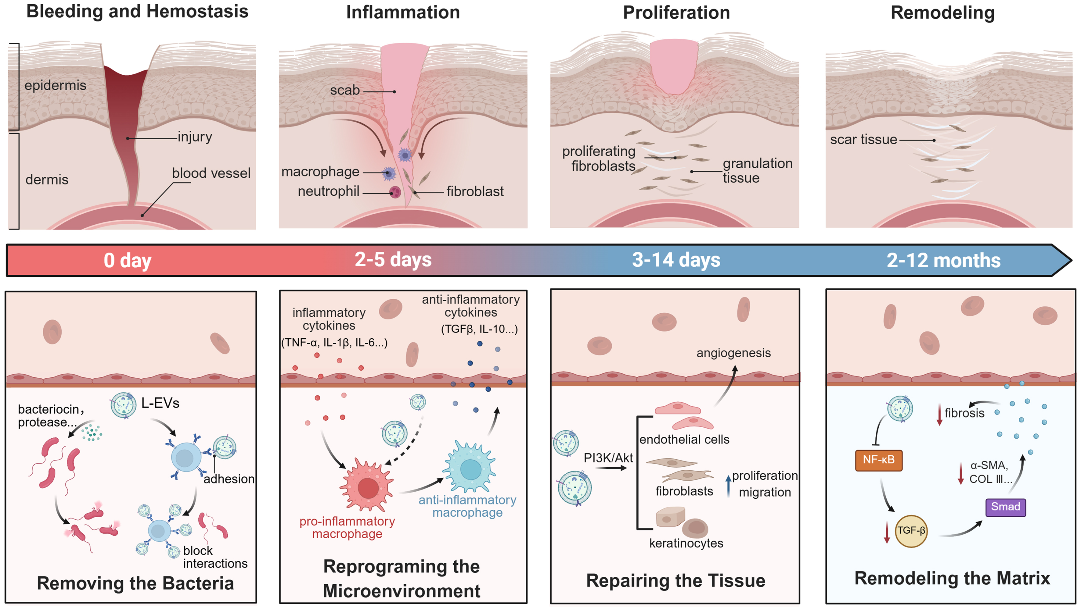
Figure 2. Illustration of the four phases during the wound healing process and the relevant functional mechanisms of L-EVs for accelerating wound repair and regeneration. The indicated time ranges are approximate estimates based on general wound-healing models. These phases are not strictly sequential and may overlap considerably depending on the wound type, size, and pathological conditions. Created in BioRender. Shi, Y. (2026). Akt: Protein kinase B; COL III: collagen type III; EV: extracellular vesicle; IL: interleukin; L-EV: Lactobacillus-derived extracellular vesicle; NF-κB: nuclear factor kappa B; PI3K: phosphoinositide 3-kinase; SMA: smooth muscle actin; TGF-β: transforming growth factor beta; TNF-α: tumor necrosis factor alpha.
The L-EV in wound management: the biological activity, bioactive cargo, mechanistic pathway, animal model, and administration route
| Lactobacillus strain | Biological activity | Bioactive cargo | Mechanistic pathway | Experimental model | Administration route | Ref. |
| Lactobacillus reuteri | Anti-inflammation, promote M2 macrophage polarization, enhance keratinocyte migration, stimulate angiogenesis | Various bioactive metabolites | Nrf2-Keap1, NF-κB, PI3K/Akt, AMPK | Diabetic mouse model, Staphylococcus aureus-infected diabetic wound model | Topical administration | [27] |
| Lactobacillus gasseri BC12 | Inhibit adhesion of opportunistic pathogens, enhance adhesion of beneficial lactobacilli | Adhesins: enolases, EF-Tu, chaperones | NR | NR | NR | [3] |
| Lactobacillus reuteri | Anti-inflammation, promote M2 macrophage polarization, improve collagen deposition, activate epithelial regeneration | 3-HPA | NR | Tongue mucosal ulcer mouse model, full-thickness wound mouse model | Hydrogel-based topical administration | [26] |
| Lactobacillus plantarum | Anti-inflammation promote M2 macrophage polarization | NR | TLR2/4, NF-κB-related immune pathways | NR | NR | [30] |
| Lactobacillus rhamnosus GG | Enhance angiogenesis, boost proliferation and migration of keratinocytes, promote re-epithelialization, improve collagen deposition | miR-21-5p | PI3K-Akt, HIF1α | Full-thickness wound mouse model | Subcutaneous injection | [35] |
| Lactobacillus plantarum | Promote proliferation and migration of fibroblasts, increase HA synthesis | NR | Reverse UV-induced photoaging-related gene expression | Healthy adult volunteers | Topical administration | [36] |
| Lactobacillus reuteri | Promote proliferation and migration of keratinocytes, enhance angiogenesis, accelerate re-epithelialization | miR-21a-5p | PI3K-Akt | Diabetic mice model | Subcutaneous injection | [37] |
| Lactobacillus casei, Lactobacillus plantarum | Anti-inflammation, promote angiogenesis and re-epithelialization, reduce dermal thickness and scar risk | NR | NR | Full-thickness wound mouse model | Hydrogel-based topical administration | [38] |
| Lactobacillus druckerii | Promote angiogenesis and re-epithelialization, suppress dermal fibrosis, reduce collagen I/III and α-SMA expression | NR | MAPK, p-JNK/p-p38 | Bleomycin-induced scleroderma mouse model, full thickness excisional burn wound mouse model | Subcutaneous injection | [40] |
Removing the bacteria
While a balanced microbiome can support wound healing, the overgrowth of pathogenic bacteria or the formation of their biofilms creates significant barriers to tissue restoration. Therefore, removing the bacteria is the primary task of the inflammatory phase. At the beginning of the inflammatory phase, neutrophils are typically recruited to the wound site, undergo phagocytosis, and release reactive oxygen species (ROS) to eliminate bacteria[21]. Whereas in most cases, additional treatments such as debridement and antibiotics are still required in clinical settings. To more efficiently clear the persistent bacteria and terminate the chronic inflammation, more and more advanced treatment modalities, such as the AIE based photodynamic and photothermal therapies, have been developed recently, facilitating the transition from inflammation to proliferation[22,23].
As most Lactobacillus have evolved antibacterial capacities to enhance competitive survival, the L-EVs have recently been found to exert antibacterial activities. These L-EVs with lipid bilayer nanostructures, on the one hand, can provide protection and enable delivery of their endogenous antibacterial substances for the direct bactericidal effects. For instance, the Lactobacillus reuteri-derived EVs contain the small molecular antibacterial agent reuterin, thus possessing bactericidal activity[24-26]. While their antibacterial activities have been preliminarily demonstrated by a series of pioneering studies, their therapeutic efficacy in managing more complex chronic wounds, such as infected diabetic wounds, remains limited. To facilitate effective chronic wound healing, additional antibacterial techniques or materials have been used in combination. For instance, Tai et al. reported a photothermal antibacterial system, which combined the Lactobacillus reuteri-derived EVs with indocyanine green (ICG) to effectively repair infected diabetic wounds[27]. On the other hand, these L-EVs can also act as adhesion analogs and block interactions between bacterial and host cells, thereby exerting indirect antibacterial effects. For instance, Lactobacillus gasseri BC12-derived EVs were capable of preventing pathogen attachment and supporting the colonization of beneficial species in vitro, suggesting that L-EVs may represent a candidate strategy in further investigation for wound healing applications[3]. Nevertheless, their in vivo therapeutic efficacy as adhesion analogs for the treatment of infected wounds is still waiting to be well evaluated.
Reprogramming the microenvironment
With the waning of the neutrophils after approximately two days of the inflammatory phase, monocytes infiltrate the wound and differentiate into macrophages, which become the dominant cells from day 2 to day 5 after injury[28]. The macrophages are highly plastic and can transition from the pro-inflammatory M1 phenotype to the pro-regenerative M2 phenotype, initiating the next proliferation phase[29].
Recent investigations have demonstrated that the L-EVs are uniquely suited to reprogram the immune macrophages and the inflammatory microenvironment of wounds. For instance, the previously mentioned Lactobacillus reuteri-derived EVs could also regulate inflammatory responses by modulating macrophage mitochondrial function through the inherent biomolecule 3-hydroxypropionaldehyde (3-HPA), promote macrophage M2 polarization, and ultimately facilitate wound healing[26]. While current study only investigated the regulatory effects of L-EVs on macrophages, without systematically examining their effects on keratinocytes, endothelial cells, or fibroblasts. Therefore, a complete mechanistic understanding of the cell network involved in wound repair has not been established. Besides, the Lactobacillus plantarum-derived EVs could induce the differentiation of human monocytic THP1 cells towards M2 macrophage polarization, downregulate inflammation-induced expression of M1 macrophage cell-surface markers, and exhibit an anti-inflammatory effect in the human skin in vitro[30]. However, it is worth noting that pro-inflammatory vs. anti-inflammatory effects vary across the different phases of wound healing. As such, promoting macrophage polarization from M1 to M2 phenotypes might only be beneficial at a specific phase. To guarantee a more effective outcome for promoting wound repair, temporally programmed therapeutic strategies should still be developed.
Repairing the tissue
Starting around day 3 and lasting for up to 2 weeks, the proliferation phase follows three sequential processes, including angiogenesis, fibroplasia, and re-epithelialization, focusing on filling the wound defect and restoring the skin barrier. Typically, angiogenesis involves the formation of new blood vessels from the pre-existing ones, supplying oxygen and nutrients to the metabolically active healing tissue[31,32]. Then the fibroblasts migrate into the wound bed to synthesize collagen and other extracellular matrix (ECM) components, forming the granulation tissue. Simultaneously, keratinocytes at the edges of the wound proliferate and migrate across the granulation tissue for re-epithelialization[33]. Myofibroblasts also begin to contract the wound, pulling the edges of the wound inward to close the wound and reduce the defect[34].
Inheriting abundant bioactive molecules from their parent Lactobacillus strain, the L-EVs have been increasingly reported to target endothelial cells, fibroblasts, as well as keratinocytes to promote tissue repair and rebuilding. For instance, the Lactobacillus rhamnosus GG-derived EVs have been found enriched in miR-21-5p, which enhances the proliferation and migration capacities of endothelial cells and subsequently promotes angiogenesis, thereby accelerating cutaneous wound healing[35]. While miR-21-5p was identified as an essential functional molecule in the presented study, other bioactive cargoes, such as proteins and lipids, have not yet been systematically examined for their contributions to wound healing, particularly through the cooperative regulatory networks with miR-21-5p. The Lactobacillus plantarum-derived EVs could reverse ultraviolet (UV)-induced photoaging-related gene expression, enhance proliferation and migration of fibroblasts, and upregulate collagen and elastin synthesis, thereby promoting cutaneous wound repair and skin rejuvenation[36]. The Lactobacillus reuteri-derived EVs have been characterized with intrinsic miR-21a-5p to enhance the function of keratinocytes and improve epidermal thickness, thereby promoting cutaneous wound healing[37]. However, in most current studies, the administration route of these L-EVs remains subcutaneous, which may not be applicable to clinical settings. Advanced delivery systems for the direct topical administration of these L-EVs should be developed in the future[38].
Remodeling the matrix
The collagen fibers deposited at the wounds during the proliferation phase are usually the disorganized type III collagen. Only until the final remodeling phase of wound healing are these collagen fibers gradually replaced by the organized type I collagen, regaining the integrity and strengthening the tensile strength of the regenerated tissues[32]. Although the myofibroblasts are essential for wound contraction, their persistent activity always leads to excessive collagen deposition, scar formation, and even fibrosis[39]. Therefore, the quality of the repaired wounds can be controlled by either modulating myofibroblast activity or driving the transition to the remodeling phase.
According to the most recent studies, L-EVs have shown promising anti-fibrotic potential that favors more natural wound regeneration. For instance, the Lactobacillus casei-derived EVs-loaded hydrogel significantly reduced the percentage of non-structured epidermis and dermis thickness within the wound site, decreasing scar formation[38]. Besides, Lactobacillus druckerii-derived EVs inhibited the expression of collagen I/III and α-SMA and cell proliferation of fibroblasts derived from hypertrophic scars, and in vivo inhibited the formation of hypertrophic scars in the scleroderma mouse model[40]. However, the absence of long-term follow-up for wound remodeling and scar maturation in these studies precluded the evaluation of the sustained anti-scarring and long-term reparative efficacy of L-EVs.
WHERE THE LACTOBACILLUS DERIVED EVS HEAD FOR FUTURE THERAPY
Despite growing research interest in L-EVs and compelling preclinical evidence of their wound-treatment capacities, several hurdles remain to be tackled for their successful clinical translation. First of all, the isolation and purification standardizations for the L-EVs are lacking. Even though the Minimum Information for Studies of Extracellular Vesicles (MISEV) recommends differential centrifugation at
Beyond the current investigation into their wound-healing mechanisms, future research on L-EVs should focus on their intelligent generation for optimal clinical efficiency. Leveraging artificial intelligence (AI) and multi-omics analyses, a specialized database of L-EVs that is similar to Vesiclepedia, ExoCarta, and EVAtlas of exosomes should be established in the future. It could guide clinicians in quickly prescribing L-EVs for the individual patient with a unique wound environment and metabolic profile, moving toward precision medicine for wound management[46]. For instance, Chouaib and colleagues have developed an Intelligent Vesicle Exocytosis Analysis (IVEA) platform, which could be extended to enable multi-label vesicle tracking and in-depth mechanistic quantification in wound therapeutics. The IVEA platform broadly supports diverse fluorescent labels for simultaneous multi-tracer tracking, while automatically extracting comprehensive kinetic parameters, such as fusion kinetics, diffusion radius, and secretion profiles, directly linking EV dynamics to specific wound repair processes like angiogenesis, collagen deposition, and macrophage polarization[47]. Besides, the diverse engineering approaches should be exploited in combination to generate L-EVs with improved properties. For instance, the L-EVs can be biologically engineered as EcN 1917 to encapsulate the VEGF[31] and then further chemically engineered to display the targeting ligand on human umbilical vein endothelial cells (HUVECs). As such, the engineered L-EVs could be endowed with endothelium-targeting and angiogenesis-promoting capacities simultaneously to achieve enhanced wound-healing efficacy. Moreover, since topical administration remains the primary route of medication for wound treatment, materials engineering approaches should be proposed and explored to generate advanced L-EVs formulations such as sprayable hydrogels and microneedle patches[48,49]. These advanced formulations could not only facilitate the controlled and sustained release of L-EVs around the wound but also improve treatment compliance of patients. Integration of AI technologies with L-EVs engineering can enable the circumvention of translational barriers in L-EVs-based wound treatment, including loading efficiency, targeted delivery, in vivo stability and pharmacokinetics, as well as immunogenicity and safety[50]. As future research continues to integrate these interdisciplinary technologies, the L-EVs are poised to become the cornerstone of the next generation of wound management modalities, exerting satisfactory clinical outcomes [Figure 3].
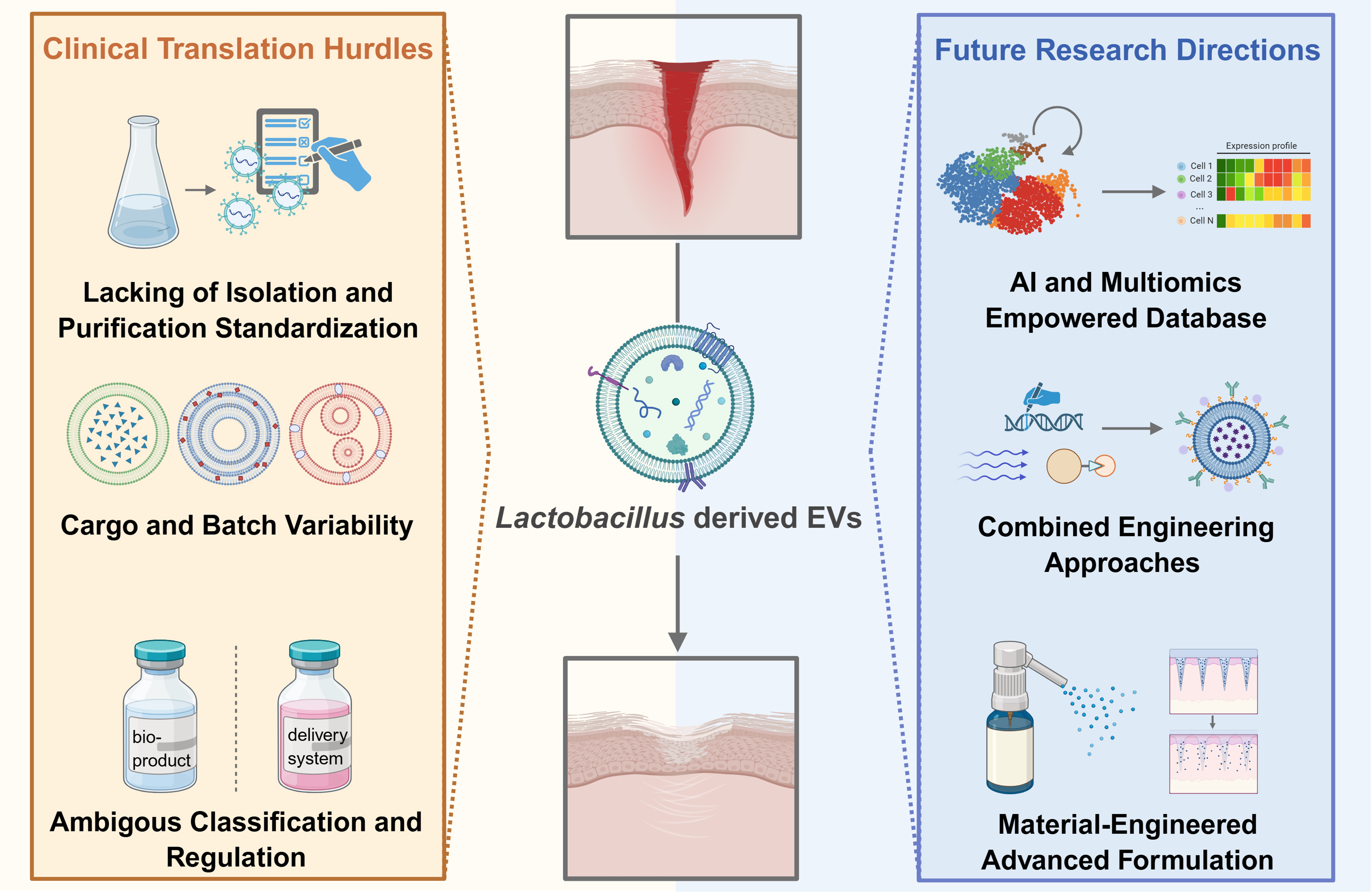
Figure 3. The clinical translation hurdles and future research directions of L-EVs for wound management. Created in BioRender. Shi, Y. (2026). AI: Artificial intelligence; EV: extracellular vesicle; L-EV: Lactobacillus-derived extracellular vesicle; multiomics: multi-omics.
DECLARATIONS
Acknowledgments
The graphic abstract was created in BioRender. Shi, Y. (2026).
Authors’ contributions
Conceptualization: Shi Y, Hu H
Literature collection and collation: Li Y, Zhang M, Wang Y
Visualization: Wang Y, Sang X, Cai W
Drafting of the manuscript: Li Y, Zhang M
Revising of the manuscript: Shi Y, Hu H
The final version of the manuscript has been approved by all authors for publication.
Availability of data and materials
Not applicable.
AI and AI-assisted tools statement
Not applicable.
Financial support and sponsorship
The work was supported by the Shanghai Pujiang Program (21PJ1404100) and the National Natural Science Foundation of China (No 22477075).
Conflicts of interest
Shi Y is an Assistant Guest Editor of the Special Topic “Extracellular Vesicles for Wound Management” of the journal Extracellular Vesicles and Circulating Nucleic Acid. Shi Y was not involved in any steps of editorial processing, notably including reviewers’ selection, manuscript handling and decision making. The other authors declare that there are no conflicts of interest.
Ethical approval and consent to participate
Not applicable.
Consent for publication
Not applicable.
Copyright
© The Author(s) 2026.
REFERENCES
1. Mejía-Caballero A, Marco ML. Lactobacilli biology, applications and host interactions. Nat Rev Microbiol. 2026;24:111-26.
2. Yang H, Hao L, Jin Y, Huang J, Zhou R, Wu C. Functional roles and engineering strategies to improve the industrial functionalities of lactic acid bacteria during food fermentation. Biotechnol Adv. 2024;74:108397.
3. Croatti V, Parolin C, Giordani B, Foschi C, Fedi S, Vitali B. Lactobacilli extracellular vesicles: potential postbiotics to support the vaginal microbiota homeostasis. Microb Cell Fact. 2022;21:237.
4. Glick VJ, Webber CA, Simmons LE, et al. Vaginal lactobacilli produce anti-inflammatory β-carboline compounds. Cell Host Microbe. 2024;32:1897-909.e7.
5. Moreno I, Codoñer FM, Vilella F, et al. Evidence that the endometrial microbiota has an effect on implantation success or failure. Am J Obstet Gynecol. 2016;215:684-703.
6. Chang H, Perkins MH, Novaes LS, et al. Stress-sensitive neural circuits change the gut microbiome via duodenal glands. Cell. 2024;187:5393-412.e30.
7. Bermúdez-Humarán LG, Chassaing B, Langella P. Exploring the interaction and impact of probiotic and commensal bacteria on vitamins, minerals and short chain fatty acids metabolism. Microb Cell Fact. 2024;23:172.
8. Christensen IB, Vedel C, Clausen ML, Kjærulff S, Agner T, Nielsen DS. Targeted screening of lactic acid bacteria with antibacterial activity toward Staphylococcus aureus clonal complex type 1 associated with atopic dermatitis. Front Microbiol. 2021;12:733847.
9. Ji HF, Li M, Han X, et al. Lactobacilli-mediated regulation of the microbial-immune axis: a review of key mechanisms, influencing factors, and application prospects. Foods. 2025;14:1763.
10. Xie Z, Li M, Qian M, Yang Z, Han X. Co-cultures of Lactobacillus acidophilus and Bacillus subtilis enhance mucosal barrier by modulating gut microbiota-derived short-chain fatty acids. Nutrients. 2022;14:4475.
11. Niu L, Li H, Guo J, et al. Gut commensal Lactobacillus strain induces a balanced trained immunity phenotype to enhance vaccine efficacy through JAK-STAT-SOCS pathway. Probiotics Antimicrob Proteins. 2026;18:4315-38.
12. Dargenio VN, Castellaneta S, Panico S, et al. Probiotics and gastrointestinal diseases. Minerva Pediatr. 2022;74:703-23.
13. Shah AB, Baiseitova A, Zahoor M, et al. Probiotic significance of Lactobacillus strains: a comprehensive review on health impacts, research gaps, and future prospects. Gut Microbes. 2024;16:2431643.
14. Haak BW, Wiersinga WJ. The differing roles of lactobacilli in critical illness. Nat Med. 2019;25:1651-3.
15. Rossi F, Amadoro C, Gasperi M, Colavita G. Lactobacilli infection case reports in the last three years and safety implications. Nutrients. 2022;14:1178.
16. Shi Z, Li X, Fan X, et al. The SlpX protein plays a crucial role in the intestinal juice tolerance of Lactobacillus acidophilus CICC6074. Food Bioscience. 2024;59:103865.
17. Li M, Mao B, Tang X, et al. Lactic acid bacteria derived extracellular vesicles: emerging bioactive nanoparticles in modulating host health. Gut Microbes. 2024;16:2427311.
18. Schulz E, Goes A, Garcia R, et al. Biocompatible bacteria-derived vesicles show inherent antimicrobial activity. J Control Release. 2018;290:46-55.
19. Li M, Zhou H, Yang C, et al. Bacterial outer membrane vesicles as a platform for biomedical applications: an update. J Control Release. 2020;323:253-68.
20. Cheng Q, Li R, He Y, Zhu Y, Kang Y, Ji X. Genetically engineered cellular nanovesicles: theories, design and perspective. Adv Funct Mater. 2024;34:2407842.
22. Dai J, Fang L, Fan Z, et al. Triphenylamine-substituted nile red derivatives with efficient reactive oxygen species generation for robust and broad-spectrum antimicrobial photodynamic therapy and abscess wound healing. Adv Funct Mater. 2025;35:2421072.
23. Li B, Wang W, Zhao L, et al. Multifunctional AIE nanosphere-based “nanobomb” for trimodal imaging-guided photothermal/photodynamic/pharmacological therapy of drug-resistant bacterial infections. ACS Nano. 2023;17:4601-18.
24. Talarico TL, Casas IA, Chung TC, Dobrogosz WJ. Production and isolation of reuterin, a growth inhibitor produced by Lactobacillus reuteri. Antimicrob Agents Chemother. 1988;32:1854-8.
25. Talarico TL, Dobrogosz WJ. Chemical characterization of an antimicrobial substance produced by Lactobacillus reuteri. Antimicrob Agents Chemother. 1989;33:674-9.
26. Chen Y, Huang X, Liu A, et al. Lactobacillus reuteri vesicles regulate mitochondrial function of macrophages to promote mucosal and cutaneous wound healing. Adv Sci. 2024;11:e2309725.
27. Tai Q, Feng H, Tang Y, et al. Inflammation-responsive DNA hydrogel integrating probiotic outer membrane vesicles for infection monitoring and precision therapy of diabetic wounds. J Control Release. 2026;391:114594.
28. Dovi JV, He LK, DiPietro LA. Accelerated wound closure in neutrophil-depleted mice. J Leukoc Biol. 2003;73:448-55.
29. Sharifiaghdam M, Shaabani E, Faridi-Majidi R, De Smedt SC, Braeckmans K, Fraire JC. Macrophages as a therapeutic target to promote diabetic wound healing. Mol Ther. 2022;30:2891-908.
30. Kim W, Lee EJ, Bae IH, et al. Lactobacillus plantarum-derived extracellular vesicles induce anti-inflammatory M2 macrophage polarization in vitro. J Extracell Vesicles. 2020;9:1793514.
31. Zheng Z, Liu X, Guo K, et al. Bioengineered probiotics derived bacterial extracellular vesicle as bioactive nanocarrier for the local VEGF expression to accelerate wound healing. J Nanobiotechnology. 2026;24:287.
32. Gurtner GC, Werner S, Barrandon Y, Longaker MT. Wound repair and regeneration. Nature. 2008;453:314-21.
33. Yao W, Wang Z, Ma H, et al. Epithelial-mesenchymal plasticity (EMP) in wound healing: exploring EMT mechanisms, regulatory network, and therapeutic opportunities. Heliyon. 2024;10:e34269.
35. Wang J, Li X, Zhao X, et al. Lactobacillus rhamnosus GG-derived extracellular vesicles promote wound healing via miR-21-5p-mediated re-epithelization and angiogenesis. J Nanobiotechnology. 2024;22:644.
36. Kang S, Jeon S, Baek H, et al. Lactobacillus-derived artificial extracellular vesicles for skin rejuvenation and prevention of photo-aging. Biomater Sci. 2025;13:2026-35.
37. Li Y, Zheng Z, Kong X, et al. Bacteria extracellular vesicles derived from Lactobacillus reuteri delivering intrinsic miR-21a-5p to accelerate diabetic wound healing. Nano Research. 2025;18:94908083.
38. Kuhn T, Aljohmani A, Frank N, et al. A cell-free, biomimetic hydrogel based on probiotic membrane vesicles ameliorates wound healing. J Control Release. 2024;365:969-80.
39. Schuster R, Younesi F, Ezzo M, Hinz B. The role of myofibroblasts in physiological and pathological tissue repair. Cold Spring Harb Perspect Biol. 2023;15:a041231.
40. Han F, Wang K, Shen K, et al. Extracellular vesicles from Lactobacillus druckerii inhibit hypertrophic scar fibrosis. J Nanobiotechnology. 2023;21:113.
41. Welsh JA, Goberdhan DCI, O’Driscoll L, et al. ; MISEV Consortium. Minimal information for studies of extracellular vesicles (MISEV2023): from basic to advanced approaches. J Extracell Vesicles. 2024;13:e12404.
42. Li Q, Ding Y, Shi Y, et al. 80 years of extracellular vesicles: from discovery to clinical translation. Extracell Vesicles Circ Nucl Acids. 2026;7:165-233.
43. Zhang X, Kong X, He X, Wang Y, Shi Y, Cai J. Nanoengineered bacterial extracellular vesicles for the photo-chemo programmed therapy to treat melanoma. Chem Eng J. 2025;522:167807.
44. Marteau P, Shanahan F. Basic aspects and pharmacology of probiotics: an overview of pharmacokinetics, mechanisms of action and side-effects. Best Pract Res Clin Gastroenterol. 2003;17:725-40.
45. Sanders ME, Akkermans LM, Haller D, et al. Safety assessment of probiotics for human use. Gut Microbes. 2010;1:164-85.
46. Tiwari A, Widodo, Krisnawati DI, Tzou KY, Kuo TR. Machine learning for extracellular vesicles enables diagnostic and therapeutic nanobiotechnology. J Nanobiotechnology. 2026;24:153.
47. Chouaib AA, Chang HF, Khamis OM, et al. Highly adaptable deep-learning platform for automated detection and analysis of vesicle exocytosis. Nat Commun. 2025;16:6450.
48. You Y, Liu M, Yan N, et al. Smart materials and devices for enhanced delivery of extracellular vesicles. Adv Mater. 2025;37:e2501081.
49. Guo K, Gong J, Zhong W, et al. Hypoxia-challenged sEVs-engineered nanofiber scaffolds accelerate diabetic wound healing via reversing cellular dysfunction of skin repair cells. Research. 2026;9:1248.
Cite This Article
 Open Access
How to Cite
Download Citation
Export Citation File:
Type of Import
Tips on Downloading Citation
Citation Manager File Format
Type of Import
Direct Import: When the Direct Import option is selected (the default state), a dialogue box will give you the option to Save or Open the downloaded citation data. Choosing Open will either launch your citation manager or give you a choice of applications with which to use the metadata. The Save option saves the file locally for later use.
Indirect Import: When the Indirect Import option is selected, the metadata is displayed and may be copied and pasted as needed.
About This Article
Special Topic
Copyright

Data & Comments
Data
0










Comments
Comments must be written in English. Spam, offensive content, impersonation, and private information will not be permitted. If any comment is reported and identified as inappropriate content by OAE staff, the comment will be removed without notice. If you have any queries or need any help, please contact us at [email protected].