


fig1
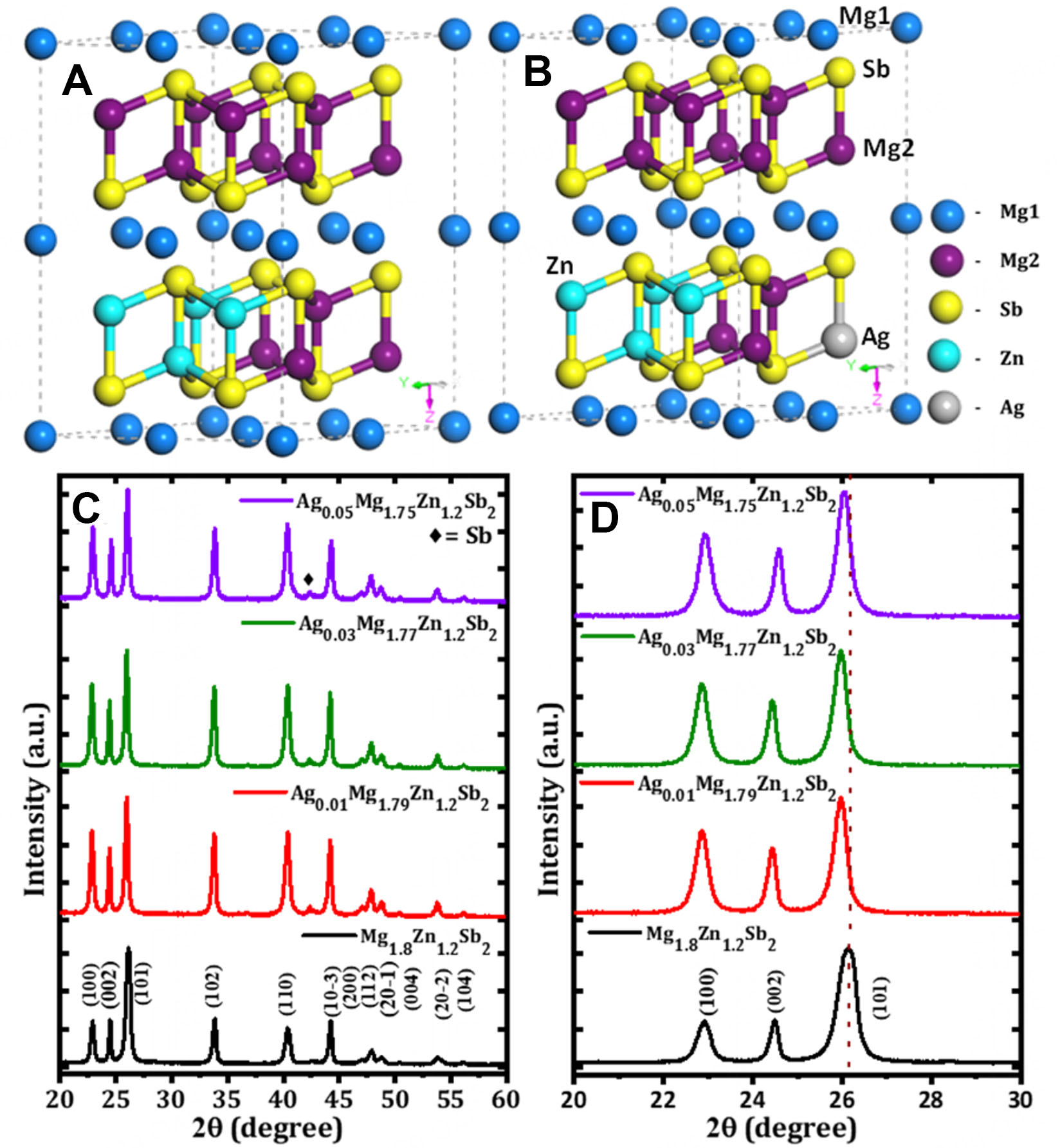
Figure 1. (A and B) crystal structures; (C and D) XRD and magnified XRD pattern of AgxMg1.8-xZn1.2Sb2 (x = 0, 0.01, 0.03 and 0.05) samples. XRD: X-ray diffraction.
Figure 1. (A and B) crystal structures; (C and D) XRD and magnified XRD pattern of AgxMg1.8-xZn1.2Sb2 (x = 0, 0.01, 0.03 and 0.05) samples. XRD: X-ray diffraction.


All published articles are preserved here permanently:
https://www.portico.org/publishers/oae/


