


fig8
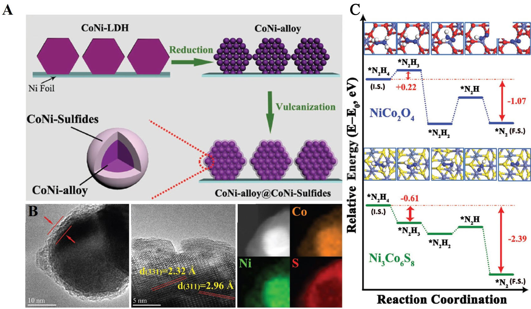
Figure 8. (A) Schematic illustration for the synthesis of CoNi-alloy@CoNi-sulfide nanoarrays (B) TEM image, crystal fringes and HAADF-STEM image and EDS mapping of CoNi-R-S; (C) The energy profiles for the reaction pathways over CoNi-sulfide and CoNi-oxide[141]. This figure is quoted with permission from Zhou et al. EDS: Energy-dispersive X-ray spectroscopy; TEM: transmission electron microscopy; HAADF-STEM: high-angle annular dark-field scanning transmission electron microscopy.


