

Hepatic SMAD4 safeguards systemic host defense in bacterial infection


 Download PDF
The immunopathogenesis of sepsis is characterized by two intertwined arms of immune dysregulation, excessive pro-inflammation and sustained immunosuppression. Over the past three decades, more than 200 trials targeting pro-inflammatory mediators and immune checkpoint molecules have failed to achieve satisfactory clinical responses[1]. This therapeutic impasse has redirected attention toward the often-overlooked non-immune mediators of septic immune dysfunction[2].
The immune responses by liver-resident Kupffer cells and natural killer (NK) cells have been extensively studied in various liver diseases[3]. Hepatocytes constitute the primary source of acute phase proteins (APPs), which are critically involved in a spectrum of chemotaxis, microbial killing, extracellular matrix (ECM) remodeling, and oxidative-stress responses. Yet the precise interactions between hepatocytes and immune cells remain poorly defined. Against this backdrop, Chen et al. report a landmark advance that addresses long-unresolved questions in hepatic innate immunity[4]. The investigators characterized hepatocyte-macrophage communication driven by a C-type lectin family (the REG3 subfamily) that plays an important role in defense against bacterial infection. Importantly, they found that transforming growth factor-beta (TGF-β)-independent activation of SMAD4 in hepatocytes is indispensable for host defense against bacterial infection[4].
In the work, Chen et al. found that Smad4 can activate the expression of a broad set of genes, e.g., Reg3b and Reg3g, which promote macrophage recruitment and bacterial clearance. More intriguingly, TGF-β superfamily ligands were dispensable in this action. Instead, interleukin 6 (IL-6) and glucocorticoids (dexamethasone or hydrocortisone) potently up-regulate expression of Reg3b and Reg3g. Transcriptional activation of Reg3b/3g depends on promoter occupancy by signal transducer and activator of transcription 3 (STAT3) and glucocorticoid receptor (GR); yet SMAD4 does not bind to the apparent Smad-binding elements (SBEs) on their promoters. The authors therefore proposed that the transcriptional activation of Reg3b and Reg3g by STAT3 and GR is contingent upon SMAD4 availability, and therefore propose a previously unrecognized role of SMAD4 in the regulation of Reg3b and Reg3g [Figure 1A].
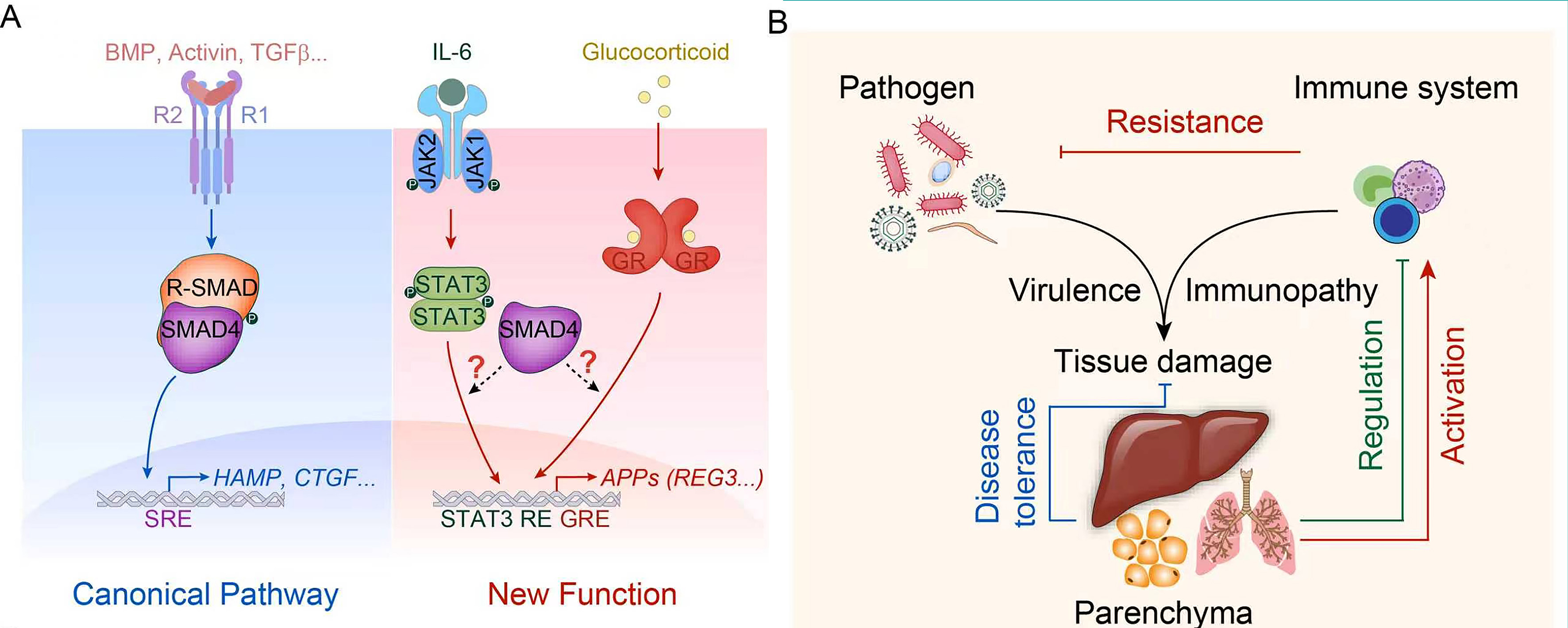
Figure 1. Canonical and non-canonical SMAD4 signaling in host defense against bacterial infection.
(A) Scheme of SMAD4 pathway. The left panel illustrates the central role of SMAD4 in the canonical TGF-β signaling pathway, in which TGF-β superfamily ligands (BMP, Activin, and TGF-β) activate the SMAD complex and drive transcriptional programs for tissue repair and iron homeostasis. The right panel depicts one new function of SMAD4, whereby SMAD4 orchestrates inflammation-elicited STAT3 and GR signaling to enable transcription of APPs such as REG3 proteins; (B) Schematic overview depicting functional relationships among pathogens, immune-cell responses, and parenchymal-tissue outputs during bacterial infection. BMP: Bone morphogenetic protein; TGF-β: transforming growth factor beta; HAMP: hepcidin antimicrobial peptide; CTGF: connective tissue growth factor; SRE: Smad-responsive element; R-SMAD: receptor-regulated Sma and Mad-related protein; IL-6: interleukin-6; STAT3: signal transducer and activator of transcription 3; JAK1: Janus kinase 1; GR: glucocorticoid receptor; APPs: acute-phase proteins; REG3: regenerating islet-derived 3; GRE: glucocorticoid-responsive element.
Profound loss of Reg3b/Reg3g in livers from Escherichia coli (E. coli)-challenged STAT3-depleted mice and RU486-treated animals indicates that both STAT3 and GR are essential for this transcriptional response, indicative of strong synergism. The relative positioning of STAT3 response elements and GR response elements at these promoters further suggests potential modes of functional interplay. Yet, the mechanism underlying SMAD4-STAT3-GR crosstalk remains puzzling. STAT3 and GR exhibit impaired DNA-binding capacity in SMAD4-deficient liver from E. coli-challenged mice. Notably, it has been demonstrated that STAT3 and GR could synergistically activate transcription, as exemplified by the α2-macroglobulin gene, through assembly of a STAT3-dependent enhanceosome[5]. How does SMAD4 affect the synergy between STAT3 and GR on the promoters of Reg3b and Reg3g? Several non-mutually exclusive proposed mechanisms can be envisaged. First, SMAD4 may maintain the epigenetic landscape at STAT3/GR-binding loci, consistent with prior work from Deng’s laboratory documenting SMAD4-dependent histone-methylation regulation[6], which may affect the recruitment of STAT3 and/or GR. In addition, it will also need confirmation through the structure-function rescue in the Smad4-null background [wild-type vs. Mad homology 1 (MH1) vs. MH2 mutants] and chromatin-state analysis of the Reg3b/3g locus in control vs. liver-specific Smad4 knockout (LKO) liver [accessibility and acetylation of histone H3 at K27 (H3K27ac)]. Secondly, whether additional SMAD proteins participate remains to be tested. SMAD3 is one candidate, as it binds GR[7] and STAT3[8]; notably, however, STAT3 can also compete with SMAD4 for SMAD3 binding. Moreover, SMAD1 has been reported to cooperate with STAT3 in driving astrocyte differentiation, although in that setting the two are bridged by p300 rather than contacting each other directly[9]. Both SMAD1 and SMAD3 form canonical heteromeric complexes with SMAD4. Whether an R-SMAD such as SMAD3 or SMAD1 would therefore bridge SMAD4 to the STAT3/GR complex, or instead sequester SMAD4 away from it, cannot be predicted a priori. Additionally, gain-of-function experiments will address a different question: because no TGF-β or bone morphogenetic protein (BMP) ligand induces Reg3b/Reg3g when applied alone, ligand addition in combination with STAT3- and GR-activating stimuli (IL-6 plus glucocorticoid) will be informative, which tests whether TGF-β superfamily ligands potentiate the transcription of Reg3b/Reg3g. Combined agonists may enable the recruitment of SMAD4 as a co-factor to chromatin alongside STAT3 and GR. Irrespective of these unresolved mechanistic details, Chen et al. have established a previously unrecognized role for hepatic SMAD4 in antibacterial defense[4].
The Reg3 family contributes to intestinal immune homeostasis and cardiac anti-infective protection[10]. This work delivers a conceptual leap in REG3-family biology by identifying infection-triggered hepatic Reg3b and Reg3g production[4]. Hepatic Reg3b and Reg3g expression was not recognized prior to this study. Remarkably, hepatocyte-specific SMAD4 deletion does not alter Reg3b/Reg3g levels in extrahepatic tissues (intestine, spleen, kidney), indicating cell- intrinsic control of the SMAD4-Reg3 regulatory axis. Future work should address whether SMAD4 governs REG3 expression in other organs, which could broaden the biological scope of these findings.
The investigators also uncovered a new function of Reg3b/Reg3g in this study. Besides the multifaceted roles previously ascribed to Reg3 proteins, including direct bactericidal activity and modulatory effects on macrophage killing/polarization. Notably, rigorous in vivo experimentation yields a key conceptual demarcation: Reg3-mediated host protection operates primarily via monocyte-macrophage recruitment rather than direct bactericidal action. Chen et al. managed to determine that Reg3b and Reg3g is essentially involved in inflammation-driven monocyte/macrophage recruitment. Nevertheless, the molecular mechanisms whereby Reg3b/Reg3g steer macrophage migration remain unresolved. Accumulating evidence suggests that exostosin-like glycosyltransferase 3 (EXTL3) is an attractive candidate receptor in mediating Reg3 signaling[11], although alternative or complementary mechanisms merit further consideration.
Iron metabolism is substantially affected during sepsis; it is recognized to be a core metabolic program in establishing nutrition immunity and disease tolerance to infectious diseases[12]. Using well-calibrated dietary iron-rescue assays, the investigators achieve a critical functional separation between SMAD4-driven iron homeostasis and its antibacterial immune activity. The investigators rigorously interrogated the contribution of iron to SMAD4-mediated host defense by administering an iron-deficient diet to correct hepatic iron levels in knockout mice to wild-type levels. In brief, they concluded that iron is not the major driver of the phenotype, since iron-corrected knockout (KO) mice still succumbed to E. coli challenge. That being said, iron accumulation remains a non-negligible factor in the phenotype of unmanipulated KO mice. Of note, diverse iron-related biological events, including cellular iron trafficking, compartmental iron distribution, and iron-dependent stress responses, exert far-reaching pathological impacts across infectious, inflammatory, and degenerative disease settings[13]. A further point of interest concerns hepcidin, which has been shown to confer protection against sepsis in mouse studies[14]. Given that SMAD4 maintains basal hepcidin expression[6], it remains possible that hepcidin and other iron-related pathways may modify the disease outcome in this model. Future studies should therefore assess the involvement of hepcidin levels and iron-distribution parameters in SMAD4-deficient mice upon bacterial infection and determine whether manipulating these pathways alters antibacterial immunity.
This study has provided a new facet of how the parenchymal cells regulate detrimental impacts during bacterial infection. Collectively, these findings represent a meaningful paradigm update, redefining hepatocytes as central immune-signaling coordinators within systemic bacterial infection. It has the potential to spark conceptual debate within the field: what exactly constitutes parenchymal manifestation during sepsis? It is now well established that the parenchyma is no longer a passive target of pathogen invasion and immune injury, but an active player in the pathogenesis of this disease [Figure 1B].
The liver serves as a representative parenchymal tissue. Its ability to sense infection and secrete APPs demonstrates its contribution to disease resistance. These APPs can either act directly on pathogens or promote immune responses, as exemplified by the REG protein family. On the other hand, the liver undergoes metabolic adaptation under septic conditions: it sustains gluconeogenesis despite sepsis-induced anorexia, thereby preserving host fitness and survival[15]. This illustrates that the liver also mediates disease tolerance. Furthermore, the liver functions as part of the immune regulatory system, dampening tissue responsiveness to commensal microbe-derived pathogen-associated molecular patterns (PAMPs). Accordingly, the roles of parenchymal tissues during sepsis require careful evaluation, which may uncover novel pathogenic mechanisms and therapeutic strategies.
Beyond bacterial infection, the newly defined SMAD4-STAT3-GR functional crosstalk opens up intriguing new conceptual avenues for broader biomedical research. IL-6-STAT3 signaling and glucocorticoid-GR cascades represent central signaling hubs operative across a broad spectrum of pathological contexts, including sterile inflammation, metabolic liver disorders, autoimmune diseases, tissue injury, and tumor-microenvironment remodeling. Whether hepatocyte SMAD4 can similarly license STAT3-GR-driven transcriptional outputs in these non-infectious pathological settings remains an open and compelling scientific question. It is plausible that this non-canonical regulatory circuitry may constitute a shared yet under-appreciated signaling module enabling parenchymal cells to integrate tissue-stress cues, cytokine inputs, and hormonal signals. Further exploration of this possibility may yield fresh mechanistic insights and substantially expand the biological boundaries of SMAD-family biology beyond infection-associated host defense.
Taken together, this mechanistic investigation opens promising translational avenues for precision immunotherapy against bacterial sepsis. This study also provides profound translational implications for sepsis precision therapy, a field that currently struggles with the lack of tissue-specific targeted interventions. Current clinical sepsis management mainly relies on broad-spectrum antibiotics, fluid resuscitation, and organ supportive treatments, while general anti-inflammatory therapies often fail to balance immune activation and immune suppression, leading to limited therapeutic effects and increased adverse reactions. The identification of the hepatic SMAD4-STAT3-GR-Reg3b/Reg3g axis fills the research gap of parenchymal cell-derived immune regulatory pathways in bacterial sepsis, providing a novel tissue-specific target for immune modulation. In addition, the study’s confirmation of iron-independent SMAD4 antibacterial function revises the traditional understanding that SMAD signaling mediates host defense mainly through nutritional immunity, refining the regulatory network of hepatic innate immunity. Nevertheless, the therapeutic potential of the hepatic SMAD4-Reg3b/Reg3g axis remains to be validated. Further exploration of the epigenetic mechanism underlying SMAD4-mediated transcriptional regulation, the validation in additional sepsis models, and the verification of EXTL3 receptor function in vivo will help transform this basic research finding into potential clinical biomarkers and therapeutic targets, offering new directions to improve survival rates in patients with Gram-negative bacterial sepsis.
DECLARATIONS
Authors’ contributions
Conception and writing: Tang, H.; Feng, X. H.
Availability of data and materials
Not applicable.
AI and AI-assisted tools statement
Not applicable.
Financial support and sponsorship
None.
Conflicts of interest
Both authors declared that there are no conflicts of interest.
Ethical approval and consent to participate
Not applicable.
Consent for publication
Not applicable.
Copyright
© The Author(s) 2026.
REFERENCES
1. Giamarellos-Bourboulis, E. J. et al. The pathophysiology of sepsis and precision-medicine-based immunotherapy. Nat. Immunol. 25, 19-28 (2024).
2. Kostallari, E. ; Schwabe, R. F.; Guillot, A. Inflammation and immunity in liver homeostasis and disease: a nexus of hepatocytes, nonparenchymal cells and immune cells. Cell. Mol. Immunol. 22, 1205-25 (2025).
4. Chen, L. et al. Hepatic Smad4 mediates antibacterial defense via macrophage recruitment. Element 1, 9 (2026).
5. Lerner, L. ; Henriksen, M. A.; Zhang, X.; Darnell, J. E., Jr. STAT3-dependent enhanceosome assembly and disassembly: synergy with GR for full transcriptional increase of the alpha 2-macroglobulin gene. Genes Dev. 17, 2564-77 (2003).
6. Wang, R. H. et al. A role of SMAD4 in iron metabolism through the positive regulation of hepcidin expression. Cell metab. 2, 399-409 (2005).
7. Song, C. Z.; Tian, X.; Gelehrter, T. D. Glucocorticoid receptor inhibits transforming growth factor-beta signaling by directly targeting the transcriptional activation function of Smad3. Proc. Natl. Acad. Sci. U S A. 96, 11776-81 (1999).
8. Wang, G. et al. STAT3 selectively interacts with Smad3 to antagonize TGF-β signalling. Oncogene 35, 4388-98 (2016).
9. Nakashima, K. et al. Synergistic signaling in fetal brain by STAT3-Smad1 complex bridged by p300. Science 284, 479-82 (1999).
10. Lörchner, H. et al. Myocardial healing requires Reg3β-dependent accumulation of macrophages in the ischemic heart. Nat. Med. 21, 353-62 (2015).
11. Zhang, S. et al. A peritumoral microenvironment engaged by Reg-EXTL3 axis fosters nerve-cancer interactions in pancreatic ductal adenocarcinoma. Neuron 114, 3381-99.e11 (2026).
12. Martins, R. et al. Disease tolerance as an inherent component of immunity. Annu. Rev. Immunol. 37, 405-37 (2019).
13. Zhang, S. ; Wang, Z.; Min, J.; Wang, F.; Chen, L. Rewiring brain iron biology: development, plasticity and disease. Element 1, 6 (2026).
14. De Domenico, I. et al. Hepcidin mediates transcriptional changes that modulate acute cytokine-induced inflammatory responses in mice. J. Clin. Invest. 120, 2395-405 (2010).
Cite This Article
 Open Access
How to Cite
Tang, H.; Feng, X.H. Hepatic SMAD4 safeguards systemic host defense in bacterial infection. Element 2026, 1, 11. https://dx.doi.org/10.20517/element.2026.29
Download Citation
If you have the appropriate software installed, you can download article citation data to the citation manager of your choice. Simply select your manager software from the list below and click on download.
Export Citation File
Type of Import
Tips on Downloading Citation
Citation Manager File Format
Type of Import
Direct Import: When the Direct Import option is selected (the default state), a dialogue box will give you the option to Save or Open the downloaded citation data. Choosing Open will either launch your citation manager or give you a choice of applications with which to use the metadata. The Save option saves the file locally for later use.
Indirect Import: When the Indirect Import option is selected, the metadata is displayed and may be copied and pasted as needed.
Article Notes
About This Article
Copyright




Prof. Feng’s research focuses on the biological functions and mechanisms of signal transduction networks, the phosphatase repertoire, and protein post-translational modifications in normal tissue and organ development, stem cell maintenance and differentiation, as well as cancer initiation and metastasis. He has made outstanding scientific achievements, particularly in the field of TGF-beta signaling. He has published more than 150 papers in internationally authoritative journals such as Nature, Cell, Molecular Cell, and PNAS, with total citations exceeding 10,300. He currently also serves as a Fellow of the American Association for the Advancement of Science (AAAS), a Council Member of the Chinese Society for Cell Biology and president of its Signal Transduction Branch, and editorial board member of Cell Discovery and Cell Research.
Data & Comments
Data
 0









Comments
Comments must be written in English. Spam, offensive content, impersonation, and private information will not be permitted. If any comment is reported and identified as inappropriate content by OAE staff, the comment will be removed without notice. If you have any queries or need any help, please contact us at [email protected].