
fig21
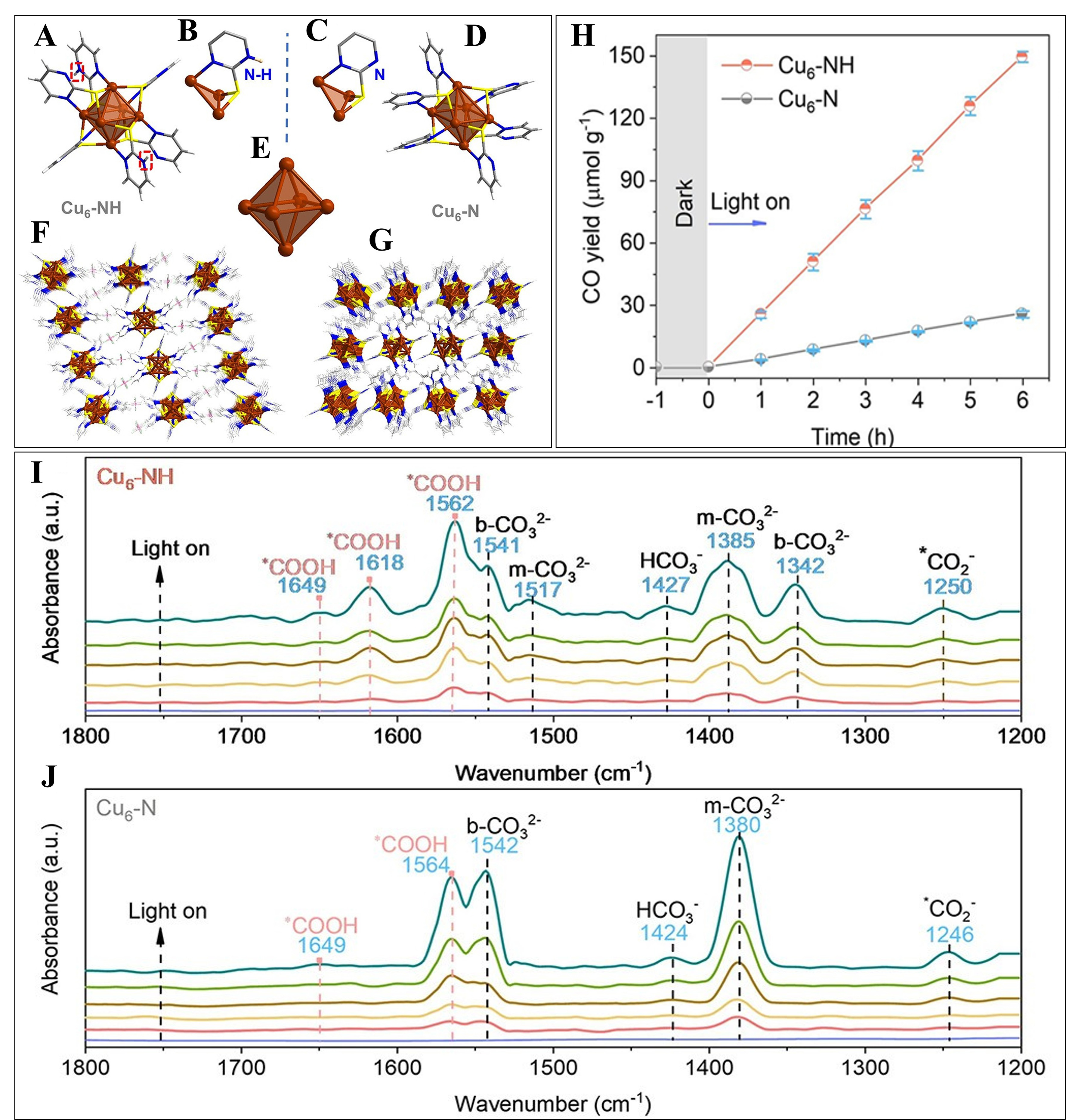
Figure 21. (A) Structure of Cu6-NH ; (B) Coordination modes of ligand in Cu6-NH ; (C) Coordination modes of ligand in Cu6-N; (D) Structure of Cu6-N; (E) Common octahedral Cu6 metal core in Cu6-NH and Cu6-N; Molecule packing diagrams of (F) Cu6-NH and (G) Cu6-N; (H) Time-dependent CO2 photoreduction performances of Cu6-NH and Cu6-N; In-situ DRIFTS spectra during CO2 photoreduction over (I) Cu6-NH and (J) Cu6-N. Color code: Cu, brown; S, yellow; C, gray; H, white; N, blue. This figure is reproduced with permission from Ref.[69], © 2023 Wiley-VCH GmbH.