
fig3
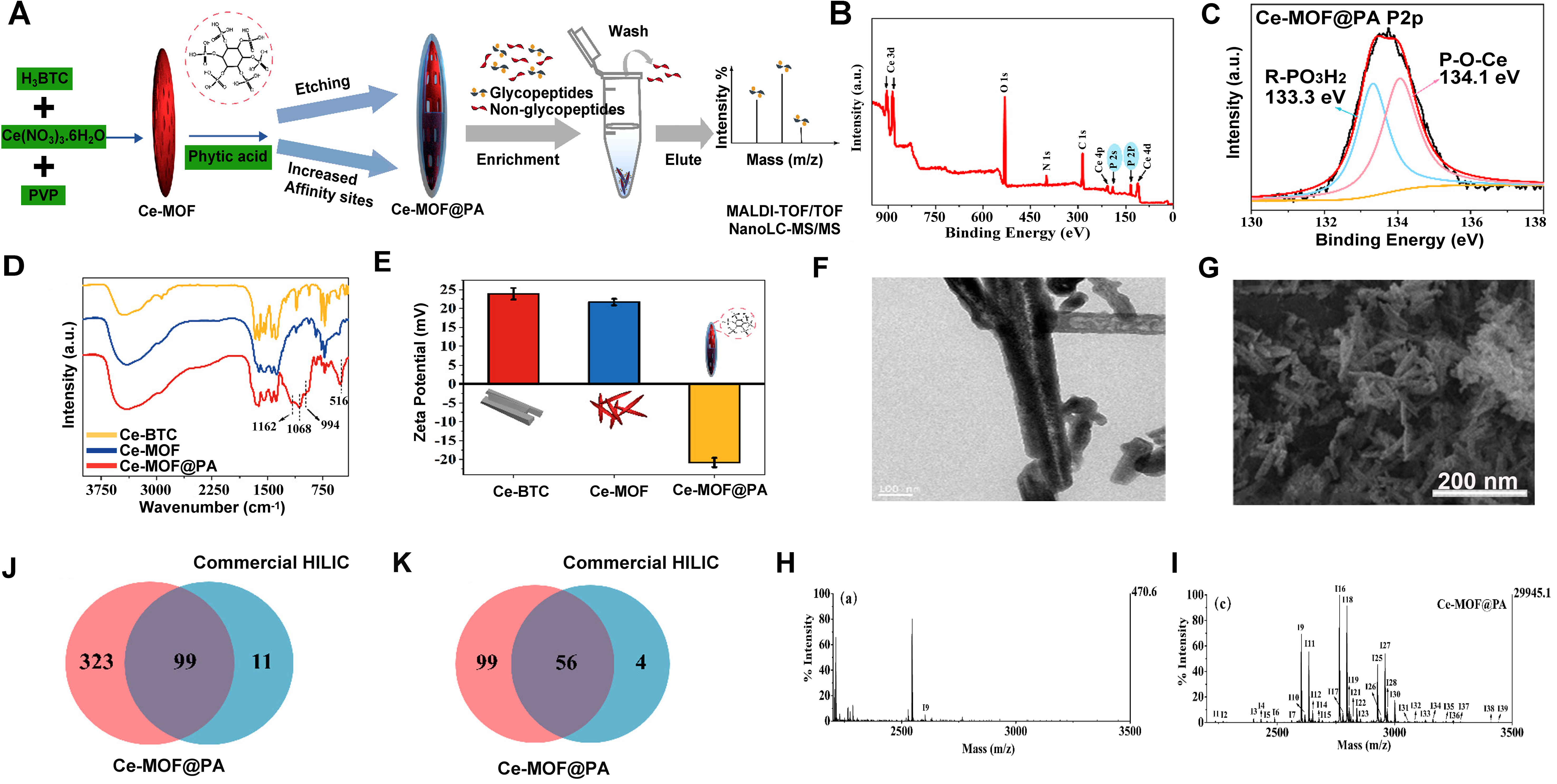
Figure 3. (A) Schematic illustration of the synthesis of Ce-MOF@PA and the enrichment procedure of glycopeptides; (B) XPS survey spectra of Ce-MOF@PA; (C) High-resolution XPS measurements of P2p for Ce-MOF@PA; (D) The FT-IR spectra of Ce-BTC, Ce-MOF, and Ce-MOF@PA; (E) Zeta potential of Ce-BTC, Ce-MOF, and Ce-MOF@PA; (F and G) SEM and TEM images of Ce-MOF@PA; MALDI-TOF MS of IgG digests (6 μg) (H) before enrichment and after enrichment by (I) Ce-MOF@PA. The peaks of glycopeptides are marked with “In”; Overlapping of identified (J) glycopeptides and (K) glycoproteins in human serum after enrichment by Ce-MOF@PA and commercial HILIC material. (A-K) are reprinted with permission from Ref.[98], Copyright © 2019 by American Chemical Society. MOF: Metal–organic framework; PA: phytic acid; XPS: X-ray diffraction; FT-IR: Fourier transform infrared spectroscopy; SEM: scanning electron microscope; TEM: transmission electron microscope; MALDI-TOF MS: matrix-assisted laser desorption/ionization time-of-flight mass spectrometry; IgG: immunoglobulin G; HILIC: hydrophilic interaction chromatography.