

fig3
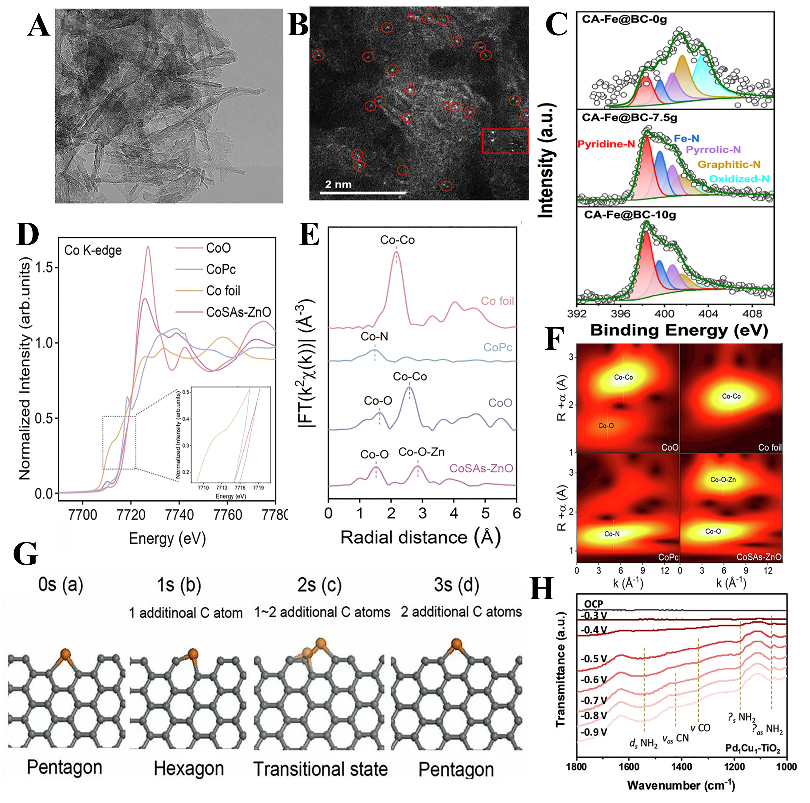
Figure 3. (A) TEM images of Fe@BC[69]; (B) AC-HAADF-STEM images of the Fe@BC catalyst (the red circles are atomic Fe species)[69]; (C) N1s XPS spectra of the CA-Fe@BC-ng catalyst[69]; (D) XANES spectra of CoSAs-ZnO at the Co K-edge (inset: magnification of local areas)[76]; (E) FT k3-weighted EXAFS spectra of CoSAs-ZnO and references[76]; (F) WT-EXAFS for K-edge for Co foil, CoO, CoPc, and CoSAs-ZnO[76]; (G) Schematic illustration for one cycle of catalytic growth of graphene edge[80]; (H) The in situ DRIFTS measurements during urea synthesis on Pd1Cu1-TiO2[82]. TEM: Transmission electron microscopy; AC-HAADF-STEM: aberration-corrected high-angle-annular-dark-field scanning transmission electron microscopy; XPS: X-ray photoelectron spectroscopy; XANES: X-ray absorption near-edge structure; FT: Fourier transform; WT-EXAFS: wavelet transform EXAFS; DRIFTS: diffuse reflectance infrared Fourier transform spectroscopy.


