

fig1
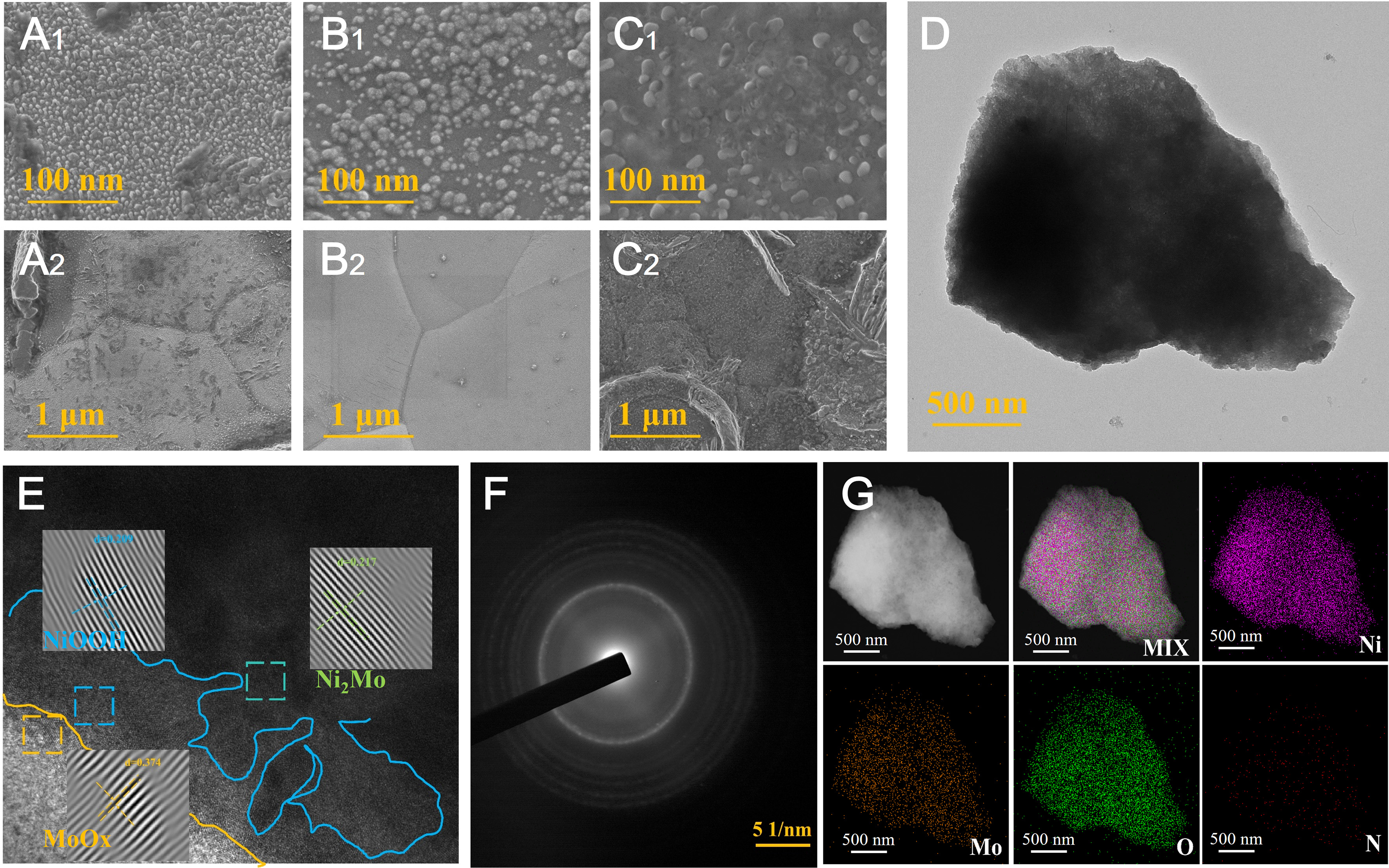
Figure 1. Morphology and structural characterization of the as-prepared catalysts. (A1 and A2) SEM images of Ni-NiOOH; (B1 and B2) SEM images of Ni2Mo/NF; (C1 and C2) SEM images of Ni2Mo-NiOOH/MoOx; (D) TEM image of Ni2Mo-NiOOH/MoOx; (E) HRTEM image of Ni2Mo-NiOOH/MoOx; (F) SAED pattern of Ni2Mo-NiOOH/MoOx; (G) EDS elemental mapping images of Ni2Mo-NiOOH/MoOx (Ni, Mo, O, N). SEM: Scanning electron microscopy; NF: nickel foam; HRTEM: high-resolution transmission electron microscopy; EDS: energy-dispersive X-ray spectroscopy; SAED: selected area electron diffraction.

