

fig1
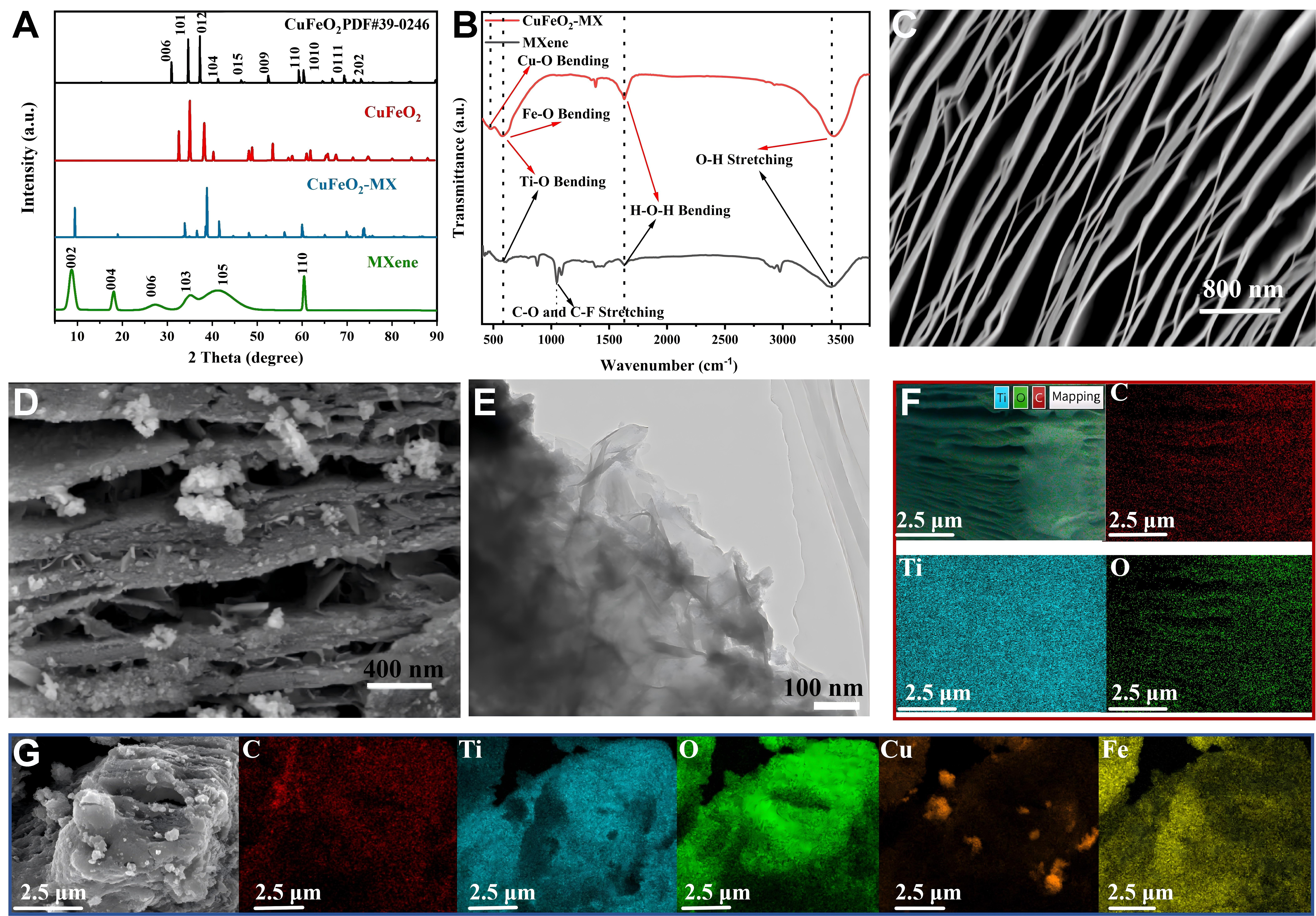
Figure 1. (A) XRD patterns of MXene, CuFeO2 and CuFeO2–MX together with the standard XRD card of CuFeO2; (B) FTIR spectra of MXene and CuFeO2–MX; SEM images of (C) MXene (D) and CuFeO2–MX; (E) TEM images of CuFeO2–MX; Elemental mapping images of (F) MXene and (G) CuFeO2–MX. XRD: X-ray diffraction; CuFeO2–MX: CuFeO2–MXene; FTIR: Fourier transform infrared; SEM: scanning electron microscopy; TEM: transmission electron microscopy.

