
fig3
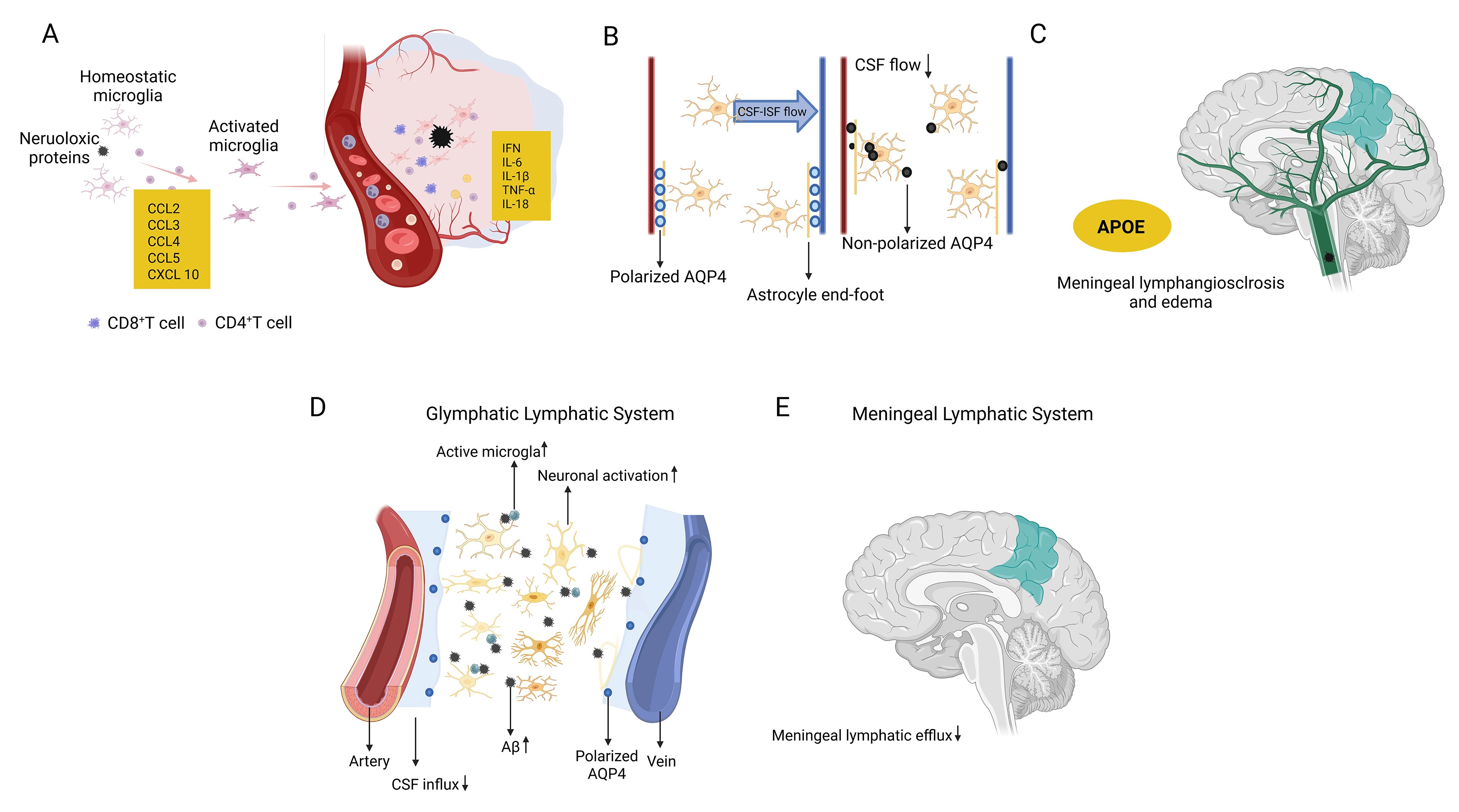
Figure 3. Glymphatic and meningeal lymphatic pathways involved in brain waste clearance in AD. (A) Homeostatic microglia become activated in response to neurotoxic protein accumulation, accompanied by the involvement of CD8+ and CD4+ T cells; (B) Aberrant loss of perivascular AQP4 polarization in astrocytes impairs CSF-ISF exchange and glymphatic flow; (C) Effects of the APOE genotype on meningeal lymphangiectasia (vessel dilation) and edema formation; (D) Schematic illustration of glymphatic and meningeal lymphatic waste clearance pathways, showing increased CSF influx associated with microglial activation and neuronal activity; (E) Illustration of the meningeal lymphatic system, including arteries, increased CSF influx, the posterior vein with AQP4, and impaired meningeal lymphatic efflux. This figure was designed by the authors using BioRender. https://BioRender.com/adw2nwf. AD: Alzheimer’s Disease; Aβ: amyloid-beta; APOE: apolipoprotein E; AQP4: aquaporin-4; CCL2: C-C motif chemokine ligand; CXCL 10: C-X-C motif chemokine ligand 10; CD8+ T cell: cluster of differentiation 8-positive T cell; CSF: cerebrospinal fluid; ISF: interstitial fluid; INF: interferon; TNF-α: tumor necrosis factor-α; IL-1β: interleukin-1β.


