
fig1
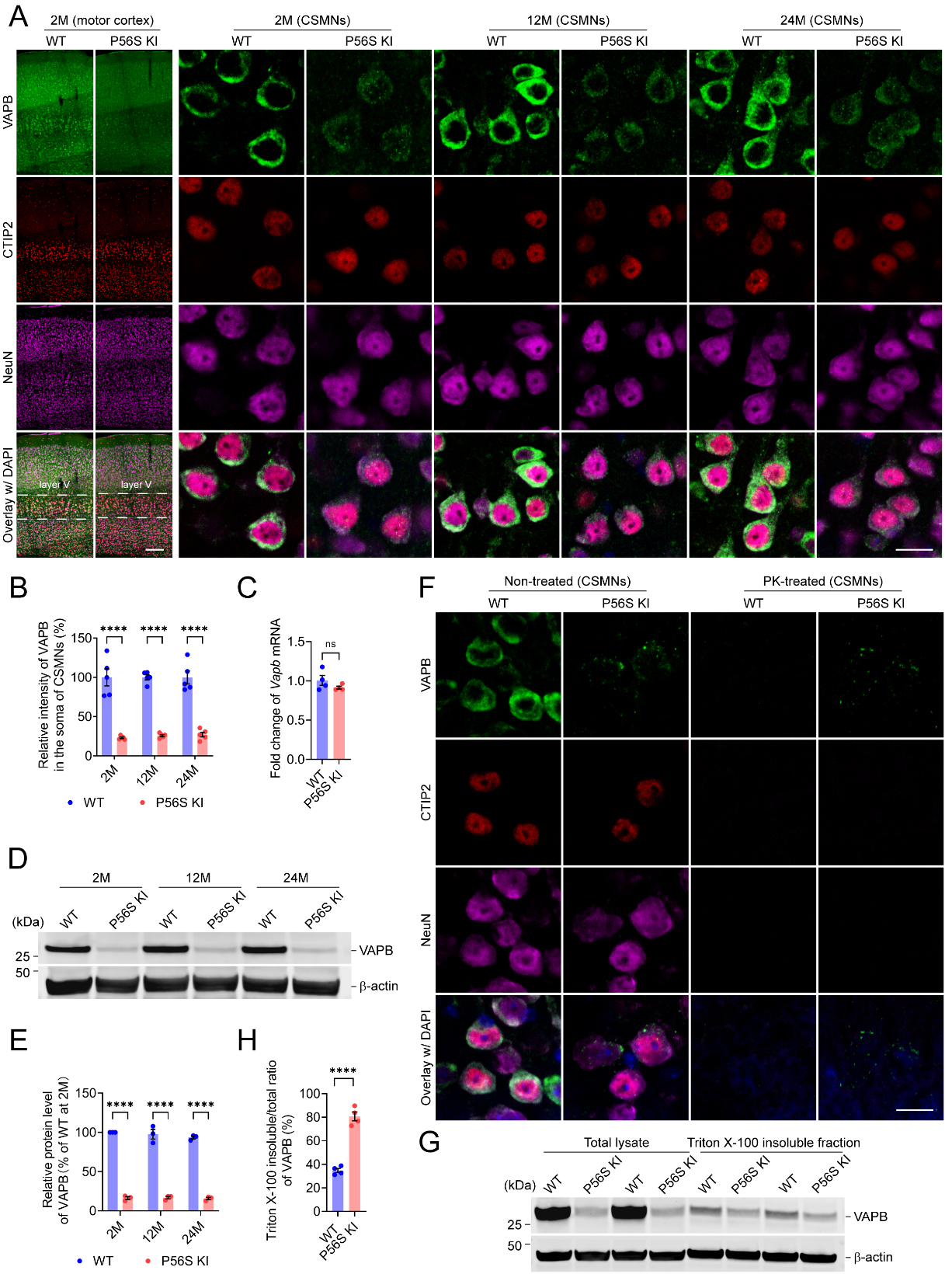
Figure 1. Downregulated protein level of mutant VAPB and PK-resistant cytoplasmic inclusions of mutant VAPB in CSMNs of P56S KI mice. (A and B) Immunostaining of VAPB, CTIP2, and NeuN in the motor cortex of WT and P56S KI mice at 2, 12, and 24 months of age (n = 5 at each time point). CSMNs located in the motor cortex layer V were visualized by CTIP2 staining. Note the reduced intensity and cytoplasmic inclusions of mutant VAPB in the CSMNs of P56S KI mice; (C) Quantitative RT-PCR of cortical Vapb mRNA of 2-month-old WT and P56S KI mice (n = 4); (D and E) Western blotting of VAPB in the cortical total lysate of WT and P56S KI mice at 2, 12, and 24 months of age (n = 3 at each time point); (F) Immunostaining of VAPB, CTIP2, and NeuN in the CSMNs of 2-month-old WT and P56S KI mice under non-treated and PK-treated conditions (for each condition, 3 mice per genotype and 5 sections per mouse were examined). Note that no staining of CTIP2, NeuN, or WT VAPB remained after PK digestion, but the PK-resistant cytoplasmic inclusions of P56S VAPB were still visible in P56S KI mice. DAPI (blue) was used to label nuclei; (G and H) Western blotting of VAPB in the total lysate and 1% Triton X-100 insoluble fraction extracted from the cortices of 2-month-old WT and P56S KI mice (n = 4). Scale bar: 200 μm (motor cortex) and 20 μm (CSMNs) in (A); 20 μm in (F). Two-way ANOVA: ****P < 0.0001 (2M, 12M, and 24M) in (B);


