
fig1
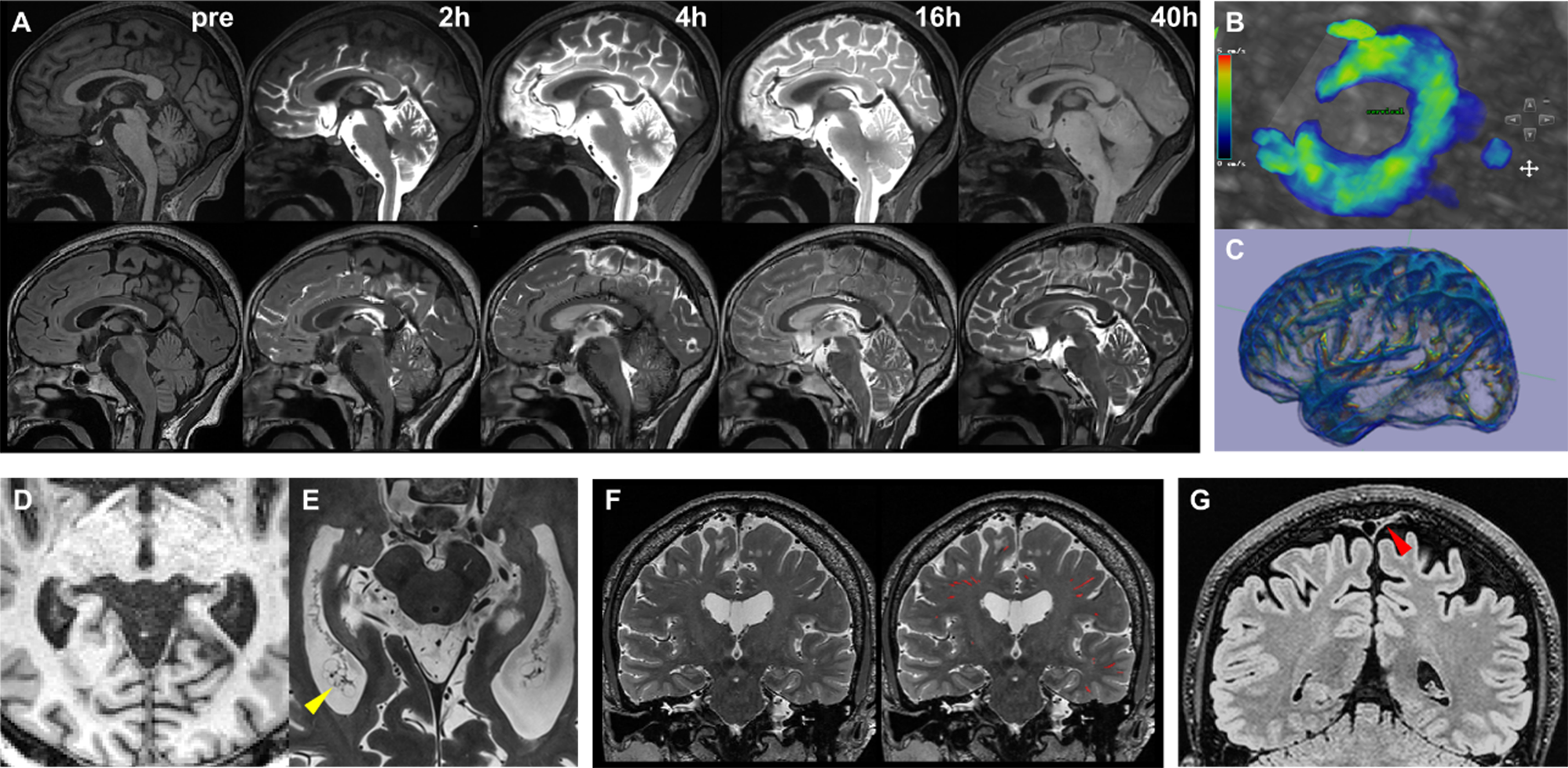
Figure 1. (A) Following intrathecal contrast administration, the contrast agent ascends through the spinal canal into the basal cisterns and progressively distributes along the cerebral convexities. Pronounced parenchymal enhancement persists up to 40 h post-injection, as shown on axial T1-weighted (upper row) and FLAIR (lower row) sequences; (B) PC-MRI quantifies CSF flow dynamics at the foramen magnum level; (C)Low b-value diffusion-weighted imaging enables whole-brain mapping of CSF flow velocity; (D) Conventional 3 Tesla T1-weighted imaging delineates only the gross morphological features of the ChP; (E) Ultra-high-resolution 7 Tesla T1-weighted imaging reveals the intricate curled architecture, surface protrusions, and pathological cystic structures (yellow arrowhead) within the ChP; (F) High-resolution 3D T2-weighted imaging visualizes PVS, enabling neural network-based segmentation and quantitative analysis; (G) Optimized high-resolution FLAIR sequences provide clear visualization of PSD mater morphology (red arrowhead). PC-MRI: Phase-contrast magnetic resonance imaging; CSF: cerebrospinal fluid; ChP: choroid plexus; PVS: perivascular spaces; PSD: parasagittal dura.


