



Minimally invasive intervention in severe acute pancreatitis: a critical, decision-oriented review of timing, route, and escalation
 0
0 
Abstract
Severe acute pancreatitis (SAP) is a heterogeneous and potentially fatal disease in which infected necrosis, persistent organ failure, and procedure-related morbidity remain major clinical challenges. Although minimally invasive intervention has largely replaced early open necrosectomy, important uncertainties persist regarding the optimal timing of intervention, the choice of initial access route, and the threshold for escalation beyond drainage alone. This review synthesizes current evidence on minimally invasive management of SAP with a particular focus on unresolved controversies and decision-making in real-world practice. Rather than viewing percutaneous, endoscopic, and minimally invasive surgical techniques as competing modalities, we propose that they should be understood as complementary tools within an individualized step-up strategy. In our view, the most meaningful clinical question is not whether minimally invasive treatment is preferable to open surgery, but how intervention should be tailored according to indication, urgency, collection maturity, anatomical distribution, solid necrotic burden, septic trajectory, and local multidisciplinary expertise. Based on these considerations, we present a practical decision-centered framework to guide route selection, timing, and escalation. Future research should move beyond broad modality comparisons toward anatomy-stratified studies, standardized escalation criteria, and long-term patient-centered outcomes.
Keywords
INTRODUCTION
Severe acute pancreatitis (SAP) is a life-threatening form of acute pancreatitis characterized by persistent organ failure, high morbidity, and substantial mortality[1]. Its clinical course is often biphasic: an early phase dominated by systemic inflammatory response syndrome (SIRS), multiple organ dysfunction, and abdominal compartment syndrome, followed by a later phase in which infected necrosis and other local complications become major drivers of deterioration and death[2]. Because SAP is characterized by substantial heterogeneity in disease severity, anatomical extent, and temporal evolution, its management remains one of the most challenging problems in pancreatic surgery and critical care.
Over the past two decades, the treatment paradigm for SAP has shifted substantially from early open necrosectomy toward delayed, minimally invasive, and stepwise intervention[3]. This transition has been driven not only by advances in imaging, endoscopy, interventional radiology, and minimally invasive surgery, but also by a deeper understanding that overly aggressive early surgery may aggravate systemic inflammation and increase procedure-related morbidity. In contrast, staged minimally invasive approaches can improve source control while reducing physiological stress and preserving pancreatic and peripancreatic tissue whenever possible. As a result, minimally invasive intervention has become a central component of contemporary SAP management[4].
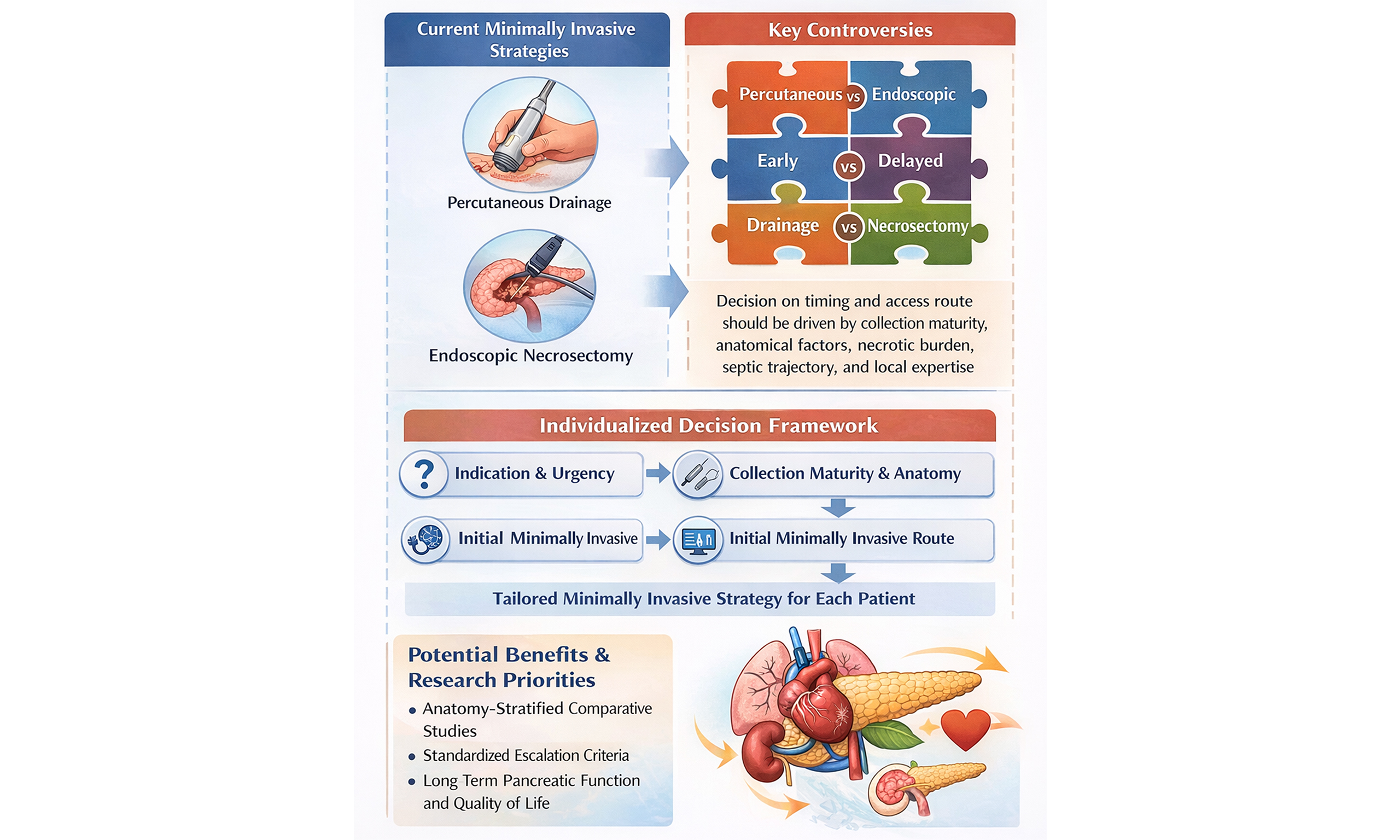
However, the key clinical question is no longer whether minimally invasive treatment should replace open surgery, but how minimally invasive strategies should be individualized in real-world practice. Although the step-up principle is now widely accepted, important uncertainties remain regarding the optimal timing of intervention, selection between endoscopic and percutaneous access, indications for escalation from drainage to necrosectomy, and the influence of anatomical factors, necrotic burden, septic trajectory, and local expertise on treatment choice[5]. Existing reviews often summarize available techniques separately, but less often address how these variables should be integrated into practical clinical decision-making. Therefore, this review aims not only to summarize current minimally invasive modalities for SAP, but also to critically appraise the available evidence, highlight unresolved controversies, and propose a pragmatic clinical decision framework for individualized intervention. A preliminary version of this study was previously published as a preprint[6].
WHY A DECISION-ORIENTED REAPPRAISAL OF MINIMALLY INVASIVE TREATMENT IS NEEDED
Although minimally invasive intervention has become a cornerstone of contemporary SAP management, the field is still often discussed in a technique-centered manner, with percutaneous, endoscopic, and surgical approaches reviewed as separate modalities. Such an approach is useful for describing procedural options, but it does not fully address the real clinical challenge: how to determine the right intervention for the right patient at the right time. In practice, treatment decisions are rarely driven by technique alone. They are shaped by the indication for source control, the maturity and anatomical distribution of necrotic collections, the proportion of solid debris, the tempo of systemic deterioration, and the expertise available within a multidisciplinary center[7]. Accordingly, a clinically meaningful review should move beyond procedural description and focus instead on how current evidence can inform individualized decision-making. The following sections therefore examine minimally invasive treatment in SAP from a decision-oriented perspective, beginning with what is firmly established, what remains controversial, and which clinical variables should guide route selection and escalation.
From open necrosectomy to minimally invasive intervention: what has changed
Open surgery historically played an important role in the management of SAP, particularly in patients with infected necrosis, uncontrolled abdominal sepsis, or abdominal compartment syndrome. In selected settings, surgical decompression and debridement could provide rapid source control, relieve intra-abdominal pressure, and remove necrotic tissue extensively. For many years, this approach represented the dominant interventional strategy for SAP, especially when critical deterioration occurred in the absence of effective alternatives. However, accumulated clinical experience and comparative studies gradually revealed the major limitations of routine open intervention. Whether performed early in the inflammatory phase or later in the necrotizing disease, open necrosectomy is associated with substantial physiological insult and may aggravate systemic inflammatory response, bleeding, enteric fistula formation, pancreatic insufficiency, and multiorgan dysfunction[8,9]. In many patients, aggressive operative debridement achieves anatomical clearance at the cost of considerable procedural morbidity. These observations prompted a re-evaluation of the long-standing assumption that extensive open surgery should be the standard response to necrotizing SAP[10].
The treatment landscape has changed fundamentally over the past two decades. Improvements in cross-sectional imaging, interventional radiology, therapeutic endoscopy, minimally invasive retroperitoneal surgery, and perioperative critical care have enabled a transition from early aggressive surgery toward staged, less invasive, and more selective intervention. Rather than pursuing immediate and complete necrosectomy in every patient, contemporary management increasingly emphasizes infection control, decompression, drainage, and escalation only when clinically necessary. This shift has not simply introduced new techniques; it has redefined the therapeutic goal of intervention from extensive tissue removal to individualized source control with minimal additional injury. As a result, minimally invasive management is now regarded not merely as a technical alternative to open surgery, but as a broader change in treatment philosophy for SAP.
What is already established in current practice
Several principles are now relatively well established in contemporary SAP management. First, invasive intervention should not be undertaken routinely in all patients with pancreatic or peripancreatic necrosis. Many patients with sterile necrosis can be managed conservatively, and even in the presence of necrotizing pancreatitis, intervention is generally reserved for infected necrosis, persistent sepsis, ongoing organ dysfunction, or local complications that cannot be controlled by supportive care alone. This indication-based approach reflects a major departure from earlier eras in which the mere presence of necrosis often prompted operative treatment[11]. Second, delayed intervention has become the preferred strategy whenever the patient’s condition permits. Allowing time for necrotic collections to organize and mature into walled-off necrosis may facilitate safer access, clearer anatomical demarcation, more effective drainage, and reduced procedural complications. Although delay is not always feasible in rapidly deteriorating patients, the general principle that “later is often better” has become a central tenet of modern practice[12]. Third, the step-up concept has emerged as the dominant therapeutic framework. In this model, treatment begins with the least invasive intervention capable of achieving clinical benefit, most commonly image-guided percutaneous drainage or endoscopic transluminal drainage, and proceeds to minimally invasive necrosectomy or surgical debridement only when the response is inadequate[13]. Importantly, the step-up approach should be understood as a treatment principle rather than a rigid procedural sequence. Its purpose is not to force all patients through identical stages, but to reduce unnecessary procedural burden while preserving the option of escalation when source control remains incomplete[14].
Finally, multidisciplinary management is now widely recognized as essential. Because SAP evolves dynamically and often involves multiple organ systems, treatment decisions increasingly depend on coordinated input from intensive care, gastroenterology, pancreatic surgery, interventional radiology, infectious disease, and nutrition teams. In this sense, contemporary practice has already moved beyond single-technique or single-specialty management. What is established today is not merely that minimally invasive techniques are available, but that successful SAP intervention depends on individualized planning within a multidisciplinary and staged framework[15].
Why existing reviews remain insufficient for clinical decision-making
Despite these advances, an important gap remains between procedural knowledge and real-world decision-making. Many existing reviews summarize minimally invasive modalities according to technique, for example, percutaneous drainage, endoscopic necrosectomy, retroperitoneal debridement, laparoscopic approaches, or adjunctive interventions. Such summaries are useful for describing procedural characteristics, indications, and historical evolution, but they often stop short of answering the questions clinicians face at the bedside[16]. In practice, the central challenge is rarely whether a given technique is technically feasible. Rather, the key questions are which patient should undergo intervention, when intervention should occur, which access route is most appropriate, and when escalation from drainage to debridement is justified. These decisions depend on variables that are often underemphasized in technique-centered reviews, including the maturity of the collection, its anatomical extension, the proportion of solid necrotic debris, the trajectory of sepsis or organ failure, the availability of local expertise, and the feasibility of repeated or combined procedures[17]. Thus, route selection in SAP is not simply a comparison between endoscopic and percutaneous methods, nor is step-up therapy a one-size-fits-all sequence.
Another limitation of the current literature is that apparent differences between modalities are frequently confounded by patient selection and center experience. Endoscopic approaches are often favored for centrally located collections adjacent to the stomach or duodenum, whereas percutaneous or retroperitoneal approaches may be more practical for lateral, deep, or complex extensions. Outcomes therefore reflect not only technique performance, but also anatomy, case mix, timing, and institutional expertise. Without acknowledging these contextual factors, comparisons between modalities may be overly simplified and potentially misleading[18].
For these reasons, a more clinically useful review must move beyond descriptive summaries of available procedures. It should critically appraise what is truly supported by current evidence, identify where uncertainty persists, and organize the field around decision-relevant variables rather than procedural categories alone. This decision-oriented perspective provides the rationale for the sections that follow, which examine indications, timing, route selection, escalation strategy, and multidisciplinary implementation as interconnected components of individualized minimally invasive management in SAP.
INDICATIONS FOR INTERVENTION IN SAP
Determining when to intervene is one of the most consequential decisions in the management of SAP. Although minimally invasive techniques have expanded the therapeutic armamentarium, intervention itself remains a double-edged therapeutic strategy: it may provide essential source control in selected patients, but may also introduce additional physiological stress, bleeding, fistula formation, secondary infection, or repeated procedural burden[19]. For this reason, the threshold for intervention in SAP should not be defined by the mere presence of necrosis or fluid collection, but by a careful assessment of whether invasive treatment is likely to alter the clinical trajectory more favorably than continued conservative management. A decision-oriented approach therefore begins with indication rather than technique. The most widely accepted trigger is infected necrosis, but clinical reality is often more complex. Some patients deteriorate despite the absence of microbiological confirmation, whereas others with extensive necrosis remain stable under noninvasive treatment[20]. Moreover, certain emergencies require prompt intervention irrespective of the maturity of the collection. The following subsections distinguish between established indications, relative indications, urgent rescue scenarios, and situations in which intervention should be deferred or avoided.
Established indications: infected pancreatic necrosis
Among all potential triggers for intervention in SAP, infected pancreatic necrosis remains the most clearly established and widely accepted indication. Once infection develops within necrotic pancreatic or peripancreatic tissue, conservative treatment alone is often insufficient to achieve durable source control, particularly in patients with persistent fever, rising inflammatory markers, clinical sepsis, or progressive organ dysfunction[21,22]. In such settings, drainage-based intervention has become the cornerstone of management, with escalation to necrosectomy reserved for inadequate response.
The diagnosis of infected necrosis may be confirmed directly, for example by positive culture obtained from fine-needle aspiration or drainage, but in contemporary practice it is often established clinically and radiologically rather than microbiologically. The presence of gas within a necrotic collection on cross-sectional imaging is strongly suggestive, although not universally present. More commonly, clinicians rely on a constellation of findings, including persistent or recurrent fever, leukocytosis, elevated inflammatory biomarkers, hemodynamic instability, failure to improve despite optimal supportive care, and exclusion of alternative sources of infection. This shift away from routine diagnostic aspiration reflects both practical considerations and the recognition that clinical context often provides sufficient justification for intervention[23,24].
Importantly, intervention for infected necrosis should not be equated with immediate necrosectomy. Current practice strongly favors drainage as the initial step in most patients, with the dual goals of source control and physiological stabilization. In a substantial proportion of cases, catheter- or endoscopy-based drainage may reduce the infectious burden sufficiently to avoid more invasive debridement altogether. Thus, the established indication is not “infected necrosis equals surgery”, but rather “infected necrosis requires active source control, preferably through a staged minimally invasive pathway”. At the same time, not every patient with suspected infected necrosis requires identical timing or access. Hemodynamic stability, collection maturity, anatomical accessibility, and local expertise all influence how intervention should be implemented. Therefore, while infected necrosis is the clearest trigger for invasive management, its presence does not eliminate the need for individualized route selection and timing.
Relative indications: persistent or progressive organ failure without proven infection
A more difficult and controversial scenario arises when patients develop persistent or worsening organ failure in the absence of definite evidence of infected necrosis. In these cases, the indication for intervention is less straightforward[25]. On the one hand, persistent systemic deterioration may reflect ongoing inflammatory toxicity from necrotic tissue, occult infection not yet radiologically apparent, or insufficient decompression of evolving collections. On the other hand, premature invasive intervention during the early inflammatory phase may exacerbate instability without addressing the underlying pathophysiology[5]. For this reason, persistent organ failure without proven infection should be regarded as a relative rather than absolute indication for intervention. The decision depends heavily on the overall clinical trajectory. Intervention may be justified when organ dysfunction fails to improve despite intensive care support, when sepsis is strongly suspected but not microbiologically confirmed, or when repeated reassessment suggests that an evolving collection is contributing meaningfully to ongoing clinical decline. In such patients, minimally invasive drainage may serve both diagnostic and therapeutic purposes by reducing inflammatory burden, decompressing a symptomatic collection, and clarifying whether occult infection is present[25,26].
However, this indication demands caution. Organ failure in early SAP is often driven predominantly by systemic inflammation rather than by a localized target amenable to intervention[27]. If a collection is immature, poorly demarcated, or not anatomically suitable for safe access, the risks of intervention may outweigh the likely benefit. In addition, progressive organ dysfunction may result from extra-pancreatic complications such as pneumonia, catheter-related infection, bowel ischemia, or cardiopulmonary decompensation, none of which would be corrected by pancreatic intervention[28]. Therefore, intervention should not become a default response to any prolonged ICU course.
A practical principle is that intervention in this setting should be considered when three conditions converge: first, the patient shows persistent or worsening clinical instability; second, imaging demonstrates a plausible target for drainage or decompression; and third, multidisciplinary reassessment indicates that non-pancreatic explanations for deterioration have been excluded or are insufficient to account for the severity of illness[29]. Even then, the goal should usually be limited and staged - most commonly drainage rather than immediate debridement. This is an area in which clinical judgment remains indispensable, and where the literature still provides less certainty than in established infected necrosis.
Urgent rescue scenarios: abdominal compartment syndrome, perforation, hemorrhage, and biliary emergencies
Although delayed intervention is preferred in most patients with SAP, some complications represent true rescue scenarios in which waiting for collection maturation is neither feasible nor safe[30]. These situations are exceptions to the general principle of postponement and require prompt action to prevent irreversible deterioration or death. Their clinical importance lies in the fact that they are driven by urgent physiology rather than the ideal timing of necrotic intervention. Abdominal compartment syndrome is one of the most important such scenarios. Markedly elevated intra-abdominal pressure may impair renal perfusion, venous return, respiratory mechanics, and visceral blood flow, thereby worsening multiorgan dysfunction[31]. When conservative decompressive measures fail, urgent procedural or surgical decompression may be necessary even in the early phase of disease. In this context, the objective is not definitive management of necrosis, but rapid reversal of life-threatening compartment physiology. The same applies to selected biliary emergencies, such as concomitant cholangitis or persistent biliary obstruction requiring urgent decompression[19]. Similarly, gastrointestinal perforation, uncontrolled hemorrhage, bowel ischemia, or visceral perforation represent emergencies that may necessitate immediate intervention irrespective of the maturity of pancreatic collections[32,33]. In these cases, intervention is triggered not by the presence of necrosis per se, but by a specific complication whose natural history is incompatible with continued delay. The route of intervention may differ accordingly: endoscopic, radiologic, surgical, or hybrid approaches may all be appropriate depending on the dominant pathology.
These rescue indications underscore an important conceptual point: the timing of intervention in SAP should not be reduced to a simple “before or after four weeks” rule. The more relevant question is whether the patient’s current complication requires immediate action for physiological stabilization or source control. When the answer is yes, earlier intervention is justified, even if the eventual necrosis-directed strategy will need to be revisited later in a more mature phase. Thus, urgent rescue scenarios are not contradictions of the delayed-intervention paradigm, but clinically necessary exceptions within a broader individualized strategy.
When not to intervene: sterile necrosis, clinical stabilization, and procedural futility
Equally important as knowing when to intervene is recognizing when intervention should be avoided. The presence of pancreatic or peripancreatic necrosis alone is not an indication for invasive treatment[34]. Many patients with sterile necrosis can be managed successfully with intensive supportive care, nutritional optimization, organ support, and close imaging follow-up. In clinically stable patients, premature drainage or debridement may expose them to unnecessary complications without providing meaningful benefit[35].
Clinical stabilization is therefore a major argument against routine intervention. A patient with extensive necrosis but improving hemodynamics, declining inflammatory markers, absence of sepsis, adequate pain control, and no significant compressive symptoms may be better served by continued observation than by invasive procedures. This principle is especially relevant because imaging severity and clinical severity do not always move in parallel[36]. Large collections may appear alarming radiologically yet remain biologically quiescent, whereas smaller collections may drive disproportionate systemic illness. Intervention should follow the patient’s clinical course, not imaging appearance alone.
Finally, the concept of procedural futility must be considered, particularly in patients with irreversible multiorgan failure, extreme frailty, or a broader clinical context in which invasive source control is unlikely to change outcome. Although this is often uncomfortable to discuss, not all technically feasible interventions are clinically justified. In some patients, the burden of repeated drainage, necrosectomy, anesthesia, and ICU escalation may exceed the realistic chance of recovery. Accordingly, the decision not to intervene can be as clinically meaningful as the decision to proceed, provided it is reached through careful multidisciplinary assessment and transparent communication with patients and families[31].
Taken together, these considerations reinforce a central message: intervention in SAP should be selective rather than reflexive. A clinically useful framework must identify not only who should undergo invasive treatment, but also who may benefit more from continued observation, delayed reassessment, or supportive care alone.
EVIDENCE SUPPORTING THE STEP-UP PRINCIPLE: WHAT IS SETTLED AND WHAT IS NOT
Once the indication for intervention has been established, the next question is not simply which procedure to perform, but how intervention should be staged. In contemporary SAP management, the step-up principle has become the dominant framework for this purpose. Rather than proceeding directly to open necrosectomy, the step-up strategy begins with the least invasive intervention capable of achieving meaningful source control and escalates only if the clinical response remains inadequate[37]. This paradigm has substantially reshaped the management of infected necrosis and other intervention-requiring complications. However, the strength of the step-up concept lies not only in its procedural sequence, but also in its underlying philosophy of minimizing treatment-related injury while preserving the capacity for escalation. Questions remain regarding the optimal initial route, the criteria for escalation, and the degree to which outcomes reflect modality choice vs. case selection and center expertise[38]. For this reason, the step-up approach should be examined not as a closed doctrine, but as a structured yet adaptable framework whose evidence base is strong in some areas and still incomplete in others[39].
Why step-up has replaced routine early open surgery
The step-up principle emerged largely in response to the limitations of routine open surgery in necrotizing pancreatitis. Traditional open necrosectomy was designed to achieve direct and extensive removal of infected or devitalized tissue, but in practice it often imposed substantial physiological burden on patients who were already critically ill[40]. The combination of severe systemic inflammation, capillary leak, organ dysfunction, nutritional depletion, and fragile retroperitoneal anatomy made open debridement particularly hazardous, especially when performed early in the disease course. Even when technically successful, surgery frequently resulted in major complications such as bleeding, enteric fistulae, incisional morbidity, repeated re-operations, and prolonged intensive care dependence[41]. By contrast, the step-up approach was built on a different therapeutic logic. It recognized that not all patients with necrotizing SAP require immediate or extensive tissue debridement, and that a meaningful proportion may improve with drainage alone once the infectious or inflammatory burden is partially controlled. This reorientation was clinically important because it shifted the aim of intervention away from anatomical completeness and toward pragmatic source control. In other words, the goal became not to remove all necrotic material at the earliest opportunity, but to stabilize the patient with the least invasive effective method and escalate only when necessary[42].
The replacement of routine open surgery by step-up strategies therefore reflects more than technical progress; it reflects a change in therapeutic priorities. Contemporary management places greater value on reducing procedure-related harm, allowing physiological recovery, preserving viable tissue, and tailoring the intensity of intervention to the patient’s evolving clinical response[43]. This is why step-up has become the preferred paradigm in most patients requiring intervention for SAP-related necrosis. Open surgery has not disappeared completely, but it is no longer viewed as the default starting point. Instead, it has become one of several escalation options within a broader staged treatment pathway[44].
Step-up as a treatment principle rather than a fixed procedural sequence
The step-up treatment strategy represents the core therapeutic concept in the management of SAP, particularly necrotizing pancreatitis. This principle is based on the natural evolution of the disease and advances in minimally invasive techniques. It emphasizes initiating treatment with the least invasive intervention once infection and inflammation are adequately controlled, and progressively escalating treatment intensity according to clinical response, thereby minimizing treatment-related complications and mortality risk[45]. The step-up strategy is not a fixed protocol, but rather a dynamic, adaptable, and escalatable therapeutic strategy. Its basic framework generally includes the following sequential levels. First, intensified medical and critical care support is provided. All SAP patients should initially receive standardized fluid resuscitation, organ function support, analgesia, infection surveillance, and enteral nutrition[46]. Although this stage does not involve invasive intervention, it constitutes the foundational platform of the step-up strategy. Second, minimally invasive drainage is employed as the preferred initial invasive intervention. When infected pancreatic necrosis or persistent sepsis is present, the least traumatic drainage method should be selected first, such as percutaneous catheter drainage (PCD) or endoscopic drainage[47]. Third, minimally invasive necrosectomy is considered as an escalation strategy. If infection control remains inadequate or clinical improvement is limited after drainage alone, treatment may be upgraded at an appropriate time to minimally invasive techniques with necrosectomy capability, such as endoscopic necrosectomy or video-assisted retroperitoneal necrosectomy. Finally, open surgery is reserved as the last option. Open necrosectomy or surgical intervention should be considered only when minimally invasive approaches have failed or when life-threatening complications occur[48].
Clinical evidence indicates that adherence to the step-up treatment principle significantly reduces the incidence of severe complications, decreases the need for open surgical intervention, shortens intensive care unit and overall hospital stay, and improves overall survival outcomes. More importantly, this principle provides a standardized and reproducible decision-making framework for minimally invasive management of SAP while allowing for individualized adaptation, and has therefore become a core strategy widely endorsed by current international guidelines and expert consensus statements[49].
Importantly, step-up should be understood as a treatment philosophy rather than a rigid procedural sequence. In practice, the initial access route, escalation threshold, and choice of necrosectomy technique vary substantially according to collection anatomy, degree of solid necrotic burden, systemic deterioration, and local expertise. Therefore, the major unresolved issue is no longer whether step-up is preferable to open necrosectomy, but how step-up should be individualized in different clinical contexts. However, step-up strategies vary across centers. The initial access route and escalation threshold are influenced by available equipment and institutional expertise. Current evidence more strongly supports a step-up approach over primary open surgery, but remains insufficient to determine which pathway is most suitable for specific patient subgroups.
The unresolved issue: which step-up pathway fits which patient
Although the general superiority of staged minimally invasive treatment over routine primary open surgery is now widely accepted, an important unresolved question remains: which step-up pathway is most appropriate for which patient? This is the point at which the strongest consensus in the field begins to give way to genuine uncertainty. The challenge is no longer whether to adopt a step-up philosophy, but how to operationalize it across highly heterogeneous anatomical and clinical presentations[41]. At present, the choice of pathway is influenced by several interrelated factors. These include the location and maturity of the collection, its proximity to the stomach or duodenum, the extent of lateral or pelvic extension, the proportion of solid necrotic debris, the urgency of source control, the patient’s physiological reserve, and the expertise available within the treating center. As a result, the question “endoscopic-first or percutaneous-first?” cannot be answered in the abstract. Each route has particular strengths and limitations, and each is favored in different anatomical contexts. Similarly, the threshold for escalation from drainage to debridement is shaped not only by persistent infection, but also by the adequacy of catheter position, the effectiveness of irrigation, the accessibility of residual necrosis, and the patient’s ongoing systemic response.
This complexity helps explain why published outcomes across different minimally invasive approaches are difficult to compare directly. Apparent superiority of one modality may partly reflect patient selection rather than intrinsic procedural advantage[18]. Endoscopic series often involve centrally located collections accessible through the gastric or duodenal wall, whereas percutaneous and retroperitoneal series may include more laterally distributed, deep, or anatomically complex necrosis. Center experience further complicates interpretation, since outcomes are strongly influenced by operator skill, multidisciplinary coordination, and the availability of repeated or hybrid procedures. What appears to be a comparison of techniques may therefore in part be a comparison of anatomy, timing, and institutional systems[50]. For these reasons, the unresolved issue is not whether step-up works, but how it should be individualized. This is precisely where future progress is needed. Better anatomy-stratified comparisons, more explicit criteria for escalation, and clinically usable decision frameworks are required to move the field beyond broad endorsement of minimally invasive care toward more precise route selection. If the main unresolved problem is not whether to use a step-up strategy, but how to tailor it, then the next task is to examine the principal controversies that shape route selection, timing, and escalation in contemporary practice.
COMPARATIVE APPRAISAL OF AVAILABLE MINIMALLY INVASIVE MODALITIES
Minimally invasive modalities in SAP should not be understood as interchangeable procedural tools. Each approach has a distinct therapeutic profile shaped by access route, anatomical reach, capacity to remove solid necrosis, procedural burden, and dependence on local expertise[51]. A meaningful appraisal therefore requires more than a technical description of how each procedure is performed. What matters clinically is which modality is best suited to which patient, under what circumstances it is likely to succeed, when it is likely to fail, and how it fits into a staged strategy of escalation. The following appraisal emphasizes these practical distinctions. Rather than ranking modalities in absolute terms, this section considers them as context-dependent options within individualized SAP management. In many cases, their value lies not in replacing one another, but in being deployed sequentially or in combination according to evolving anatomy and clinical response.
PCD: strengths, limitations, and best-use scenarios
PCD remains one of the most widely used minimally invasive interventions in SAP and continues to play a central role in step-up treatment strategies. Its most important strength is accessibility. Under image guidance, percutaneous drainage can often be established rapidly and relatively safely, even in critically ill patients who are poor candidates for more invasive procedures[52,53]. This makes PCD particularly attractive when urgent decompression or early source control is needed, especially in patients with hemodynamic instability, respiratory compromise, or evolving sepsis. From an anatomical perspective, PCD is especially valuable for collections that are laterally located, extend into the paracolic gutters or pelvis, involve deep retroperitoneal compartments, or are otherwise poorly accessible from the upper gastrointestinal lumen[54]. It may also be useful when disease is multiloculated and different compartments require separate access routes. In these situations, PCD offers practical versatility that is difficult to match with a purely transluminal endoscopic approach. Another important advantage is that it can serve both therapeutic and diagnostic purposes, allowing decompression, culture acquisition, irrigation, and radiological reassessment through an established route[55].
However, the limitations of PCD are equally important. Drainage is often less effective when collections contain a high burden of solid necrotic debris rather than primarily fluid content[56]. In such cases, catheter placement alone may provide only partial source control and may need repeated upsizing, repositioning, or adjunctive irrigation. External drains may also be uncomfortable for patients, prone to clogging or dislodgement, and associated with prolonged catheter dependence. In addition, persistent pancreatic fistula burden and skin-site complications may complicate longer treatment courses[57]. These limitations mean that PCD should not be viewed as a definitive answer in all cases, even when it is the most logical initial step. Failure of PCD is often less about the concept of drainage itself than about a mismatch between catheter-based treatment and disease morphology. Laterally extensive collections with significant solid debris, multiple undrained loculations, or persistent systemic sepsis despite technically adequate drainage are common scenarios in which escalation becomes necessary. Even so, one of the major strengths of PCD is precisely that it creates a bridge to further intervention. It may stabilize patients, reduce the inflammatory burden, and establish a tract for later retroperitoneal debridement if needed. Thus, its role within step-up treatment is not merely as a standalone modality, but as a highly flexible access strategy that can initiate staged source control in a wide range of anatomical settings[58,59].
The evidence base for PCD is strong in the context of step-up treatment and as an alternative to routine primary open surgery. Nonetheless, comparative interpretation requires caution because patients selected for PCD often differ anatomically and physiologically from those selected for endoscopic approaches. Its strengths are clearest in anatomically complex or lateral disease, but its outcomes cannot be generalized without reference to case mix and escalation strategy.
Endoscopic drainage and necrosectomy: advantages, anatomical requirements, and risks
Endoscopic transluminal drainage, with or without subsequent endoscopic necrosectomy, has become a major component of modern SAP management, particularly in high-volume centers with advanced therapeutic endoscopy expertise. Its principal appeal lies in the possibility of achieving internal drainage and debridement through the stomach or duodenum without the need for external drains or large incisions[60,61]. For centrally located collections that are mature and closely opposed to the gastrointestinal wall, this approach can offer a relatively direct, controlled, and repeatable route for source control.
One of the major advantages of endoscopic management is that it integrates drainage and escalation within the same anatomical corridor. After creating transluminal access, clinicians may proceed from stent-based decompression to repeated necrosectomy sessions if residual solid necrosis continues to drive symptoms or sepsis[62]. This internal pathway may reduce external fistula burden, improve patient comfort, and facilitate serial interventions without repeated percutaneous punctures or operative re-entries. In selected patients, particularly those with walled-off necrosis adjacent to the stomach or duodenum, endoscopic treatment may therefore represent a highly efficient step-up pathway[63,64].
Its limitations, however, are strongly anatomy-dependent. Endoscopic access requires close apposition between the gastrointestinal lumen and the target collection, and it is less suitable for extensive lateral extension, deep retroperitoneal spread, pelvic components, or disease distributed across multiple remote compartments[65]. Moreover, collections with very high solid debris burden may still require repeated necrosectomy sessions, which can be procedurally demanding and resource-intensive. Endoscopic treatment also depends heavily on local experience, not only for initial access but for timing, stent management, complication rescue, and repeated debridement planning[66]. Risks include bleeding, perforation, stent-related adverse events, air embolic phenomena in rare cases, secondary infection, and procedural burden from repeated necrosectomy sessions. These risks do not negate its value, but they underline that endoscopic treatment is not a universally low-complexity solution. Its success depends on careful patient selection, mature collection formation, and the ability to manage complications promptly within an experienced multidisciplinary environment[67,68].
Within the step-up framework, endoscopic drainage is best viewed as a route-specific pathway rather than a universally preferred modality. It is especially compelling when the anatomy is favorable and internal access can reasonably achieve both decompression and subsequent debridement if required. It is less compelling when anatomy is unfavorable or when disease distribution exceeds what can be addressed through a transluminal corridor. The evidence supporting endoscopic management is increasingly strong, particularly for selected central walled-off collections, but interpretation remains limited by anatomy-driven selection bias and center-expertise effects. Accordingly, endoscopic superiority should not be assumed outside contexts in which its access advantages are actually present.
Laparoscopic necrosectomy: where does it still fit?
Laparoscopic necrosectomy occupies a more selective and somewhat less standardized role in contemporary SAP management. Compared with catheter-based, endoscopic, or retroperitoneal approaches, laparoscopy may offer broader operative visualization and the possibility of more deliberate debridement while still avoiding some of the morbidity associated with open laparotomy[69]. In theory, it can serve as a minimally invasive surgical alternative when less invasive access routes are inadequate, inaccessible, or unlikely to provide effective source control[70].
Its potential advantages are most relevant in patients who require more active debridement than drainage can provide, but whose disease pattern still permits a minimally invasive operative approach. Laparoscopy may be considered in selected cases of localized necrosis, organized collections accessible from the lesser sac or adjacent spaces, or when other minimally invasive routes have failed to achieve adequate control[71]. In experienced hands, it may allow irrigation, debridement, and strategic access under direct vision with less abdominal wall trauma than open surgery[72].
However, laparoscopy also has important limitations that have prevented it from becoming the dominant minimally invasive strategy in SAP. Necrotizing pancreatitis often presents with dense inflammation, friable tissues, distorted anatomy, bowel edema, and severe physiological instability, all of which may reduce the safety and practicality of laparoscopic manipulation. Access can be challenging, exposure may be incomplete, and the operative burden may still be considerable in patients with ongoing systemic illness. As a result, laparoscopy is often less attractive as an initial strategy than drainage-based or route-limited step-up approaches, which impose lower physiological stress and can be more easily repeated. Another issue is that the evidence base for laparoscopic necrosectomy is less robust and less standardized than for step-up strategies built around PCD or endoscopic drainage. Much of the available literature consists of case series, selected-center experiences, or heterogeneous cohorts in which laparoscopy is used in carefully chosen patients. This makes generalization difficult. Accordingly, laparoscopic necrosectomy should probably be regarded not as a first-line modality for most patients with necrotizing SAP, but as a selective option within the escalation spectrum when anatomy, expertise, and failure of less invasive methods make it reasonable[73].
Retroperitoneal approaches: MARPN and VARD in the step-up pathway
Retroperitoneal approaches, including minimal-access retroperitoneal pancreatic necrosectomy (MARPN) and video-assisted retroperitoneal debridement (VARD), occupy an important intermediate position between catheter-based drainage and more invasive operative intervention[74]. Their major value lies in allowing direct debridement of necrotic tissue through a limited retroperitoneal tract, often using a previously established percutaneous access route. In appropriately selected patients, these techniques preserve the step-up principle by escalating beyond drainage without reverting immediately to formal open necrosectomy[75].
The strengths of retroperitoneal approaches include more direct mechanical access to solid necrosis than drainage alone, compatibility with existing percutaneous tracts, and reduced surgical trauma compared with open necrosectomy. They may be especially useful when repeated catheter optimization has failed or when the burden of necrotic debris exceeds what can reasonably be managed by drainage alone[76]. By maintaining treatment through a flank or retroperitoneal route, these approaches may also avoid contamination of the peritoneal cavity and limit the morbidity associated with transabdominal surgery[77].
Their limitations are primarily anatomical and technical. Retroperitoneal access is best suited to disease that can be reached through a safe flank corridor and may be much less effective for centrally located or widely distributed collections. Visualization and maneuverability are more limited than in open surgery, and multiple procedures may still be necessary[60]. Bleeding, incomplete debridement, injury to adjacent structures, and the need for repeated intervention remain important concerns. Moreover, successful performance of MARPN or VARD requires dedicated experience that is not uniformly available across centers.
Within a step-up pathway, retroperitoneal approaches are best understood as escalation tools for a specific anatomical subset of SAP rather than as standalone competitors to all other modalities. Their greatest contribution is in anatomically favorable lateral or posterior disease, after initial drainage has demonstrated the need for more definitive solid necrosis removal[78]. The evidence supporting their use is strongest in this context, but direct comparisons with endoscopic pathways remain difficult because the underlying anatomy often differs substantially between treated populations. As such, their value is considerable but inherently context-specific.
Continuous regional arterial infusion: adjunctive value and current limitations
Continuous regional arterial infusion (CRAI) occupies a distinctly different position from the other modalities discussed above because it is not primarily a drainage or debridement technique. Rather, it has been explored as an adjunctive interventional strategy aimed at delivering therapeutic agents directly to the pancreatic arterial supply in an effort to attenuate local inflammation, infection-related progression, or tissue injury[79]. Its theoretical rationale has made it attractive in certain settings, particularly in East Asian practice patterns, but its role in contemporary SAP management remains controversial.
The potential appeal of CRAI lies in its attempt to intervene earlier in the disease process without immediate mechanical entry into necrotic compartments. In theory, regional delivery of protease inhibitors, antibiotics, or other agents may help modify the local inflammatory environment, limit necrosis progression, or improve drug concentration in ischemic pancreatic tissue[80]. This concept is particularly attractive in severe early disease, when patients may not yet have mature collections suitable for drainage but remain at high risk of deterioration. However, the clinical limitations of CRAI are substantial. First, its mechanism and therapeutic target differ fundamentally from the source-control logic that underpins drainage-based step-up strategies. Even if regional infusion modifies local inflammation, it does not directly evacuate infected material, decompress collections, or remove solid necrosis. Second, the evidence supporting CRAI is less consistent, less internationally reproducible, and more context-specific than that supporting drainage, endoscopy, or retroperitoneal debridement[81]. Third, CRAI requires specialized interventional capability and introduces catheter-related procedural risks without clearly established universal indications.
For these reasons, CRAI is best understood as a possible adjunct rather than a core modality in the minimally invasive management of SAP. Its future relevance will depend on whether more standardized and reproducible evidence can clarify which patients, if any, derive consistent benefit beyond optimal modern supportive care and staged intervention[82]. It reflects the broader ambition to intervene earlier and less invasively in severe disease, but it has not achieved the same level of evidentiary support or practical centrality as route-based minimally invasive source-control strategies.
To move beyond a purely descriptive summary of techniques, the major minimally invasive modalities can be compared according to anatomical suitability, role within the step-up pathway, procedural strengths, limitations, and evidence boundaries [Table 1][83].
Comparative characteristics of minimally invasive treatment modalities for SAP
| Modality | Best anatomical/clinical scenario | Primary role in the step-up pathway | Major strengths | Major limitations |
| PCD[45,66] | Collections with a substantial liquid component; laterally located collections; deep retroperitoneal or pancreatic extension; patients requiring urgent source control when endoscopic access is not feasible | Preferred initial drainage modality in many step-up strategies | Widely available; can be performed early when necessary; effective for sepsis control and decompression; useful bridge to retroperitoneal debridement | Limited efficacy in collections with high solid necrotic burden; catheter malposition or insufficient drainage may lead to failure; often requires repeated upsizing or repositioning |
| Endoscopic transluminal drainage with endoscopic necrosectomy[13,45,66] | Centrally located walled-off necrosis adjacent to the stomach or duodenum; mature collections with a defined wall | Initial drainage and/or escalation to necrosectomy in selected patients | Internal drainage; avoids external fistula; less invasive than surgery; favorable outcomes in appropriately selected central WON | Requires advanced expertise and specialized equipment; limited applicability in lateral/deep collections; bleeding risk; usually unsuitable when access route is not opposed to the gastrointestinal lumen |
| ERCP with sphincterotomy/biliary drainage[13,45,66] | Biliary SAP with cholangitis, persistent biliary obstruction, or urgent need for decompression | Etiology-directed adjunct rather than direct escalation therapy | Rapid biliary decompression; prevents ongoing biliary/pancreatic injury | Not a primary treatment for pancreatic necrosis itself; procedure-related pancreatitis and technical risks |
| LPN[13,45,66] | Patients with significant solid necrotic burden or when debridement is needed and minimally invasive drainage alone is insufficient | Escalation strategy after failure of drainage alone | Direct visualization; more effective debridement of solid necrosis; less wound morbidity than open surgery | More invasive than drainage/endoscopy; potential risk of contamination spread; requires surgical expertise |
| MARPN[13,45,66] | Retroperitoneally accessible necrosis, especially left-sided collections after prior drainage or tract establishment | Escalation option in retroperitoneal step-up pathways | Avoids full laparotomy; allows staged necrosectomy; suitable for retroperitoneal disease | Technically demanding; often requires multiple procedures and ongoing irrigation; limited by anatomical route |
| VARD[45,66] | Infected necrosis accessible after a retroperitoneal drainage tract, especially after failed PCD | Classic escalation technique after drainage failure | Familiar surgical visualization; effective for solid necrosis through existing tract avoids formal laparotomy | Timing of escalation remains controversial; risk of bleeding or injury; depends on tract maturity and experience |
| CRAI[45,66] | Selected centers using adjunctive regional therapy, usually early in disease course or as supportive intervention in severe disease | Adjunctive/strategic therapy rather than main interventional route | Increases regional drug delivery; potential benefit in selected settings | Limited availability; heterogeneous evidence base; catheter-related complications; not a definitive source-control technique |
TIMING OF INTERVENTION: BEYOND THE “FOUR-WEEK RULE”
Timing remains one of the most consequential and most frequently debated aspects of minimally invasive intervention in SAP[84]. Even when the indication for intervention is clear and the access route appears technically feasible, the question of when to act often determines whether the chosen strategy succeeds with limited morbidity or becomes complicated by bleeding, incomplete source control, repeated procedures, and worsening systemic instability[85]. For this reason, timing should not be regarded as a secondary logistical consideration, but as a central determinant of therapeutic effectiveness.
Why delayed intervention remains the default strategy
Delayed intervention remains the preferred strategy in most patients with SAP because it generally improves both technical feasibility and clinical safety. As necrotic collections mature, they tend to develop clearer boundaries and a more organized wall, allowing clinicians to identify a more discrete target for drainage or debridement[86]. This maturation facilitates access planning, reduces the likelihood of entering poorly demarcated inflammatory tissue, and often improves the effectiveness of intervention by increasing the fluid component and reducing procedural trauma[87].
The benefits of postponement are not merely anatomical. Delay may also permit partial resolution of systemic inflammation, stabilization of organ function, correction of coagulopathy, optimization of nutrition, and more careful multidisciplinary planning[35]. In critically ill patients, this window can be clinically decisive. Rather than intervening during the peak of inflammatory instability, clinicians may be able to undertake drainage or debridement under more controlled conditions with lower cumulative morbidity. This is one of the main reasons why delayed management has become closely linked to the success of step-up strategies[84]. Another important point is that time itself may be therapeutic in selected patients. Some patients with necrotic collections improve with intensive supportive care, antibiotics when appropriate, nutritional management, and close monitoring, without requiring immediate source-control procedures[88]. In these individuals, early intervention would not only be unnecessary but potentially harmful. Thus, delayed intervention should not be understood as passive waiting; it is an active strategy based on the recognition that the biological evolution of necrotizing pancreatitis may create a more favorable window for treatment.
For these reasons, delay remains the default approach whenever the patient’s condition allows it. However, this principle is strongest when the patient is clinically stable enough to tolerate continued observation and when no urgent complication forces deviation from the preferred timing window.
How timing should be individualized in real-world practice
The main limitation of the traditional “four-week rule” is that it may encourage an overly rigid interpretation of what is actually a dynamic clinical process[12]. While this rule captures the broad observation that collections become more amenable to intervention over time, it does not adequately reflect the biological variability of SAP. Some patients develop relatively well-organized collections earlier than expected, whereas others remain poor candidates for definitive intervention even after the nominal threshold has passed. Time alone therefore cannot substitute for individualized reassessment.
In real-world practice, timing should be guided by the interaction of three domains: the patient’s systemic trajectory, the local anatomical target, and the feasibility of a low-trauma intervention[83]. The systemic trajectory includes hemodynamics, organ function, inflammatory progression, infectious burden, nutritional tolerance, and overall clinical momentum. The anatomical target includes collection maturity, location, wall formation, internal debris burden, and whether meaningful access can be achieved through an appropriate route. Feasibility refers to whether intervention can be performed in a way that is likely to help more than harm, given the available expertise and expected need for repeated procedures[89].
A practical approach to timing is to treat chronological delay as a default preference, then adjust according to repeated bedside and imaging reassessment. This preserves the value of the delayed-intervention principle without converting it into a rule that overrides clinical judgment. In this sense, optimal timing is individualized not because the evidence is weak, but because the disease itself is biologically heterogeneous.
Reassessment after initial drainage: when is escalation justified?
Timing does not stop being important once intervention has begun. After initial drainage, clinicians face a second timing question that is often just as difficult as the first: how long should one wait for drainage to work before deciding that escalation is necessary? This decision is central to the success of the step-up approach, yet it remains insufficiently standardized across the literature and across centers[90].
The first principle is that radiographic persistence of necrotic material after drainage does not, by itself, justify escalation. Drainage is intended to improve clinical status, not necessarily to eliminate all visible necrosis immediately. Many patients continue to show substantial residual collections on imaging even as fever resolves, organ function stabilizes, and inflammatory markers improve. In such cases, continued drainage, catheter optimization, irrigation, or observation may be entirely appropriate. Escalation should be driven primarily by ongoing clinical failure rather than by incomplete radiographic clearance alone[91]. Escalation becomes more justified when there is evidence that drainage is not achieving functional source control. Persistent sepsis, recurrent fever after transient improvement, ongoing or worsening organ dysfunction, uncontrolled pain, inability to tolerate enteral nutrition, unresolved mass effect, or repeated need for vasopressor or ventilatory support may all indicate that drainage alone is insufficient[92]. Even then, the decision should not be automatic. Before proceeding to necrosectomy or more invasive debridement, clinicians should reassess whether the current drainage strategy has truly been optimized. Questions of catheter position, diameter, number of drains, irrigation adequacy, undrained compartments, and anatomical mismatching are often crucial. What appears to be “failure of drainage” may sometimes be failure of the specific drainage setup[93].
The timing of escalation should therefore be understood as response-based rather than protocol-based. Some patients require relatively early progression because systemic instability persists despite technically adequate drainage. Others benefit from additional time once catheter access has been optimized. This is one of the clearest areas in which expert multidisciplinary reassessment adds value, because the decision depends on integrating clinical evolution, repeat imaging, procedural feasibility, and route-specific debridement options[4].
Ultimately, escalation is justified when three conditions are present: first, the patient continues to suffer clinically significant consequences of unresolved necrosis or infection; second, initial drainage has been optimized but remains insufficient; and third, a more invasive step is likely to address a residual burden that drainage alone cannot reasonably control. Framing escalation this way helps prevent both premature intervention and harmful delay, while preserving the adaptive logic of the step-up strategy[85].
THE ROLE OF MULTIDISCIPLINARY CARE IN IMPLEMENTING INDIVIDUALIZED STRATEGIES
A decision framework is only clinically meaningful if it can be translated into coordinated real-world care. In SAP, this translation is especially demanding because intervention is rarely a single isolated event. Patients often require dynamic reassessment, repeated imaging, serial procedures, organ support, nutritional management, anti-infective stewardship, and contingency planning for escalation or rescue[94]. As a result, the success of minimally invasive treatment depends not only on the technical selection of the route but also on the capacity of the treating system to deliver staged, adaptive, and multidisciplinary care over time. This is particularly important in a field where no single minimally invasive pathway is universally optimal. Because route selection, timing, and escalation must all be individualized, the management of SAP is inherently collaborative[29]. The value of multidisciplinary care lies not merely in assembling multiple specialists, but in integrating different perspectives into a coherent treatment pathway that can evolve with the patient’s condition[95].
The management of SAP is highly complex and dynamic, involving rapid disease progression, multiple organ system involvement, and frequent changes in clinical status. Effective minimally invasive treatment therefore requires close collaboration among multiple disciplines rather than reliance on a single specialty[96]. Multi-Disciplinary Team (MDT)-based management of SAP typically involves specialists in intensive care medicine, gastroenterology, pancreatic surgery, interventional radiology, infectious diseases, anesthesiology, and clinical nutrition. Each discipline plays a distinct yet complementary role throughout the disease course. Intensive care specialists are responsible for hemodynamic stabilization, respiratory support, renal replacement therapy, and overall organ function management[5]. Gastroenterologists contribute expertise in endoscopic diagnosis and intervention, particularly in biliary decompression, endoscopic drainage, and endoscopic necrosectomy. Pancreatic surgeons provide surgical judgment regarding escalation of intervention, selection of minimally invasive or open approaches, and management of procedure-related complications. Interventional radiologists are essential for image-guided percutaneous drainage, angiographic evaluation, and endovascular management of bleeding complications. Infectious disease specialists guide antimicrobial selection, therapy duration, and surveillance for secondary infections. Anesthesiologists ensure procedural safety, especially in critically ill patients undergoing repeated interventions. Clinical nutrition specialists design and adjust enteral or parenteral nutritional strategies to support metabolic demands and promote recovery. MDT-based management allows comprehensive evaluation of disease severity, timely identification of complications, and rational selection of interventional modalities[31]. Through regular multidisciplinary discussions, treatment strategies can be dynamically adjusted according to changes in clinical condition and treatment response. This collaborative approach helps avoid premature or unnecessary invasive procedures while ensuring timely source control when indicated. Accumulating clinical evidence indicates that MDT management significantly improves outcomes in SAP patients. Compared with fragmented or single-discipline management, MDT-based care is associated with reduced mortality, lower rates of severe complications, shorter intensive care unit and hospital stay, and improved long-term quality of life. Accordingly, MDT collaboration has become a central component of modern SAP management and an essential safeguard for the successful implementation of minimally invasive treatment strategies[97].
CONCLUSION
In conclusion, minimally invasive intervention has fundamentally reshaped the management of SAP, but its value lies not simply in replacing open surgery with less invasive techniques. The more important challenge is to determine when to intervene, which route to choose, and when escalation is justified in a disease characterized by substantial clinical and anatomical heterogeneity. Current evidence supports delayed and step-up strategies in many patients, yet does not establish a single universally optimal pathway for all cases. Rather, minimally invasive treatment in SAP should be guided by a balanced assessment of indication, urgency, collection maturity, anatomical accessibility, solid necrotic burden, response to initial drainage, and multidisciplinary expertise. Future progress in the field will depend on more anatomy-stratified comparative studies, standardized criteria for escalation, and better evaluation of long-term pancreatic function and quality of life. A more critical, evidence-based, and decision-oriented approach is therefore essential to further standardize and individualize the management of SAP.
DECLARATIONS
Acknowledgments
The authors would like to thank all the researchers whose work has been cited in this review. We also extend our appreciation to our colleagues and research teams for their valuable discussions and support throughout the preparation of this work.
Authors’ contributions
Conceptualization, investigation and original draft: Li H, Wang P, Wang Z
Reference literature search and screening: Han J, Jin Y
Writing - review and editing: Li H, Wang P, Wang Z
Methodology, resources and software: Han J, Jin Y
Visualization: Jin Y, Wang Z
Availability of data and materials
Not applicable.
AI and AI-assisted tools statement
During the preparation of this manuscript, the AI tool ChatGPT (version GPT-5.2 Instant, released 2026-02-10) was used solely for language editing. The tool did not influence the study design, data collection, analysis, interpretation, or the scientific content of the work. All authors take full responsibility for the accuracy, integrity, and final content of the manuscript.
Financial support and sponsorship
This work was supported by the National Natural Science Foundation of China grants (grant number 82370651) and the Heilongjiang Provincial Natural Science Foundation (Provincial Key Construction Project, grant number ZD2025H001).
Conflicts of interest
All authors declared that there are no conflicts of interest.
Ethical approval and consent to participate
Not applicable.
Consent for publication
Not applicable.
Copyright
© The Author(s) 2026.
REFERENCES
1. Iannuzzi JP, King JA, Leong JH, et al. Global incidence of acute pancreatitis is increasing over time: a systematic review and meta-analysis. Gastroenterology. 2022;162:122-34.
2. Mehta V, Kumar R, Parkash S, et al. Role of percutaneous catheter drainage as primary treatment of necrotizing pancreatitis. Turk J Gastroenterol. 2019;30:184-7.
4. Tenner S, Vege SS, Sheth SG, et al. American college of gastroenterology guidelines: management of acute pancreatitis. Am J Gastroenterol. 2024;119:419-37.
5. Leppäniemi A, Tolonen M, Tarasconi A, et al. 2019 WSES guidelines for the management of severe acute pancreatitis. World J Emerg Surg. 2019;14:27.
6. Li H, Wang P, Han J, Jin Y, Wang Z. Minimally invasive intervention in severe acute pancreatitis: a critical, decision-oriented review of timing, route, and escalation. ScieRxiv. 2026; 2026040234. Available from: https://doi.org/10.20517/scierxiv202604.0234.v1. [accessed 15 Jul 2026].
7. Palumbo R, Schuster KM. Contemporary management of acute pancreatitis: what you need to know. J Trauma Acute Care Surg. 2024;96:156-65.
8. Mier J, León EL, Castillo A, Robledo F, Blanco R. Early versus late necrosectomy in severe necrotizing pancreatitis. Am J Surg. 1997;173:71-5.
9. Matteucci M, Ranucci MC, Guarino S, et al. Minimally invasive approach versus open approach in the management of necrotizing acute pancreatitis: a systematic review and meta-analysis. Ann Hepatobiliary Pancreat Surg. 2025;29:226-39.
10. Bang JY, Arnoletti JP, Holt BA, et al. An endoscopic transluminal approach, compared with minimally invasive surgery, reduces complications and costs for patients with necrotizing pancreatitis. Gastroenterology. 2019;156:1027-40.e3.
11. Gao L, Zhang H, Li G, et al.; Chinese Acute Pancreatitis Clinical Trials Group (CAPCTG). The clinical outcome from early versus delayed minimally invasive intervention for infected pancreatic necrosis: a systematic review and meta-analysis. J Gastroenterol. 2022;57:397-406.
12. Li G, Yang J, Huang MF, et al. Involvement of transverse mesocolon is associated with development of colonic fistula in patients with acute necrotizing pancreatitis. Pancreatology. 2023;23:314-20.
13. van Brunschot S, van Grinsven J, van Santvoort HC, et al.; Dutch Pancreatitis Study Group. Endoscopic or surgical step-up approach for infected necrotising pancreatitis: a multicentre randomised trial. Lancet. 2018;391:51-8.
14. Valentin C, Le Cosquer G, Tuyeras G, et al. Step-up approach for the treatment of infected necrotising pancreatitis: real life data from a single-centre experience with long-term follow-up. BMC Gastroenterol. 2024;24:213.
15. Baron TH, DiMaio CJ, Wang AY, Morgan KA. American Gastroenterological Association clinical practice update: management of pancreatic necrosis. Gastroenterology. 2020;158:67-75.e1.
16. Hollemans RA, Bakker OJ, Boermeester MA, et al.; Dutch Pancreatitis Study Group. Superiority of step-up approach vs open necrosectomy in long-term follow-up of patients with necrotizing pancreatitis. Gastroenterology. 2019;156:1016-26.
17. Paramythiotis D, Karlafti E, Tsavdaris D, et al. When to intervene in acute necrotizing pancreatitis: a narrative review of the optimal timing for intervention strategies. Medicina. 2024;60:1592.
18. Tang P, Ali K, Khizar H, et al. Endoscopic versus minimally invasive surgical approach for infected necrotizing pancreatitis: a systematic review and meta-analysis of randomized controlled trials. Ann Med. 2023;55:2276816.
19. Hamesch K, Hollenbach M, Guilabert L, Lahmer T, Koch A. Practical management of severe acute pancreatitis. Eur J Intern Med. 2025;133:1-13.
20. Büchler MW, Gloor B, Müller CA, Friess H, Seiler CA, Uhl W. Acute necrotizing pancreatitis: treatment strategy according to the status of infection. Ann Surg. 2000;232:619-26.
21. Boxhoorn L, van Dijk SM, van Grinsven J, et al.; Dutch Pancreatitis Study Group. Immediate versus postponed intervention for infected necrotizing pancreatitis. N Engl J Med. 2021;385:1372-81.
22. Sahu SK, Giri S, Das S, et al. Approach to the diagnosis and management of infected pancreatic necrosis: a narrative review. Cureus. 2025;17:e83020.
23. Shah J, Fernandez Y Viesca M, Jagodzinski R, Arvanitakis M. Infected pancreatic necrosis - current trends in management. Indian J Gastroenterol. 2024;43:578-91.
24. Banks PA, Bollen TL, Dervenis C, et al.; Acute Pancreatitis Classification Working Group. Classification of acute pancreatitis - 2012: revision of the Atlanta classification and definitions by international consensus. Gut. 2013;62:102-11.
25. Bu M, Zhang Y, Xie X, et al. Influencing factor analysis and clinical efficacy of early intervention in severe acute pancreatitis with persistent organ failure. Sci Rep. 2025;15:16230.
26. Rainho A, Sridharan M, Strand DS. Pancreatic necrosis: a scoping review. Minerva Gastroenterol. 2025;71:48-64.
27. Garg PK, Singh VP. Organ failure due to systemic injury in acute pancreatitis. Gastroenterology. 2019;156:2008-23.
28. Brown LA, Hore TA, Phillips AR, Windsor JA, Petrov MS. A systematic review of the extra-pancreatic infectious complications in acute pancreatitis. Pancreatology. 2014;14:436-43.
29. Ben-Ami Shor D, Ritter E, Borkovsky T, Santo E. The multidisciplinary approach to acute necrotizing pancreatitis. J Clin Med. 2025;14:2904.
30. Kirkpatrick AW, Roberts DJ, De Waele J, et al.; Pediatric Guidelines Sub-Committee for the World Society of the Abdominal Compartment Syndrome. Intra-abdominal hypertension and the abdominal compartment syndrome: updated consensus definitions and clinical practice guidelines from the World Society of the Abdominal Compartment Syndrome. Intensive Care Med. 2013;39:1190-206.
31. Boone B, Zureikat A, Hughes SJ, et al. Abdominal compartment syndrome is an early, lethal complication of acute pancreatitis. Am Surg. 2013;79:601-7.
32. Noh BG, Yoon M, Park YM, et al. Successful resolution of gastric perforation caused by a severe complication of pancreatic walled-off necrosis: a case report. World J Clin Cases. 2023;11:6298-303.
33. Xu J, Tian X, Zhang C, Wang M, Li Y. Management of abdominal compartment syndrome in severe acute pancreatitis patients with early continuous veno-venous hemofiltration. Hepatogastroenterology. 2013;60:1749-52.
34. Valverde-López F, Han S, Coughlin S, et al.; Collaborative Alliance for Pancreatic Education and Research. Outcomes of endoscopic management of sterile walled-off pancreatic necrosis: a systematic review. Pancreas. 2021;50:1357-62.
35. Zhang H, Gao L, Mao WJ, et al. Early versus delayed intervention in necrotizing acute pancreatitis complicated by persistent organ failure. Hepatobiliary Pancreat Dis Int. 2022;21:63-8.
36. Ignatavicius P, Gulla A, Cernauskis K, Barauskas G, Dambrauskas Z. How severe is moderately severe acute pancreatitis? Clinical validation of revised 2012 Atlanta Classification. World J Gastroenterol. 2017;23:7785-90.
37. Hu WM, Hua TR, Zhang YL, et al. Prognostic significance of organ failure and infected pancreatic necrosis in acute pancreatitis: an updated systematic review and meta-analysis. J Dig Dis. 2023;24:648-59.
38. Troncone E, Amendola R, Gadaleta F, et al. Indications, techniques and future perspectives of walled-off necrosis management. Diagnostics. 2024;14:381.
39. Szeliga J, Jagielski M, Sobocki J, et al. Diagnostic and therapeutic management of severe acute pancreatitis. Evidence based medicine (EBM) clinical practice guidelines. Wideochir Inne Tech Maloinwazyjne. 2025;20:1-29.
40. Mo Q, Liu W. Minimally invasive step-up surgery demonstrates superior suitability over open surgery in severe acute pancreatitis. Am J Transl Res. 2025;17:7039-46.
41. Mihoc T, Pirvu C, Dobrescu A, et al. Risk factors influencing mortality in open necrosectomy for acute pancreatitis: a comparative analysis. J Clin Med. 2024;13:7151.
42. Masood M, Vedamurthy A, Krishnamoorthi R, Irani S, Fotoohi M, Kozarek R. Interventional management of acute pancreatitis and its complications. J Clin Med. 2025;14:6683.
43. IAP/APA/EPC/IPC/JPS Working Group. International Association of Pancreatology revised guidelines on Acute Pancreatitis 2025: supported and endorsed by the American Pancreatic Association, European Pancreatic Club, Indian Pancreas Club, and Japan Pancreas Society. Pancreatology. 2025;25:770-814.
44. Subasinghe D, Jayasinghe R, Fernandopulle N, Dassanayake V, Sivaganesh S. Surgery for necrotizing acute pancreatitis: surgical approach, morbidity and challenges encountered: experience from a tertiary care hepatopancreatobiliary unit in Sri Lanka. Front Surg. 2026;13:1709496.
45. Working Group IAP/APA Acute Pancreatitis Guidelines. IAP/APA evidence-based guidelines for the management of acute pancreatitis. Pancreatology. 2013;13:e1-15.
46. Tenner S, Baillie J, DeWitt J, Vege SS; American College of Gastroenterology. American College of Gastroenterology guideline: management of acute pancreatitis. Am J Gastroenterol. 2013;108:1400-15.
47. Basha J, Maharshi TS, Lakhtakia S. Endoscopic step-up approach in management of necrotizing pancreatitis. Gastroenterol Clin North Am. 2025;54:37-51.
48. Bang JY, Lakhtakia S, Thakkar S, et al.; United States Pancreatic Disease Study Group. Upfront endoscopic necrosectomy or step-up endoscopic approach for infected necrotising pancreatitis (DESTIN): a single-blinded, multicentre, randomised trial. Lancet Gastroenterol Hepatol. 2024;9:22-33.
49. Onnekink AM, Boxhoorn L, Timmerhuis HC, et al.; Dutch Pancreatitis Study Group. Endoscopic versus surgical step-up approach for infected necrotizing pancreatitis (ExTENSION): long-term follow-up of a randomized trial. Gastroenterology. 2022;163:712-22.e14.
50. Ning C, Sun Z, Shen D, et al. Is contemporary open pancreatic necrosectomy still useful in the minimally invasive era? Surgery. 2024;175:1394-401.
51. Mastronardi M, Moghnie G, Crociato S, et al. Endoscopic versus surgical management for infected necrotizing pancreatitis and walled-off necrosis: a systematic review of randomized controlled trials. Medicina. 2025;61:2149.
52. Zerem E, Imamovic G, Omerović S, Imširović B. Randomized controlled trial on sterile fluid collections management in acute pancreatitis: should they be removed? Surg Endosc. 2009;23:2770-7.
53. Liu Y, Wang X, Wang H, Tian Y, Fu X. Application and value of percutaneous catheter drainage in contemporary surgical treatment of pancreatic necrosis. Dig Dis Sci. 2025;70:2977-89.
54. Samanta J, Dhar J, Muktesh G, et al. Endoscopic drainage versus percutaneous drainage for the management of infected walled-off necrosis: a comparative analysis. Expert Rev Gastroenterol Hepatol. 2022;16:297-305.
55. Khan MA, Hammad T, Khan Z, et al. Endoscopic versus percutaneous management for symptomatic pancreatic fluid collections: a systematic review and meta-analysis. Endosc Int Open. 2018;6:E474-83.
56. Freeny PC, Hauptmann E, Althaus SJ, Traverso LW, Sinanan M. Percutaneous CT-guided catheter drainage of infected acute necrotizing pancreatitis: techniques and results. AJR Am J Roentgenol. 1998;170:969-75.
57. Gupta P, Madhusudhan KS, Padmanabhan A, Khera PS. Indian College of Radiology and Imaging Consensus guidelines on interventions in pancreatitis. Indian J Radiol Imaging. 2022;32:339-54.
58. Earjala JK, Muthukumarasamy T, Govindaraj Raman SK, et al. Step-up management in acute pancreatitis: a tertiary care center’s experience from Southern India. Cureus. 2024;16:e58971.
59. Portelli M, Jones CD. Severe acute pancreatitis: pathogenesis, diagnosis and surgical management. Hepatobiliary Pancreat Dis Int. 2017;16:155-9.
60. Rana SS. Endoscopic treatment of pancreatic necrosis: still searching for perfection! J Gastroenterol Hepatol. 2023;38:1252-8.
61. Arvanitakis M, Dumonceau JM, Albert J, et al. Endoscopic management of acute necrotizing pancreatitis: European Society of Gastrointestinal Endoscopy (ESGE) evidence-based multidisciplinary guidelines. Endoscopy. 2018;50:524-46.
62. Mukai S, Sofuni A, Tsuchiya T, et al. Endoscopic step-up approach for walled-off necrosis after acute pancreatitis. DEN Open. 2026;6:e70188.
63. Nakai Y, Saito T, Hamada T, et al. Controversies in endoscopic ultrasonography-guided management of walled-off necrosis. Dig Endosc. 2025;37:29-39.
64. Nemoto Y, Attam R, Arain MA, et al. Interventions for walled off necrosis using an algorithm based endoscopic step-up approach: outcomes in a large cohort of patients. Pancreatology. 2017;17:663-8.
65. Tsiotos GG, Luque-de León E, Sarr MG. Long-term outcome of necrotizing pancreatitis treated by necrosectomy. Br J Surg. 1998;85:1650-3.
66. van Santvoort HC, Besselink MG, Bakker OJ, et al.; Dutch Pancreatitis Study Group. A step-up approach or open necrosectomy for necrotizing pancreatitis. N Engl J Med. 2010;362:1491-502.
67. Vuthaluru AR, Mehta V, Goyal O, Mehta P, Goyal MK. The evolving role of endoscopic ultrasound-guided lumen-apposing metal stents in the management of peri-pancreatic fluid collections. Ther Adv Gastroenterol. 2025;18:17562848251353627.
68. Kleemann T, Freund R, Braden B, et al. An international survey on the geographical differences in practice patterns and training of endoscopic ultrasound. J Transl Int Med. 2025;13:48-64.
69. Wan R, Hua Y, Tong Y, Yu X, Shen B, Yu H. Efficiency of laparoscopic retroperitoneal pancreatic necrosectomy for treating infected pancreatic necrosis with duodenal fistula: a single-center retrospective cohort study. BMC Gastroenterol. 2024;24:477.
70. Tonsi AF, Bacchion M, Crippa S, Malleo G, Bassi C. Acute pancreatitis at the beginning of the 21st century: the state of the art. World J Gastroenterol. 2009;15:2945-59.
71. Wani SV, Patankar RV, Mathur SK. Minimally invasive approach to pancreatic necrosectomy. J Laparoendosc Adv Surg Tech A. 2011;21:131-6.
72. Bello B, Matthews JB. Minimally invasive treatment of pancreatic necrosis. World J Gastroenterol. 2012;18:6829-35.
73. Feng L, Guo J, Wang S, et al. Endoscopic transmural drainage and necrosectomy in acute necrotizing pancreatitis: a review. J Transl Int Med. 2021;9:168-76.
74. Maatman TK, Zyromski NJ. Surgical step-up approach in management of necrotizing pancreatitis. Gastroenterol Clin North Am. 2025;54:53-74.
75. Hozaka Y, Kurahara H, Mataki Y, et al. Successful treatment for severe pancreatitis with colonic perforation using video-assisted retroperitoneal debridement: a case report. Int J Surg Case Rep. 2018;52:23-7.
76. Hu Y, Jiang X, Li C, Cui Y. Outcomes from different minimally invasive approaches for infected necrotizing pancreatitis. Medicine. 2019;98:e16111.
77. Eickhoff RM, Steinbusch J, Seppelt P, et al. [Video-assisted retroperitoneal debridement : Minimally invasive treatment and long-term results for necrotizing pancreatitis]. Chirurg. 2017;88:785-91.
78. Zhu S, Ning C, Sun Z, et al. Minimal access retroperitoneal pancreatic necrosectomy for infected pancreatic necrosis: a single-center of 15 years’ experience. Surgery. 2025;183:109392.
79. Piaścik M, Rydzewska G, Milewski J, et al. The results of severe acute pancreatitis treatment with continuous regional arterial infusion of protease inhibitor and antibiotic: a randomized controlled study. Pancreas. 2010;39:863-7.
80. Matsumoto K, Miyake Y, Nakatsu M, et al. Usefulness of early-phase peritoneal lavage for treating severe acute pancreatitis. Intern Med. 2014;53:1-6.
81. Hirota M, Shimosegawa T, Kitamura K, et al. Continuous regional arterial infusion versus intravenous administration of the protease inhibitor nafamostat mesilate for predicted severe acute pancreatitis: a multicenter, randomized, open-label, phase 2 trial. J Gastroenterol. 2020;55:342-52.
82. Chen HM, Chen JC, Hwang TL, Jan YY, Chen MF. Prospective and randomized study of gabexate mesilate for the treatment of severe acute pancreatitis with organ dysfunction. Hepatogastroenterology. 2000;47:1147-50.
83. Cerrato Delgado S, Valverde Martinez A, Gómez Sánchez T, Fierro Aguilar A, Pacheco García JM. Step-up approach in severe necrotizing pancreatitis: combination of video-assisted retroperitoneal debridement and endoscopic necrosectomy. Cir Esp. 2022;100:52-5.
84. Beij A, Verdonk RC, van Santvoort HC, de-Madaria E, Voermans RP. Acute pancreatitis: an update of evidence-based management and recent trends in treatment strategies. United European Gastroenterol J. 2025;13:97-106.
85. Vaidya K, Shinde RK, Nagtode T, Jivani A, Goel S, Samuel J. Role of necrosectomy in necrotizing pancreatitis: a narrative review. Cureus. 2024;16:e70470.
86. Bomman S, Canakis A, Masood M, et al. Early versus late drainage of pancreatic necrotic fluid collections: a systematic review and meta-analysis. Ann Gastroenterol. 2025;38:221-9.
87. Ke L, Li G, Mao W, et al.; Chinese Acute Pancreatitis Clinical Trials Group (CAPCTG). Early versus delayed catheter drainage for patients with necrotizing pancreatitis and early persistent organ failure (TIMING): a multicenter randomized controlled trial. Intensive Care Med. 2025;51:1431-41.
88. van Santvoort HC, Bakker OJ, Bollen TL, et al.; Dutch Pancreatitis Study Group. A conservative and minimally invasive approach to necrotizing pancreatitis improves outcome. Gastroenterology. 2011;141:1254-63.
89. Heckler M, Hackert T, Hu K, Halloran CM, Büchler MW, Neoptolemos JP. Severe acute pancreatitis: surgical indications and treatment. Langenbecks Arch Surg. 2021;406:521-35.
90. Sagar AJ, Khan M, Tapuria N. Evidence-based approach to the surgical management of acute pancreatitis. Surg J. 2022;8:e322-35.
91. Li G, Li S, Cao L, et al. Nomogram development and validation for predicting minimally invasive step-up approach failure in infected necrotizing pancreatitis patients: a retrospective cohort study. Int J Surg. 2023;109:1677-87.
92. Purschke B, Bolm L, Meyer MN, Sato H. Interventional strategies in infected necrotizing pancreatitis: indications, timing, and outcomes. World J Gastroenterol. 2022;28:3383-97.
93. Zerem E, Imamović G, Sušić A, Haračić B. Step-up approach to infected necrotising pancreatitis: a 20-year experience of percutaneous drainage in a single centre. Dig Liver Dis. 2011;43:478-83.
94. Pancreas Study Group, Chinese Society of Gastroenterology, Chinese Medical Association. Practice guidance for diagnosis and treatment of pancreatitis-related splanchnic vein thrombosis (Shenyang, 2020). J Dig Dis. 2021;22:2-8.
95. Trikudanathan G, Arain M, Attam R, Freeman ML. Interventions for necrotizing pancreatitis: an overview of current approaches. Expert Rev Gastroenterol Hepatol. 2013;7:463-75.
96. Yin J, Chen Z, Niu W, et al. Using a multidisciplinary team for the staged management and optimally minimally invasive treatment of severe acute pancreatitis. Biosci Trends. 2021;15:180-7.
Cite This Article
 Open Access
How to Cite
Download Citation
Export Citation File:
Type of Import
Tips on Downloading Citation
Citation Manager File Format
Type of Import
Direct Import: When the Direct Import option is selected (the default state), a dialogue box will give you the option to Save or Open the downloaded citation data. Choosing Open will either launch your citation manager or give you a choice of applications with which to use the metadata. The Save option saves the file locally for later use.
Indirect Import: When the Indirect Import option is selected, the metadata is displayed and may be copied and pasted as needed.
About This Article
Copyright

Data & Comments
Data
0









Comments
Comments must be written in English. Spam, offensive content, impersonation, and private information will not be permitted. If any comment is reported and identified as inappropriate content by OAE staff, the comment will be removed without notice. If you have any queries or need any help, please contact us at [email protected].