fig5

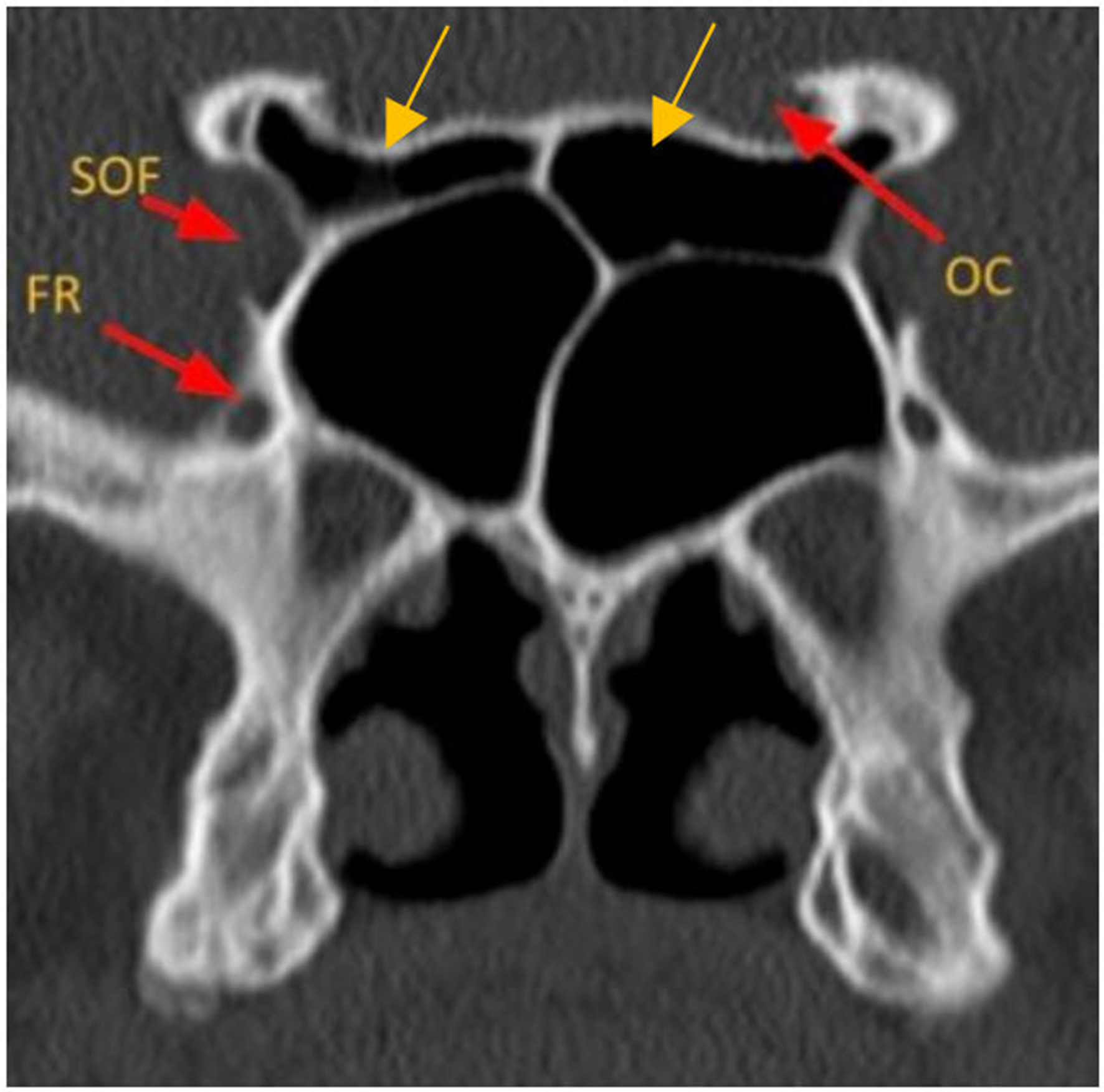
Figure 5. Coronal CT image of skull base foramina. SOF: Superior orbital fissure; FR: foramen rotundum; OC: optic canal. Onodi air cells (yellow arrows), anatomical variant. CT: Computed tomography.

Figure 5. Coronal CT image of skull base foramina. SOF: Superior orbital fissure; FR: foramen rotundum; OC: optic canal. Onodi air cells (yellow arrows), anatomical variant. CT: Computed tomography.


All published articles are preserved here permanently:
https://www.portico.org/publishers/oae/