fig4

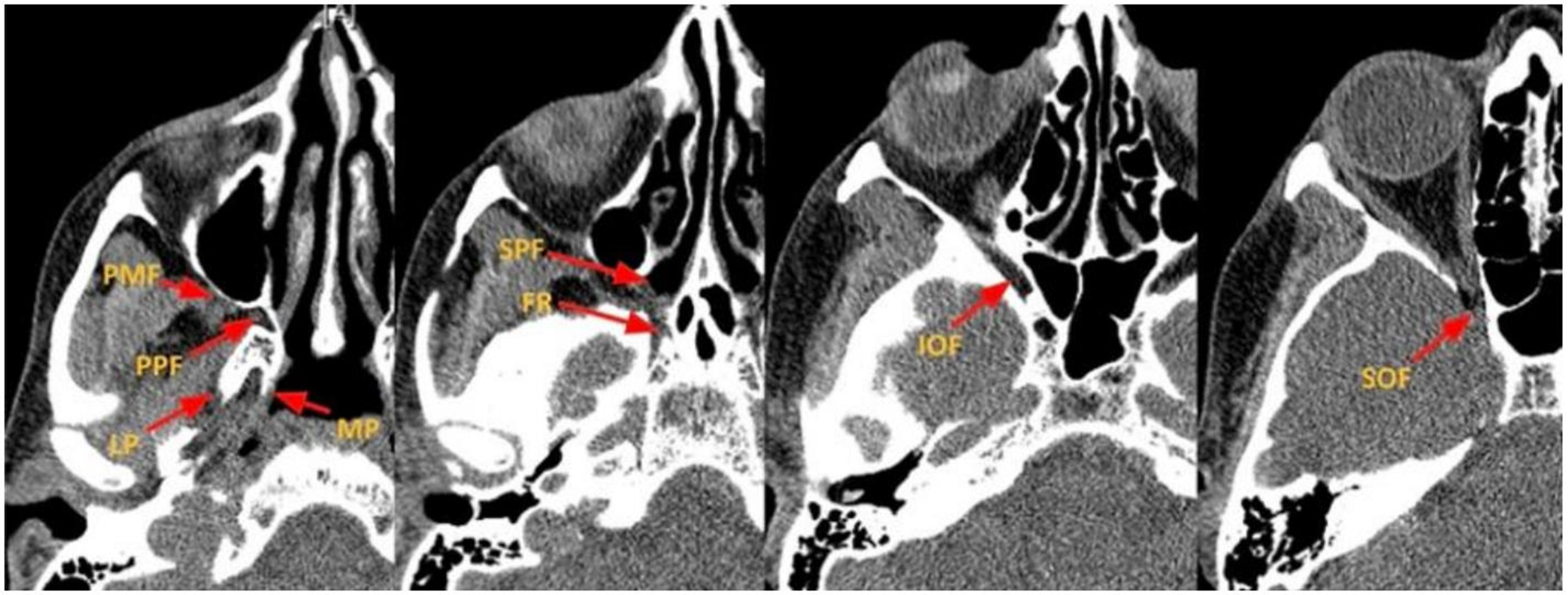
Figure 4. Axial CT images depicting the key components of the PPF and skull base foramina. PPF: Pterygopalatine fossa; PMF: pterygomaxillary fissure; LP: lateral plate of the pterygoid; MP: medial plate of the pterygoid; SPF: sphenopalatine foramen; FR: foramen Rotundum; IOF: inferior orbital fissure; SOF: superior orbital fissure; CT: computed tomography.


