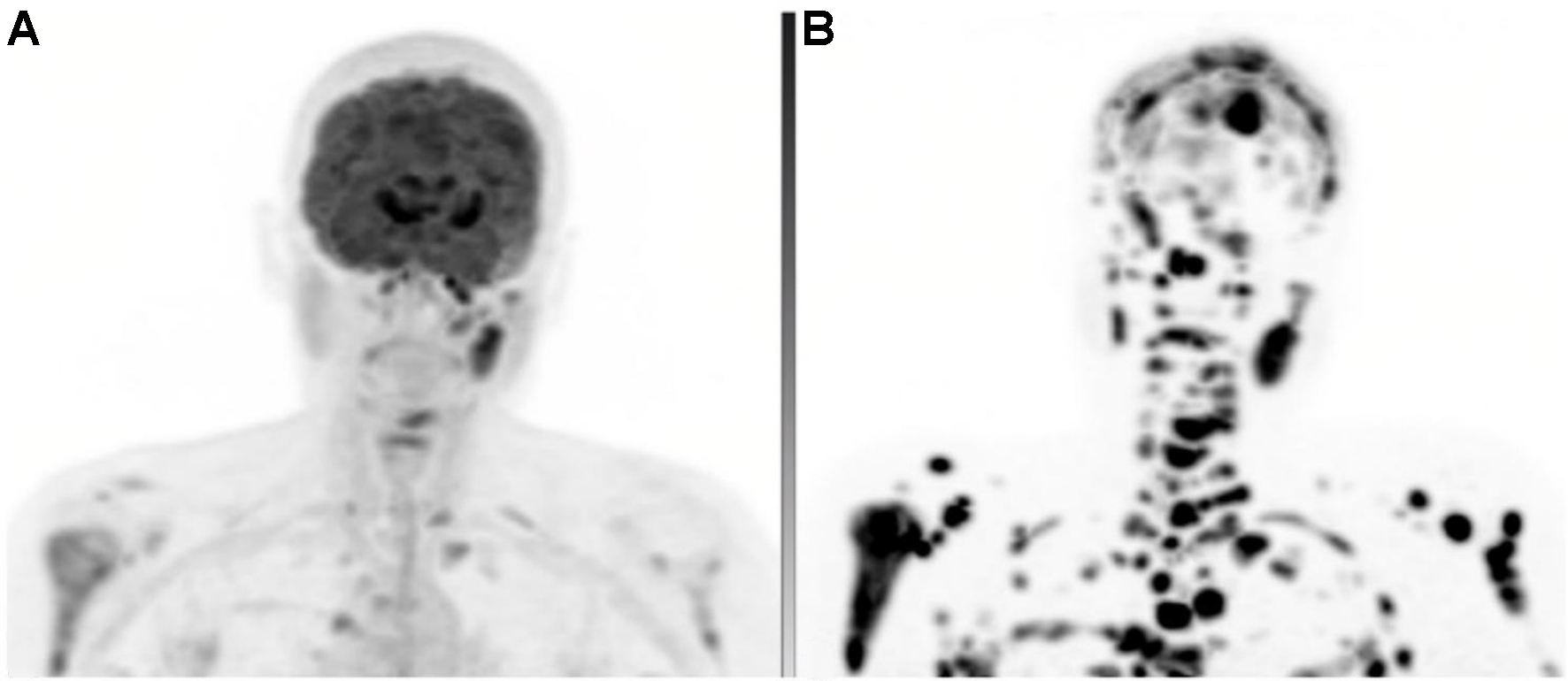
fig30

Figure 30. 56-year-old female with metastatic ENB. (A) FDG PET MIP demonstrating multiple metastatic lymph nodes and osseous lesions; (B) DOTATATE PET MIP obtained 1 month later revealing a much greater extent of metastatic disease, including numerous osseous and intracranial sites not appreciated on FDG PET. The underlying CT appearances were grossly similar, excluding the possibility that these differences were due to disease progression. ENB: Esthesioneuroblastoma; MIP: maximum intensity projection; CT: computed tomography.


