fig3

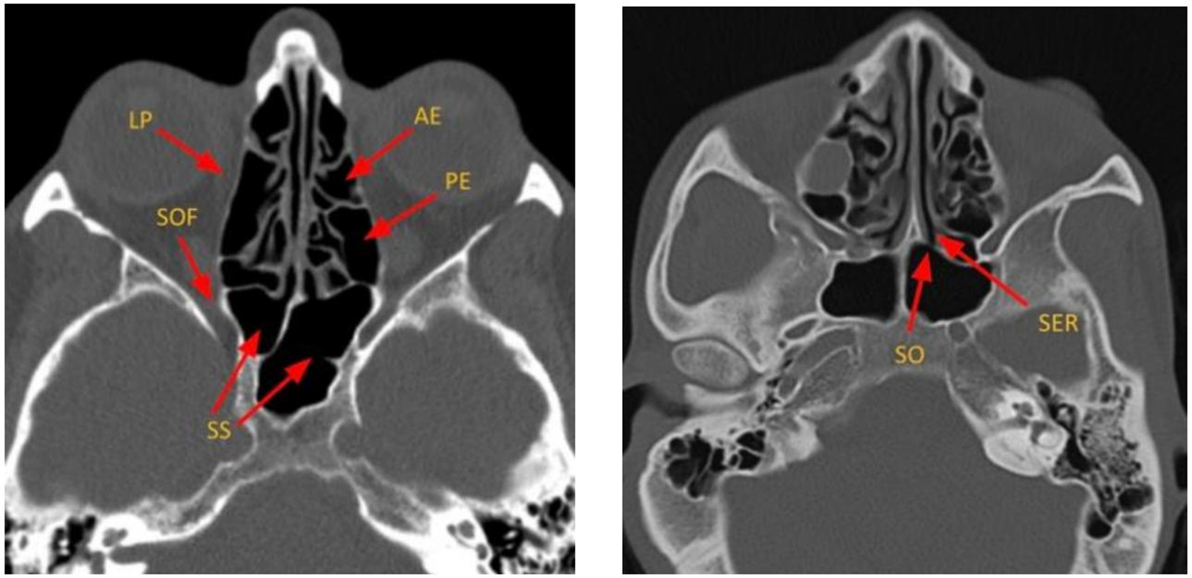
Figure 3. Axial CT images depicting the key components of ethmoid air cells and sphenoid sinuses. LP: Lamina papyracea; SOF: superior orbital fissure; SS: sphenoid sinus; SER: sphenoethmoidal recess; SO: sphenoid ostium; AE: anterior ethmoid air cells; PE: posterior ethmoid air cells; CT: computed tomography.


